Paget's Disease Candidate: Decoding the Radiograph in Hip Exam
Key Takeaway
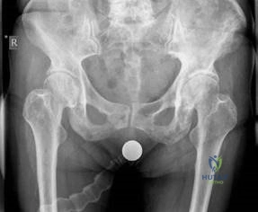
This article provides essential research regarding Paget's Disease Candidate: Decoding the Radiograph in Hip Exam. Paget's disease is a metabolic bone disorder of unknown cause, characterized by disorganized increases in osteoclastic bone resorption and compensatory osteoblastic new bone formation. This leads to accelerated, chaotic bone remodeling, making the bone biomechanically weak, prone to deformity and fracture. A radiograph showing coarsened trabecular patterns, thickened cortex, and increased density of affected areas suggests a Paget's disease candidate.
You are presented with an AP radiograph of the pelvis of a 68-year-old woman with chronic hip pain. Based on the imaging features, what is your diagnosis and how do you systematically describe this radiograph?
Candidate: This is an AP radiograph of the pelvis showing a coarsened trabecular pattern and thickened cortices in the left hemipelvis and proximal femur. There is increased bony density and a generalized enlargement of the bone contours. Both the iliopectineal (Brim sign) and ilioischiatic lines are thickened. The findings are characteristic of Paget’s disease. My differential would include sclerotic metastasis, fibrous dysplasia, and chronic renal osteodystrophy.
Failing to mention the "Brim sign," omitting specific cortical/trabecular changes, or jumping straight to "Paget's" without describing the radiological morphology. Candidates often miss the secondary signs like protrusio acetabuli or the distinction between lytic and blastic phases.
A structured "describe, diagnose, and differentiate" approach. Systematically identify: 1) Cortical thickening/trabecular coarsening, 2) Bone expansion/enlargement, 3) Specific signs (Brim sign/iliopectineal thickening), 4) The diagnosis (Paget’s), and 5) A concise differential (Prostatic mets, fibrous dysplasia, renal osteodystrophy, Paget's sarcoma).
The patient requires a Total Hip Arthroplasty (THA). What are the specific technical surgical challenges and planning considerations unique to the Pagetoid hip?
Candidate: Challenges include increased vascularity leading to higher blood loss, necessitating availability of blood products and potential use of cell salvage. Mechanically, the bone is sclerotic and brittle, making reaming and broaching difficult; I might need burrs. Anatomical deformities like varus proximal femur and acetabular protrusio require careful component positioning, potentially using bone graft or modular stems.
Ignoring the "systemic" aspect of disease activity. Failing to mention pre-operative medical optimization (bisphosphonates) to reduce vascularity and bone turnover is a critical oversight in the FRCS exam.
Categorize into: 1) Medical (Pre-op bisphosphonates to reduce AlkPhos/vascularity), 2) Hematological (Increased blood loss risk), 3) Anatomical (Protrusio, varus deformity), and 4) Technical (Hard sclerotic bone requiring burrs, risk of intra-op fracture, cementation vs. cementless debate). Mention the need to rule out sarcomatous change if pain is unrelenting.
There is controversy regarding the choice of fixation (cemented vs. uncemented) in Paget's disease. How do you defend your choice?
Candidate: Traditionally, cemented fixation was preferred due to concerns regarding ingrowth. However, modern evidence, such as studies by Parvizi and Lusty, suggests that cementless components achieve stable ingrowth in Pagetoid bone, provided the healing potential is normal. I would consider cementless for younger patients or where sclerotic bone makes cement interdigitation poor, using adjuvant acetabular screws.
Taking an dogmatic stance ("I would always cement"). The examiner wants to see that you are aware of the modern shift towards cementless implants and the literature supporting them.
Demonstrate knowledge of the literature. Explain that while cemented fixation was standard, studies demonstrate successful biological fixation of porous implants. Acknowledge the trade-offs: Cementless is preferred in extremely sclerotic bone where cement interdigitation is inadequate or when concurrent osteotomy is planned to correct deformity.
