Charcot Neuroarthropathy: Diagnosis, Surgical Management & Rocker Bottom Deformity
Key Takeaway
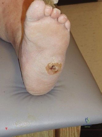
Surgical intervention for Charcot neuroarthropathy is indicated for unstable deformities, severe rocker bottom leading to chronic ulceration, recurrent infection (osteomyelitis), or a non-plantigrade foot preventing bracing. The goal is to achieve a stable, braceable foot. Contraindications include uncontrolled infection, severe PAD, poor medical condition, and non-compliance.
You are presented with a 62-year-old patient with type 2 diabetes who presents with a red, swollen, and warm foot. How would you systematically approach the clinical assessment and initial differential diagnosis to ensure you do not mismanage this patient?
Candidate: I would perform a thorough history and physical exam, focusing on signs of infection vs. Charcot. I would use the elevation test, check for ulcers, and order imaging. My primary differential is acute Charcot versus osteomyelitis, but I must also consider cellulitis, gout, and septic arthritis.
Candidates often jump straight to suggesting an MRI. A poor answer fails to emphasize the clinical distinction (the "Elevation Test") and ignores the patient's systemic status, such as glycemic control, vascular inflow (ABI/Toe pressures), and the presence of sensory neuropathy which is the fundamental driver of the neurotraumatic/neurovascular theories.
A high-scoring answer systematically covers: 1. Clinical differentiation: Mentioning the "Elevation Test" (erythema resolves in Charcot, persists in infection). 2. Systemic assessment: Vascular status (ABI/TBI/TcPO2) to ensure healing potential and neurological assessment. 3. Differential diagnosis: Charcot (Eichenholtz 0/I) vs. Osteomyelitis vs. Cellulitis vs. Gout. 4. Imaging strategy: Plain radiographs for baseline, followed by MRI (Gold Standard) to differentiate Charcot joint-based marrow edema from contiguous osteomyelitis.
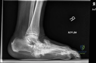
Look at this radiograph. What are the key findings, and how does this impact your decision-making regarding the Brodsky and Eichenholtz classifications?
Candidate: The image shows midfoot collapse with fragmentation and subluxation at the TMT joints. This fits the Brodsky Type 1 classification. Given the active destruction, it appears to be Eichenholtz Stage I.
Failing to mention the pathomechanical implications—specifically that this represents a 'rocker bottom' deformity—and failing to discuss the surgical implications of the Stage (i.e., operating in Stage I is generally contra-indicated unless there is catastrophic instability).
Structure the response: 1. Radiographic Identification: Describe the TMT dislocation, fragmentation, and loss of the medial longitudinal arch. 2. Classification: Identify as Brodsky Type 1 (Midfoot). 3. Staging: Identify as Eichenholtz Stage I (Developmental/Fragmentation). 4. Management Implications: Note that Stage I requires non-operative management (Total Contact Casting) because the bone is highly osteopenic/hyperemic. Surgery here risks hardware failure and massive tissue morbidity unless the deformity is limb-threateningly unstable.
You have decided to reconstruct a chronic, stable Eichenholtz Stage III rocker-bottom foot. What are the principles of "Superconstruct" fixation you would employ to ensure success?
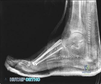
Candidate: I would use "Superconstructs" as defined by Sammarco. This involves extending the fusion beyond the area of damage, doing adequate bone resection to prevent tension, using very strong hardware, and placing it to maximize biomechanical advantage, often using intramedullary beaming or locking plates.
Forgetting to mention the necessity of addressing the gastrocnemius-soleus equinus contracture. A "superconstruct" will fail if the underlying deforming force (equinus) remains, as the midfoot will simply continue to be crushed by the dorsiflexion-moment during the stance phase.
1. Address the Equinus: MANDATORY TAL or gastrocnemius recession. 2. The 4 Principles of Superconstructs: (a) Extend fusion to healthy bone; (b) Resect bone to avoid closure tension; (c) Use the strongest available implants (beams/plates); (d) Optimize hardware position. 3. Technique: Describe intramedullary 'beaming' (screws from the metatarsal base into the talus/calcaneus) to act as an internal splint. 4. Soft tissue: Mention respect for the soft tissue envelope/skin bridges.
