Sacral Insufficiency Fractures: Comprehensive Diagnosis, Management & Biomechanics
Key Takeaway
Sacral insufficiency fractures (SIFs) are fragility fractures in osteoporotic bone, common in elderly patients. They often cause insidious back or buttock pain. Standard X-rays frequently appear normal, making diagnosis challenging. A high index of suspicion and advanced imaging such as CT or MRI are crucial for confirmation, preventing delayed treatment and morbidity.
An 82-year-old female presents to your clinic with a 3-month history of worsening low back and buttock pain. She has a history of rheumatoid arthritis and long-term steroid use. Plain radiographs were reported as "unremarkable." How would you approach the diagnosis, and what is your primary clinical suspicion?
Candidate: Given her age, chronic steroid use, and inflammatory arthropathy, my primary suspicion is a sacral insufficiency fracture. I would conduct a thorough physical examination, including a neurological screen. Since radiographs are often insensitive, I would order an MRI of the pelvis to confirm the diagnosis, as it is the gold standard for identifying bone marrow edema.
Failing to mention the specific risk factors provided (steroids, RA) or assuming a normal X-ray means "nothing is wrong." Candidates often jump to requesting a CT scan first, missing the superior sensitivity of MRI for marrow edema in early-stage insufficiency fractures.
Start with a high index of clinical suspicion due to the "fragility triad": age, comorbidities (RA/steroids), and activity-related pain. State clearly that plain radiographs have low sensitivity. Propose MRI (STIR/T2) as the gold standard to identify bone marrow edema. Mention that a CT scan is reserved for surgical planning to assess fracture geometry and cortical breach once the diagnosis is confirmed.
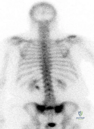
Following up on your diagnosis, the MRI confirms a fracture. The patient undergoes a CT scan which reveals this pattern.
How do you classify this fracture, and what are the specific clinical implications of this pattern?
Candidate: This is a classic "H-pattern" sacral insufficiency fracture. It involves bilateral vertical alar fractures connected by a transverse line across the sacral body. This pattern is inherently unstable, often failing conservative management, and carries a higher risk of non-union and neurological compromise compared to isolated unilateral alar fractures.
Describing the fracture only as a "sacral fracture" without acknowledging the H-pattern. Failing to recognize that the H-pattern is effectively a "spinopelvic dissociation" equivalent in osteoporotic bone, which requires a more aggressive surgical approach than a simple unilateral zone I fracture.
Identify it as an H-pattern fracture. Explicitly label it as biomechanically unstable. Explain that it indicates failure of the posterior tension band. Mention the Denis classification (Zone I-III) but highlight that the H-pattern transcends these by linking both sides and the sacral canal, thus significantly increasing the risk of cauda equina impingement and requiring stabilization (e.g., S2AI screws or lumbopelvic fixation).
The patient remains in intractable pain despite 8 weeks of bed rest and analgesia. You decide to proceed with surgery. Describe your surgical plan and the radiographic landmarks for optimal screw placement.
Candidate: Given the instability, I would opt for percutaneous stabilization. I would use S2-Alar-Iliac (S2AI) screws or iliosacral screws with cement augmentation, given her poor bone quality. For the iliosacral trajectory, I use inlet and outlet views: the inlet view ensures the screw stays within the sacral ala and body without violating the anterior cortex or canal, while the outlet view ensures the screw is cephalad to the neural foramina.
Ignoring the "poor bone quality" aspect. Failing to mention neuromonitoring or the importance of cement augmentation in osteoporotic bone. Forgetting the specific fluoroscopic views (Inlet/Outlet) required to ensure the screw doesn't impinge on the S1 nerve root.
Start with patient positioning (prone on a radiolucent table). Emphasize the multidisciplinary approach. Detail the use of S2AI screws for better purchase in the ilium. Explain the use of intraoperative neuromonitoring (EMG/SSEP). Stress the "three-view" check: AP for overall alignment, Lateral for the sacral body/canal protection, and Inlet/Outlet for the narrow corridors within the sacral alae. Mention cement augmentation (sacroplasty) as a critical step to achieve "purchase" in brittle, osteoporotic bone.
