Calcaneal Fractures: Epidemiology, Surgical Anatomy, Biomechanics, and Advanced Diagnostics
Key Takeaway
Calcaneal fractures are challenging high-energy tarsal injuries, critical for hindfoot mechanics. Diagnosis necessitates clinical evaluation, plain radiographs assessing Böhler's and Gissane's angles, and CT scans. Mastery of surgical anatomy, fracture biomechanics, and soft tissue status is paramount for optimal treatment and minimizing long-term disability, including post-traumatic arthritis.
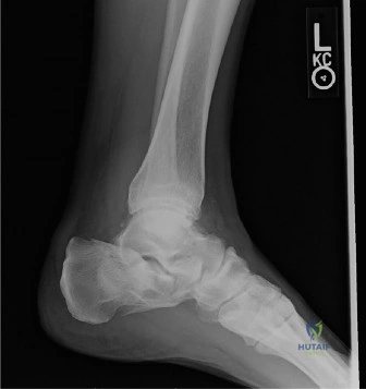
A 35-year-old male presents to the ED following a fall from a 3-meter ladder. He is complaining of severe right heel pain. On inspection, there is significant swelling and ecchymosis extending to the sole of the foot (Mondor’s sign). How would you approach the clinical assessment and initial management of this patient?
Candidate: I would perform a primary and secondary trauma survey. Specifically for the foot, I would assess the neurovascular status, check for skin compromise, and look for signs of compartment syndrome. I’d order plain radiographs (AP, lateral, and axial) and a CT scan of the calcaneus. I'd manage with elevation, ice, and immobilization until the swelling subsides for potential surgery.
Candidates often focus solely on the foot. The examiner is testing "Trauma Safety." Failing to mention the 10-15% association with thoracolumbar spine fractures—a "must-rule-out"—is a critical omission that suggests a lack of systemic trauma awareness.
Adopt an ATLS approach: 1. Rule out associated injuries: Specifically the thoracolumbar spine (the "Don Juan Syndrome"), contralateral calcaneus, and pelvic/pilon fractures. 2. Soft tissue assessment: Document the "wrinkle sign" and ecchymosis; explain that surgery is contraindicated until the soft tissue envelope is stable. 3. Neurovascular: Palpate pulses and assess for tarsal tunnel compression. 4. Imaging: Standard radiographs followed by a CT scan as the gold standard for surgical planning.
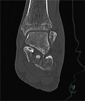
You are reviewing the CT imaging for this patient. How do you utilize the Sanders classification to guide your surgical strategy and prognosis?
Candidate: The Sanders classification is based on the number of articular fracture lines visible on the coronal CT through the posterior facet. Type I is non-displaced. Type II is two-part, III is three-part, and IV is highly comminuted. It helps me decide if surgery is needed and tells me that a higher Sanders grade usually has a worse prognosis for post-traumatic arthritis.
Failing to mention the sustentaculum tali fragment. A high-scoring candidate recognizes that this fragment is often undisplaced and serves as the "anchor" for reduction.
Structure the answer: 1. Technical: Sanders classifies based on the coronal CT image at the widest point of the posterior facet. 2. Anatomy: Mention the sustentaculum tali (medial) fragment is usually fixed to the talus, acting as the key reference point for reduction. 3. Clinical Utility: Higher Sanders grades correlate with increased risk of post-traumatic subtalar arthritis, but the decision to operate depends more on the degree of displacement, Böhler’s angle restoration, and the patient's functional demands rather than the Sanders grade alone.
During your surgery using the Extensile Lateral Approach, what are the primary "landmarks" and risks you must respect to ensure a successful outcome and minimize complications?
Candidate: I would use the L-shaped incision. I need to protect the sural nerve and the peroneal tendons. I’ll make a full-thickness flap to avoid necrosis. I must aim to restore Böhler's and Gissane's angles and reduce the posterior facet anatomically.
Candidates often forget to mention the timing of the surgery in relation to the "wrinkle test." They also fail to emphasize that the lateral wall often needs to be "out-fractured" or removed to visualize the joint.
Organize by "Soft Tissue" vs "Reduction": 1. Soft Tissue Safety: Wait for the wrinkle test (7-14 days). Use a true full-thickness flap (skin, subcutaneous fat, and periosteum) to keep the blood supply intact. Protect the sural nerve. 2. Reduction Strategy: Temporarily remove the lateral wall to visualize the posterior facet. Use the sustentaculum tali as your fixed medial starting point. 3. Restoration: Aim for Böhler's angle (20–40°) and Gissane's angle (120–145°) to prevent impingement and ensure a correct foot geometry.
