Extraarticular Calcaneal Fractures: Comprehensive Surgical Management and Techniques
Key Takeaway
Extraarticular calcaneal fractures, encompassing the tuberosity, sustentaculum tali, and anterior process, require precise diagnostic and therapeutic strategies. While less common than intraarticular variants, they present unique biomechanical challenges, such as Achilles tendon avulsion forces and soft tissue compromise. This guide details evidence-based surgical indications, operative approaches, and fixation techniques—including tension band constructs and medial approaches—to optimize functional outcomes and prevent complications like tarsal tunnel syndrome.
Comprehensive Introduction and Patho-Epidemiology
Calcaneal fractures represent the most frequently encountered tarsal bone fractures, accounting for approximately 60% of all major tarsal injuries and 2% of all adult fractures. Traditionally, orthopedic literature has dichotomized these injuries into intraarticular variants—which involve the posterior facet of the subtalar joint and demand meticulous articular reconstruction—and extraarticular variants. Extraarticular calcaneal fractures, which constitute approximately 25% to 30% of all calcaneal fractures, are defined by their complete sparing of the posterior subtalar articulation. However, this nomenclature can be deceptively reassuring; these injuries frequently involve other critical articulations, such as the calcaneocuboid joint or the middle facet of the subtalar joint, and they present formidable biomechanical and soft-tissue challenges that demand a high level of surgical acumen.
The patho-epidemiology of extraarticular calcaneal fractures demonstrates a distinct bimodal distribution, reflecting two divergent mechanisms of injury and patient demographics. The first peak occurs in younger, active patients, typically males in their third to fifth decades of life. In this cohort, the mechanism of injury generally involves high-energy trauma, such as motor vehicle collisions, falls from a significant height, or severe twisting injuries during athletic participation. These high-energy vectors often result in avulsion fractures of the anterior process due to extreme tension on the bifurcate ligament, or severe axial loads combined with inversion that shear the sustentaculum tali. The sheer force of these injuries frequently results in concomitant osseous and ligamentous trauma to the ipsilateral lower extremity, necessitating a comprehensive secondary survey of the foot and ankle.
Conversely, the second epidemiological peak is observed in the elderly population, particularly among postmenopausal women and individuals with compromised bone mineral density. In these osteoporotic patients, extraarticular calcaneal fractures are predominantly insufficiency or low-energy avulsion injuries. The classic presentation is a calcaneal tuberosity avulsion fracture resulting from a seemingly innocuous misstep or a sudden, forceful contraction of the triceps surae complex. Furthermore, patients with peripheral neuropathy—most notably due to long-standing diabetes mellitus—are at a significantly elevated risk for these fractures. Neuropathic arthropathy (Charcot foot) can lead to spontaneous avulsion fractures of the tuberosity, driven by the massive, unopposed tensile forces of the Achilles tendon acting upon structurally compromised, osteopenic bone.
Despite their classification as "extraarticular," these fractures encompass a highly heterogeneous group of pathoanatomic patterns, primarily localized to three distinct anatomic regions: the calcaneal tuberosity, the sustentaculum tali, and the anterior process of the calcaneus. Each of these specific subtypes presents a unique set of biomechanical disruptions and risks of severe, limb-threatening complications. From the acute surgical emergency of posterior skin necrosis in displaced tuberosity beak fractures to the insidious development of chronic tarsal tunnel syndrome in missed sustentacular fractures, the treating orthopedic surgeon must maintain a high index of suspicion. Mastery of the underlying patho-epidemiology is the foundational step in preventing the profound, long-term morbidity historically associated with the mismanagement of these complex injuries.
Detailed Surgical Anatomy and Biomechanics
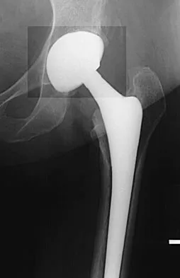
A profound understanding of the complex osteology, ligamentous attachments, and vascular supply of the calcaneus is an absolute prerequisite for the successful surgical management of extraarticular fractures. The calcaneus is the largest of the tarsal bones, serving as the primary osseous foundation of the posterior column of the foot. It functions as a critical lever arm for the gastrocnemius-soleus complex and acts as the primary weight-bearing structure during the heel-strike phase of the gait cycle. The internal architecture of the calcaneus is highly specialized, consisting of a thin cortical shell enveloping a trabecular network that is precisely oriented to withstand both immense compressive loads from the talus and extreme tensile forces from the Achilles tendon and plantar fascia. The intersection of these trabecular patterns creates a central area of relative radiolucency known as the "neutral triangle" or Ward's triangle of the heel, which is structurally weaker and frequently the site of fracture propagation.
The calcaneal tuberosity forms the posterior extremity of the bone and serves as the insertion site for the Achilles tendon. The insertion is not uniform; the tendon inserts predominantly into the middle third of the posterior surface, while the superior third is separated from the tendon by the retrocalcaneal bursa, and the inferior third extends into the plantar fascia origin. Biomechanically, the tuberosity acts as a massive lever arm. When a fracture occurs here, the unopposed pull of the triceps surae complex (capable of generating forces exceeding several times body weight) rapidly displaces the avulsed fragment superiorly. This displacement obliterates the retrocalcaneal space and exerts devastating pressure on the posterior heel skin. The vascular supply to this posterior skin is tenuous, relying on a watershed area between the calcaneal branches of the peroneal artery laterally and the posterior tibial artery medially. Excessive tension from a displaced "beak" fracture rapidly induces ischemia, leading to full-thickness skin necrosis if not emergently decompressed.
The sustentaculum tali is a dense, horizontal, shelf-like medial projection of the calcaneus that provides critical support to the middle facet of the subtalar joint. In the context of complex hindfoot trauma, it is universally referred to as the "constant fragment" because it rarely displaces from the talus, remaining securely tethered by the robust talocalcaneal interosseous ligaments and the superficial deltoid ligament complex. Directly inferior to the sustentaculum tali runs the flexor hallucis longus (FHL) tendon within a dedicated fibro-osseous groove. Immediately medial to this structure lies the critical neurovascular bundle containing the posterior tibial artery and the tibial nerve, all contained within the tight confines of the flexor retinaculum (tarsal tunnel). Biomechanically, the sustentaculum acts as a medial buttress; loss of its integrity through fracture leads to medial column collapse, valgus hindfoot deformity, and direct mechanical impingement on the FHL and tibial nerve, culminating in acute or chronic tarsal tunnel syndrome.
The anterior process of the calcaneus is a saddle-shaped prominence that projects distally to articulate with the cuboid, forming the lateral column of the transverse tarsal (Chopart) joint. It serves as the vital origin for the bifurcate ligament, a Y-shaped structure comprising the calcaneocuboid and calcaneonavicular bands, which is essential for midfoot stability during inversion and eversion. Additionally, the lateral aspect of the anterior process provides the origin for the extensor digitorum brevis muscle. Biomechanically, fractures of the anterior process typically result from extreme plantarflexion and inversion, which place catastrophic tensile failure loads on the bifurcate ligament, resulting in an avulsion fracture. Alternatively, forced abduction can cause a compression or "nutcracker" fracture against the cuboid. Disruption of the anterior process compromises lateral column length and stability, leading to profound midfoot dysfunction and early-onset calcaneocuboid arthrosis if anatomic reduction is not achieved.
Exhaustive Indications and Contraindications
The decision-making process for operative versus non-operative management of extraarticular calcaneal fractures is highly nuanced, requiring the surgeon to synthesize fracture morphology, degree of displacement, soft-tissue integrity, and host factors. While historical perspectives often favored conservative management for extraarticular patterns, contemporary orthopedic principles strongly advocate for surgical intervention when specific biomechanical parameters are violated. The primary goals of operative management are the restoration of the Achilles lever arm, the decompression of neurovascular structures, the re-establishment of lateral column length, and the preservation of adjacent joint congruity.
Absolute indications for surgical intervention are primarily dictated by soft-tissue emergencies and catastrophic biomechanical failure. The most critical absolute indication is the presence of a displaced calcaneal tuberosity "beak" fracture (Type II) that causes skin tenting, blanching, or impending necrosis of the posterior heel. This scenario represents a true orthopedic emergency requiring immediate reduction, either in the emergency department or the operating theater, to prevent the conversion of a closed fracture into a disastrous open injury requiring free tissue transfer. Other absolute indications include open fractures, fractures associated with acute compartment syndrome of the foot, and sustentaculum tali fractures presenting with acute, progressive tarsal tunnel syndrome due to direct mechanical compression of the tibial nerve by the displaced osseous fragment.
Relative indications for operative management are broader and depend heavily on the functional demands of the patient. For tuberosity fractures, displacement greater than 2 to 3 mm, loss of Achilles tendon continuity or resting tension, and a significant decrease in the calcaneal pitch angle warrant surgical fixation. For sustentaculum tali fractures, any displacement greater than 2 mm or any incongruity of the middle facet of the subtalar joint is an indication for open reduction and internal fixation (ORIF) to prevent late-onset arthrosis and chronic FHL impingement. Regarding anterior process fractures, surgical indications include large Type 2 avulsion fractures with significant displacement, and Type 3 fractures involving more than 25% of the calcaneocuboid articular surface or demonstrating articular step-off greater than 2 mm.
Contraindications to surgical intervention must be rigorously evaluated, as the complications of calcaneal surgery in a compromised host can be devastating. Absolute contraindications include patients who are non-ambulatory prior to the injury, patients who are medically unstable for anesthesia, and the presence of active, untreated local infection or severe peripheral arterial disease (e.g., Ankle-Brachial Index < 0.5 or absent pedal pulses without reconstructable options). Relative contraindications require careful shared decision-making and include poorly controlled diabetes mellitus (HbA1c > 8.0%), active heavy tobacco use (which exponentially increases the risk of wound necrosis and nonunion), severe baseline peripheral neuropathy, and profound patient non-compliance, as strict adherence to prolonged non-weight-bearing protocols is mandatory for successful outcomes.
| Factor Category | Indications for Operative Management (ORIF) | Contraindications to Operative Management |
|---|---|---|
| Tuberosity Fractures | Skin tenting/blanching (EMERGENT), displacement > 2mm, loss of Achilles tension, open fracture. | Severe peripheral vascular disease (ABI < 0.5), non-ambulatory baseline status. |
| Sustentaculum Tali | Displacement > 2mm, middle facet articular incongruity, acute tarsal tunnel syndrome, FHL impingement. | Active local soft-tissue infection, severe medical comorbidities precluding anesthesia. |
| Anterior Process | Articular step-off > 2mm at calcaneocuboid joint, large displaced avulsion fragment, symptomatic nonunion (late). | Small, non-displaced avulsion fragments (Type 1), severe osteopenia precluding hardware purchase (relative). |
| Host Factors | High functional demands, healthy soft-tissue envelope (positive wrinkle test), compliant patient. | Active heavy smoking (relative), uncontrolled diabetes (HbA1c > 8.0%), severe neuropathy (Charcot). |
Pre-Operative Planning, Templating, and Patient Positioning
Meticulous pre-operative planning is the cornerstone of successful surgical execution in extraarticular calcaneal fractures. The initial evaluation mandates a comprehensive radiographic series of the foot and ankle, including weight-bearing (if tolerated and not contraindicated by displacement) anteroposterior (AP), lateral, and oblique views. Standard AP and lateral views are often insufficient for fully characterizing these complex injuries due to extensive bony overlap. For tuberosity fractures, a Harris axial view is critical for assessing the degree of widening, varus/valgus malalignment of the tuberosity, and extension of the fracture into the subtalar joint. For anterior process fractures, a specialized oblique view of the foot is mandatory, as the anterior process is frequently obscured by the talar head and navicular on standard projections.
In contemporary orthopedic practice, a fine-cut computed tomography (CT) scan (1.0 to 2.0 mm slice thickness) with multiplanar and three-dimensional (3D) reconstructions is considered the gold standard and is virtually mandatory for all but the most trivial extraarticular calcaneal fractures. CT imaging allows for the precise delineation of fracture morphology, the identification of occult intraarticular extension, and the assessment of bone stock quality. Pre-operative templating utilizing the CT scan is essential for determining the optimal trajectory of fixation hardware. For instance, templating the sustentaculum tali requires measuring the exact distance from the lateral calcaneal wall to the medial cortex of the sustentaculum to select appropriate lag screw lengths, ensuring that screws do not penetrate the medial cortex and injure the neurovascular bundle.
Patient positioning is highly dependent on the specific fracture pattern and the chosen surgical approach. For calcaneal tuberosity avulsion fractures, the patient is almost universally placed in the prone position. This orientation provides unobstructed, direct access to the posterior heel, the Achilles tendon, and the posterior aspect of the triceps surae complex. The operating table should be slightly flexed at the knees, or pillows placed under the shins, to allow the ankle to rest in passive plantarflexion, thereby releasing tension on the Achilles tendon. A thigh tourniquet is applied, and the entire lower extremity is prepped and draped free to permit intraoperative manipulation of the foot and ankle. Fluoroscopy must be positioned to allow seamless acquisition of lateral and axial views without compromising the sterile field.
For isolated fractures of the sustentaculum tali or anterior process, the patient is typically placed in the supine position. For medial approaches to the sustentaculum, a large bump is placed beneath the contralateral hip to allow the operative leg to externally rotate, providing excellent visualization of the medial hindfoot. Conversely, for lateral approaches to the anterior process, a bump is placed under the ipsilateral hip to internally rotate the leg, bringing the lateral column into profile. In all cases, the soft-tissue envelope must be rigorously assessed prior to making an incision. The presence of massive edema, fracture blisters, or the absence of the "wrinkle test" dictates that surgery must be delayed until the soft tissues have adequately recovered, typically 10 to 14 days post-injury, unless an emergent indication (e.g., skin blanching in a beak fracture) forces immediate intervention.
Step-by-Step Surgical Approach and Fixation Technique
Tuberosity Fracture Fixation Techniques
The surgical management of calcaneal tuberosity fractures requires a robust construct capable of neutralizing the massive tensile forces of the Achilles tendon. With the patient prone, a longitudinal incision is utilized, placed slightly medial or lateral to the midline of the Achilles tendon to prevent direct shoe-wear irritation over the resultant scar. The paratenon is meticulously incised and preserved for later repair to prevent tendon adhesions. The fracture hematoma is evacuated, and the fracture margins are debrided of any interposed soft tissue or organizing clot.
Reduction is achieved by maximizing ankle plantarflexion to relax the gastrocnemius-soleus complex. A 5.0-mm Schanz pin is frequently inserted percutaneously into the avulsed proximal fragment to serve as a joystick, allowing the surgeon to pull the fragment distally and correct any rotational malalignment. Pointed reduction forceps are then applied to compress the fracture, and provisional fixation is achieved with multiple 1.6-mm or 2.0-mm Kirschner wires (K-wires). Definitive fixation in healthy bone is typically achieved using two or three large-fragment (4.5-mm or 6.5-mm) partially threaded cannulated cancellous screws. The trajectory is critical: screws must be directed from posterosuperior to anteroinferior, aiming for the dense trabecular bone of the plantar calcaneal body to maximize pull-out strength.
In osteoporotic bone, screw fixation alone is notoriously prone to catastrophic failure. In these instances, augmentation is mandatory. A highly effective technique is the lateral tension band construct. Heavy braided non-absorbable suture (e.g., #2 or #5 FiberWire) or heavy stainless steel wire is passed through the distal substance of the Achilles tendon, just proximal to its insertion. A transverse drill hole is created in the intact plantar-anterior calcaneus. The suture or wire is then routed in a figure-of-eight fashion through this tunnel and tied securely over the posterior aspect of the tuberosity. This biomechanically superior construct converts the tensile forces of the Achilles tendon into dynamic compressive forces at the fracture site. Furthermore, if profound gastrocnemius tightness precludes anatomic reduction without excessive tension, an adjunctive gastrocnemius recession (Strayer procedure) is highly recommended to protect the osteosynthesis.
Sustentaculum Tali Surgical Approach
Fixation of the sustentaculum tali is technically demanding due to the critical medial neurovascular anatomy. With the patient supine and externally rotated, a medial approach is utilized. The incision begins approximately 2 cm inferior to the tip of the medial malleolus and extends longitudinally from the navicular tuberosity posteriorly toward the heel pad. The superficial fascia is divided, and the flexor retinaculum is carefully incised.
The immediate priority is the identification and protection of the posterior tibial artery and the tibial nerve, which are mobilized together and retracted gently with vessel loops. Deep dissection proceeds by identifying the interval between the flexor digitorum longus (FDL) dorsally and the flexor hallucis longus (FHL) plantarward. The fracture is typically visualized directly within this interval. The fracture site is debrided using a dental pick, and the sustentacular fragment is reduced anatomically using a small elevator or a periosteal elevator.
Provisional fixation is achieved with a K-wire directed from medial to lateral. Definitive fixation relies on lag screw principles. Two 3.5-mm or 4.0-mm partially threaded lag screws are inserted. The trajectory is paramount: screws must be directed from medial to lateral, and slightly superiorly, aiming into the dense cortical bone of the lateral calcaneal wall. The surgeon must utilize intraoperative fluoroscopy (specifically Broden's views and Harris axial views) to confirm that the screws do not violate the posterior facet of the subtalar joint and that the screw heads are adequately countersunk to prevent postoperative irritation of the FHL tendon.
Anterior Process Management Strategies
Surgical management of the anterior process depends on the size of the fragment and the chronicity of the injury. For acute, large displaced avulsion fractures (Type 2) or those involving the calcaneocuboid joint (Type 3), a modified Ollier incision or a direct lateral approach over the calcaneocuboid joint is employed. The sural nerve, which courses laterally, must be meticulously identified and protected.
The origin of the extensor digitorum brevis muscle is partially reflected distally to expose the anterior process and the calcaneocuboid articulation. The joint is inspected for osteochondral debris, and the fracture is anatomically reduced. Fixation is typically achieved using 2.0-mm or 2.7-mm mini-fragment cortical lag screws. If the fragment is too small or comminuted to accept a screw, threaded K-wires or a small hook plate may be utilized.
In cases of missed anterior process fractures presenting as symptomatic nonunions months after the initial injury, the surgical strategy shifts. If the calcaneocuboid joint remains free of arthritic changes, surgical excision of the ununited osseous fragment and direct repair or reconstruction of the bifurcate ligament yields excellent functional results. However, if delayed diagnosis has resulted in post-traumatic calcaneocuboid arthrosis, simple excision will fail to relieve pain. In these salvage scenarios, a localized calcaneocuboid arthrodesis is required, utilizing rigid plate fixation and autologous bone grafting to restore lateral column stability.
Complications, Incidence Rates, and Salvage Management
The surgical management of extraarticular calcaneal fractures is fraught with a high complication profile, largely attributable to the tenuous soft-tissue envelope of the foot and the immense biomechanical forces acting upon the hindfoot. The most devastating acute complication is posterior heel skin necrosis, which occurs with alarming frequency in delayed presentations of Type II tuberosity beak fractures. The skin in this region lacks a robust subcutaneous fat pad and relies on a fragile vascular watershed. If necrosis occurs, it rapidly converts a closed osteosynthesis into an open, infected wound. Management of this catastrophic complication requires aggressive serial debridement, removal of infected hardware, and complex soft-tissue reconstruction, typically necessitating a reverse sural artery fasciocutaneous flap or a free tissue transfer (e.g., anterolateral thigh flap) in conjunction with plastic surgery colleagues.
Nerve injuries are another significant source of postoperative morbidity. In medial approaches for sustentaculum tali fractures, iatrogenic injury to the tibial nerve or its calcaneal branches can result in profound medial heel numbness or painful neuromas. Conversely, non-operative management of displaced sustentacular fractures frequently leads to malunion. The medially displaced osseous mass mechanically encroaches upon the tarsal tunnel, resulting in chronic, intractable tarsal tunnel syndrome. Salvage of this condition requires a technically demanding late excision of the malunited fragment, extensive tarsal tunnel release, and neurolysis of the tibial nerve, though functional outcomes are consistently inferior to acute anatomic fixation. In lateral approaches for anterior process fractures, the sural nerve is at high risk of iatrogenic transection or traction neuropraxia.
Hardware failure and loss of reduction are particularly common in osteoporotic patients sustaining tuberosity avulsion fractures. If simple screw fixation is utilized without tension band augmentation, the pull of the Achilles tendon can easily cause the screws to plow through the soft cancellous bone, resulting in superior migration of the fragment and catastrophic loss of plantarflexion power. Salvage of hardware failure requires revision ORIF utilizing robust suture anchors, Achilles tendon advancement, and mandatory gastrocnemius recession to neutralize deforming forces. Lastly, post-traumatic arthritis is a frequent long-term complication, particularly at the calcaneocuboid joint following anterior process fractures. If symptomatic, this is managed definitively with a localized calcaneocuboid arthrodesis.
| Complication | Estimated Incidence | Primary Etiology / Risk Factor | Salvage Management Strategy |
|---|---|---|---|
| Posterior Skin Necrosis | 10% - 15% (Tuberosity Type II) | Delayed reduction of beak fracture, smoking, severe peripheral vascular disease. | Serial debridement, hardware removal, reverse sural artery flap or free tissue transfer. |
| Hardware Failure / Pull-out | 5% - 12% (Osteoporotic bone) | Failure to neutralize Achilles tension, lack of tension band augmentation. | Revision ORIF with heavy suture anchors, Achilles advancement, Strayer procedure. |
| Tarsal Tunnel Syndrome | 15% - 20% (Missed Sustentacular) | Malunion of sustentaculum tali causing direct mechanical compression of tibial nerve. | Extensive tarsal tunnel release, excision of malunited fragment, tibial neurolysis. |
| Calcaneocuboid Arthrosis | 20% - 30% (Type 3 Anterior Process) | Intraarticular step-off > 2mm, missed acute diagnosis leading to joint incongruity. | Calcaneocuboid joint arthrodesis with rigid plate fixation and autogenous bone grafting. |
| Sural Nerve Neuroma | 3% - 5% (Lateral Approaches) | Iatrogenic injury during lateral incision for anterior process or percutaneous pinning. | Gabapentinoids, local corticosteroid injections; surgical excision and burying of nerve stump. |
Phased Post-Operative Rehabilitation Protocols
The post-operative rehabilitation following the fixation of extraarticular calcaneal fractures is a protracted, highly structured process that demands strict patient compliance. The protocol is generally divided into four distinct phases, though specific timelines must be tailored to the fracture subtype, the security of the intraoperative fixation, and the patient's bone quality. The overarching goal of rehabilitation is to protect the osteosynthesis from premature mechanical loading while simultaneously preventing debilitating stiffness of the subtalar and transverse tarsal joints.
Phase 1: Maximum Protection and Wound Healing (Weeks 0 to 2)
Immediately following surgery, the primary focus is on strict elevation, edema control, and wound healing. The patient is placed in a well-padded, short-leg posterior splint. For tuberosity fractures, it is absolutely critical that the splint is molded with the ankle in 15 to 20 degrees of plantarflexion to minimize tension on the Achilles tendon and the posterior skin incision. For sustentacular and anterior process fractures, the ankle is typically splinted in a neutral position. The patient is strictly non-weight-bearing (NWB) on the operative extremity. Deep venous thrombosis (DVT) prophylaxis is initiated per institutional guidelines, particularly given the NWB status. At the two-week mark, sutures are removed, and the soft-tissue envelope is meticulously inspected.
Phase 2: Early Mobilization and Continued Protection (Weeks 2 to 6)
Once the surgical incisions have completely healed, the patient is transitioned from a rigid splint to a removable controlled ankle motion (CAM) boot. For tuberosity fractures, the CAM boot is initially fitted with a large heel lift (e.g., 2 to 3 cm) to maintain relative plantarflexion. The patient remains strictly NWB. The critical transition in this phase is the initiation of early, controlled range of motion (ROM) exercises. Patients are instructed to remove the boot multiple times daily to perform gentle active and active-assisted ROM of the ankle, subtalar, and midtarsal joints. However, for tuberosity fractures, active dorsiflexion past neutral is strictly prohibited to prevent catastrophic hardware pull-out.
Phase 3: Progressive Weight-Bearing and Strengthening (Weeks 6 to 12)
At the six-week postoperative mark, follow-up radiographs are obtained to assess for early signs of osseous bridging and the maintenance of hardware position. If radiographic healing is progressing satisfactorily, the patient initiates a graduated weight-bearing protocol. Weight-bearing is typically advanced by 25% of body weight per week, utilizing crutches or a walker, while remaining in the CAM boot. For tuberosity fractures, the heel lifts within the boot are sequentially removed (e.g., one layer every 1 to 2 weeks) to slowly stretch the gastrocnemius-soleus complex and allow the ankle to reach neutral dorsiflexion. Formal physical therapy is initiated, focusing on isometric strengthening of the triceps surae, peroneal tendons, and posterior tibial tendon, alongside proprioceptive retraining.
Phase 4: Return to Function and Advanced Rehabilitation (Weeks 12 and Beyond)
By the 12-week mark, the patient is typically transitioned out of the CAM boot and into supportive, rigid-soled athletic shoe wear. A custom orthotic with medial arch support may be prescribed, particularly following sustentaculum tali fractures, to support the medial column. Physical therapy intensifies to include dynamic strengthening, closed kinetic chain exercises, and gait normalization. Return to high-impact activities, heavy manual labor, or competitive sports is generally not permitted until 5 to 6 months post-operatively, contingent upon full radiographic union, symmetric calf muscle strength, and the absence of pain during plyometric loading. Patients must be counseled that maximal medical improvement following complex hindfoot trauma often requires 12 to 18 months.
Summary of Landmark Literature and Clinical Guidelines
The evolution of surgical management for extraarticular calcaneal fractures is deeply rooted in several landmark biomechanical and clinical studies. Historically, the literature was heavily skewed toward the management of intraarticular fractures, often relegating extra
