Mastering Elbow Dislocation Trauma for FRCS Exam
Key Takeaway
We review everything you need to understand about Mastering Elbow Dislocation Trauma for FRCS Exam. Elbow dislocation trauma, commonly posterior, involves displacement of the joint requiring prompt management. Initial steps include thorough clinical and neurovascular assessment, followed by reduction, ideally under sedation, using specific maneuvers to realign the olecranon with the distal humerus. Post-reduction, immobilization in an above elbow backslab and follow-up imaging are crucial to confirm stability.
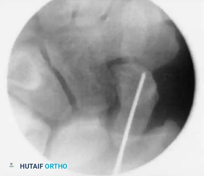
A 32-year-old male presents with a persistent painful elbow following a fall onto an outstretched hand 3 months ago. Clinical examination reveals a "clunk" on terminal extension during pronation. Radiographs are suspicious for an occult injury. You are presented with this follow-up imaging.
Candidate: "Based on the history of a 'clunk' and the radiographs, the patient likely has Posterolateral Rotatory Instability (PLRI) secondary to a chronic LUCL injury. I would confirm this with a physical examination using the posterolateral pivot-shift test and then consider reconstruction."
Failing to mention the "Horii-Morrey Circle of Instability." Candidates often treat the LUCL injury as an isolated event rather than part of the progressive sequence of soft tissue disruption that leads to PLRI, and they often forget to mention assessing for concomitant occult coronoid pathology.
Systematically address: 1. Diagnosis: Chronic PLRI secondary to LUCL insufficiency. 2. Pathology: Describe the "Horii-Morrey Circle" where injury progresses from lateral to medial (LUCL -> Capsular -> MUCL). 3. Clinical Assessment: Confirm with Pivot Shift, Tabletop Relocation, and Push-up test. 4. Management: If conservative therapy (strengthening/bracing) fails, propose anatomic reconstruction of the LUCL using a graft (e.g., palmaris longus or triceps strip), ensuring the graft is placed at the isometric point on the lateral humerus.
The patient is now scheduled for an open reduction of a complex injury pattern. What are the key surgical "must-haves" for a stable elbow in a Terrible Triad injury?
Candidate: "You need to fix the radial head, fix the coronoid, and repair the LUCL."
A list is not a structured answer. The candidate fails to prioritize the importance of each component or mention the "Order of Operations." They often forget to mention testing for stability after fixing each component, which is critical in an OR setting.
"I follow a structured 'inside-out' priority sequence: 1. Coronoid: Restore the anterior buttress (essential for stability). 2. Radial Head: Restore the lateral column length and articulation. 3. LUCL: Re-establish the lateral tension band. 4. Intra-operative Testing: Test stability at every stage. If the elbow remains unstable after all three, I would consider a hinged external fixator or a coronoid augment/graft if the coronoid deficiency is severe."
