Spine Cases: Consider the Patient for Accurate Diagnosis
Key Takeaway
Discover the latest medical recommendations for Spine Cases: Consider the Patient for Accurate Diagnosis. Spondylolysis in adolescent athletes, like c the patient, is a common cause of low back pain, often linked to repetitive hyperextension activities and involving a pars fracture or stress reaction. Initial treatment typically consists of 3-6 months of nonoperative care, including a rigid lumbar brace (e.g., Boston flexion type) to manage pain and support healing, while avoiding extension exercises.
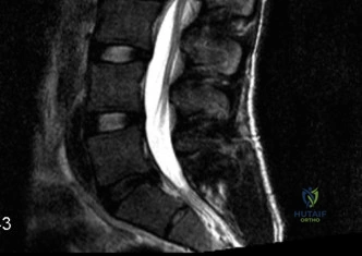
A 15-year-old high-performance linebacker presents with 6 months of worsening mechanical low back pain, localized to the midline. He denies radicular symptoms. The patient is tender over the L5-S1 junction. Given the demographics and activity, describe the likely underlying pathology and the diagnostic steps you would take to confirm it.
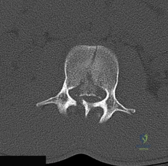
Candidate: The patient likely has spondylolysis at L5. I would order plain radiographs, specifically including oblique views to look for the "Scotty dog" sign. If these are negative but clinical suspicion remains high, I would proceed to an MRI or SPECT scan to identify early stress reactions in the pars.
Failing to emphasize the importance of weight-bearing films or neglecting to mention the staging of the lesion. Candidates often jump straight to "surgery" without discussing the importance of identifying whether the defect is acute/stress-reaction (potentially reversible) or chronic established pseudoarthrosis.
I would suspect isthmic spondylolysis due to repetitive hyperextension/axial loading. Diagnostics: 1. Standing AP and Lateral radiographs (to rule out listhesis). 2. Oblique views (to visualize the 'Scotty Dog' collar). 3. Crucial next step: MRI (STIR sequences) is preferred over SPECT to avoid radiation; it identifies high-signal marrow edema consistent with a stress reaction. This differentiates between an acute injury (high potential for healing with bracing) versus a chronic fibrous non-union (where bracing is futile).
The patient returns at age 20 with a Grade 2 Meyerding slip and L5 radiculopathy. You note a high pelvic incidence. Explain the relationship between spinopelvic parameters and the progression of this deformity.
Candidate: High pelvic incidence means the sacral slope is steep. This increases the anterior shear forces on the L5-S1 segment. In a patient with a pars defect, this force causes the L5 vertebra to slide forward, leading to the spondylolisthesis seen here.
Ignoring the "Pelvic Incidence = Sacral Slope + Pelvic Tilt" equation and failing to link the sagittal profile to the mechanical instability. A failing candidate describes the slip without mentioning why the geometry of this patient specifically led to progression.
The progression is driven by the PI (Pelvic Incidence). High PI correlates with a higher Sacral Slope and a greater lumbosacral shear angle. Biomechanical impact: The high PI dictates a vertical sacrum and forces the L5 segment into a state of chronic anterior shear. Once the posterior tension band (pars) fails, there is no structural restraint to oppose this shear, leading to the Grade 2 slip. The high PI makes these patients 'progression-prone,' necessitating fusion rather than just decompression.
Given the patient's age (20), Grade 2 listhesis, and neurological deficit (EHL weakness), describe your surgical strategy. Why would you choose a TLIF over a simple decompression?
Candidate: I would perform a decompression and instrumented fusion. I would do a TLIF because it allows me to clear out the disc space and restore the height, which helps the nerves. A decompression alone would make him unstable.
Failing to explicitly name the "Gill laminectomy" and not explaining why it is contra-indicated as an isolated procedure. Failing to mention "indirect decompression" through disc height restoration.
The patient has symptomatic isthmic spondylolisthesis with radiculopathy. 1. Strategy: Posterior decompression, TLIF, and instrumentation. 2. Why TLIF: It achieves 360-degree fusion. It addresses the foraminal stenosis by restoring disc height (indirect decompression) and allows for the resection of the fibrocartilaginous mass at the pars defect (direct decompression). 3. Avoidance of Gill-only: A simple Gill laminectomy without fusion in this patient would remove the remaining structural support of the posterior arch, resulting in catastrophic progression of the slip and likely further neurological deterioration.
