Comprehensive Diagnosis and Surgical Evaluation of Spinal Infections
Key Takeaway
Spinal infections present a complex diagnostic challenge, primarily manifesting as severe, mechanically exacerbated back pain. Early detection relies on a high index of clinical suspicion combined with advanced imaging, particularly MRI, and acute-phase reactants like ESR and CRP. Definitive diagnosis requires tissue culture via image-guided or open surgical biopsy. Delayed diagnosis can lead to devastating neurological compromise, necessitating urgent surgical decompression and stabilization.
Comprehensive Introduction and Patho-Epidemiology
The diagnosis and surgical management of spinal infections—encompassing pyogenic vertebral osteomyelitis, spondylodiscitis, and epidural abscesses—demand a meticulous, multidisciplinary approach combining the expertise of orthopedic spine surgeons, neurosurgeons, and infectious disease specialists. Because the clinical presentation of spinal infections is notoriously insidious, a high index of suspicion remains paramount. Delayed diagnosis is a frequent clinical reality that invariably results in extensive biomechanical destruction of the anterior column, progressive sagittal deformity, and, in severe cases, devastating and irreversible neurological compromise. According to seminal studies by Ross and Fleming, localized, relentless back pain is the primary presenting symptom in 85% of patients. This pain is typically mechanical in nature, severely exacerbated by changes in position, ambulation, and weight-bearing activities, and ranges in intensity from a mild, deep-seated aching to extreme, intractable agony that awakens the patient from sleep.
The pathophysiology of pyogenic spinal infections is primarily driven by hematogenous dissemination of bacterial pathogens, though direct inoculation from spinal procedures and contiguous spread from adjacent soft-tissue infections also occur. In the adult spine, the intervertebral disc is avascular; therefore, hematogenous seeding typically begins in the highly vascularized metaphyseal region of the vertebral body, adjacent to the cartilaginous endplate. The primary nutrient arteries bifurcate into small, tortuous subchondral arterioles where blood flow becomes sluggish, creating an ideal microenvironment for bacterial stasis and colonization. Once an infectious nidus is established in the subchondral bone, the resulting inflammatory cascade and localized ischemia lead to rapid bone necrosis and micro-trabecular collapse. The infection subsequently breaches the cartilaginous endplate, invading the avascular intervertebral disc space, which acts as a relatively defenseless reservoir for bacterial proliferation. From the disc space, the infection can easily spread to the adjacent vertebral body, resulting in the classic radiographic appearance of contiguous two-level spondylodiscitis.
Epidemiologically, the incidence of spinal infections has seen a marked increase over the past two decades. This rise is multifactorial, driven by an aging population with multiple medical comorbidities, the widespread use of immunosuppressive therapies, an escalating epidemic of intravenous drug use (IVDU), and the exponential increase in invasive spinal procedures. Staphylococcus aureus remains the most frequently isolated pathogen, responsible for over 50% of pyogenic spinal infections, with Methicillin-resistant S. aureus (MRSA) presenting a formidable challenge requiring prolonged, targeted antimicrobial therapy. Gram-negative bacilli, such as Escherichia coli and Pseudomonas aeruginosa, are increasingly prevalent, particularly in the elderly, immunocompromised hosts, and patients with concurrent genitourinary tract infections. Furthermore, clinicians must remain vigilant for granulomatous infections, particularly Mycobacterium tuberculosis (Pott's disease) and Brucella species, which present with a more indolent clinical course but possess a profound capacity for extensive osteoarticular destruction and massive paraspinal abscess formation.
Constitutional symptoms, such as fever, chills, and night sweats, are remarkably inconsistent and may be absent in up to 50% of patients, particularly in the elderly, diabetic, or profoundly immunocompromised populations. Clinicians must be acutely vigilant for signs of abscess pointing, which may manifest at a significant distance from the primary infectious focus. A classic clinical pearl involves the paraspinal abscess, which commonly presents as a swelling in the groin below the Poupart ligament (inguinal ligament). This phenomenon occurs due to the anatomical tracking of purulent material along the fascial planes of the psoas muscle. Consequently, patients may present with an involuntary hip flexion contracture and a positive psoas sign, often misleading the initial diagnostic workup toward intra-abdominal or primary hip pathology.
Detailed Surgical Anatomy and Biomechanics
A profound mastery of spinal vascular anatomy and biomechanics is essential for understanding the pathogenesis of spinal infections and for executing safe, effective surgical interventions. The arterial supply to the spinal column is segmental. In the thoracic and lumbar regions, paired segmental arteries arise directly from the descending aorta and traverse the waist of the vertebral bodies. These vessels give rise to equatorial branches that supply the vertebral body and primary nutrient arteries that penetrate the posterior cortex. The venous drainage of the spine is equally critical; the valveless epidural venous plexus of Batson allows for retrograde flow of venous blood during periods of increased intra-abdominal or intrathoracic pressure. This unique anatomical configuration facilitates the paradoxical spread of pelvic and genitourinary infections directly to the spinal column, bypassing the pulmonary filtration system.
Biomechanically, the anterior column of the spine—comprising the vertebral bodies and the intervertebral discs—is the primary load-bearing structure, responsible for supporting approximately 80% of the physiological axial load. Pyogenic spondylodiscitis exhibits a distinct predilection for destroying this critical anterior structural support. As the infection progresses, the trabecular architecture of the vertebral body is compromised, and the intervertebral disc height is obliterated. This extensive anterior destruction shifts the instantaneous axis of rotation (IAR) posteriorly. As the anterior column collapses, the spine hinges on the relatively intact posterior elements, specifically the facet joints and posterior ligamentous complex. This biomechanical failure inevitably leads to a progressive, rigid focal kyphosis, which not only alters the global sagittal vertical axis (SVA) but also places immense tension on the posterior neural elements.
The anatomical proximity of the infectious process to the delicate neural elements dictates the neurological presentation and the urgency of surgical intervention. The spinal canal and the epidural space are separated from the infected vertebral body only by the posterior longitudinal ligament (PLL) and a thin layer of cortical bone. When purulent material or necrotic bone fragments breach the posterior cortex, an epidural abscess forms, directly compressing the thecal sac and spinal cord. When paralysis occurs from spinal cord compression, Eismont et al. noted that central cord syndrome is present in two-thirds of patients, while anterior cord syndrome is found in one-third. Neurological symptoms are exquisitely anatomically dependent; they become significantly more frequent at higher spinal levels (cervical and thoracic) due to the critically narrow dimensions of the spinal canal in these regions. Conversely, neurological deficits are least common in the thoracolumbar and lumbar regions, where the spinal canal is more capacious and the cauda equina is relatively more resilient to gradual compression.
Surgical reconstruction of the infected spine must be fundamentally grounded in these biomechanical principles. Because the pathology is primarily an anterior column deficiency, surgical intervention must focus on aggressive anterior debridement (corpectomy) to eradicate the infectious nidus, followed by robust structural grafting to restore anterior load-bearing capacity and correct the kyphotic deformity. However, relying solely on an anterior construct in the setting of infection and altered biomechanics is fraught with the risk of graft subsidence and construct failure. Therefore, modern surgical management mandates the addition of posterior tension-band stabilization using rigid pedicle screw instrumentation, which neutralizes the posterior hinging forces, provides immediate biomechanical stability, and creates an optimal mechanical environment for bony arthrodesis.
Exhaustive Indications and Contraindications
The decision-making process regarding the surgical management of spinal infections is highly complex, requiring a careful synthesis of the patient's neurological status, biomechanical stability, systemic physiological reserve, and response to conservative therapy. While medical management with targeted, prolonged intravenous antibiotics remains the first-line treatment for uncomplicated, hemodynamically stable spinal infections, surgical intervention is frequently and urgently required to prevent catastrophic morbidity. The definitive diagnosis ultimately relies on the isolation and culture of the offending organism from infected tissue, which often necessitates surgical or image-guided intervention when blood cultures remain sterile.
The primary and most absolute indication for emergent surgical intervention is the presence of a progressive neurological deficit. This typically results from the formation of a ventral epidural abscess or the retropulsion of necrotic bone fragments into the spinal canal, leading to direct mechanical compression of the spinal cord or cauda equina. Immediate surgical decompression is mandatory to halt the progression of ischemic neural injury and maximize the potential for neurological recovery. A second critical indication is biomechanical instability or the development of a progressive kyphotic deformity secondary to extensive anterior column destruction. Allowing a severe kyphotic deformity to progress unhindered will result in intractable mechanical back pain, altered global sagittal balance, and delayed, insidious neurological compromise. Furthermore, surgery is heavily indicated in cases of medical failure, defined as persistent, severe localized pain and continuously elevated inflammatory markers (CRP and ESR) despite a minimum of four to six weeks of culture-directed intravenous antimicrobial therapy.
Contraindications to surgical intervention must be carefully weighed against the natural history of the untreated infection. Absolute contraindications are relatively rare but include patients who are physiologically incapable of surviving general anesthesia or massive surgical trauma due to profound cardiopulmonary failure, end-stage multiorgan dysfunction, or severe, uncorrectable coagulopathy. Relative contraindications involve patients with diffuse, multi-level osteomyelitis without significant focal deformity or neural compression, where the surgical morbidity of a massive multi-level reconstruction outweighs the potential benefits. In such cases, optimization of medical management, prolonged suppressive antibiotic therapy, and rigid external orthosis may be the most prudent course of action.
| Clinical Parameter | Indications for Surgical Intervention | Contraindications to Surgical Intervention |
|---|---|---|
| Neurological Status | Progressive motor deficit, cauda equina syndrome, acute myelopathy secondary to epidural abscess. | Intact neurological examination with no evidence of impending neural compression. |
| Biomechanical Stability | Severe anterior column destruction, progressive focal kyphosis, segmental instability. | Stable spinal alignment, intact posterior elements, minimal loss of vertebral body height. |
| Medical Management | Failure of targeted IV antibiotics (persistently elevated CRP/ESR, intractable pain after 4-6 weeks). | Rapid clinical and serological improvement following the initiation of empiric or targeted IV antibiotics. |
| Diagnostic Necessity | Need for definitive open tissue biopsy when blood cultures and percutaneous biopsies are repeatedly negative. | Causative organism identified via blood cultures; patient responding well to corresponding antimicrobial therapy. |
| Systemic Health | Patient physiologically optimized and capable of tolerating major reconstructive spine surgery. | Profound medical comorbidities, uncorrectable coagulopathy, severe hemodynamic instability (Absolute). |
Pre-Operative Planning, Templating, and Patient Positioning
Thorough pre-operative planning is the cornerstone of successful surgical outcomes in the management of spinal infections. This process begins with an exhaustive clinical and serological evaluation. Laboratory diagnostics are essential for both the initial identification and the longitudinal monitoring of the infectious process. The Erythrocyte Sedimentation Rate (ESR) is highly sensitive, elevated in up to 97% of acute cases, but lacks specificity. C-Reactive Protein (CRP) is a superior early indicator of infection due to its rapid kinetic response; it normalizes quickly with the resolution of infection and is the primary biomarker used to track the efficacy of treatment. Furthermore, in immunocompromised patients, particularly those with HIV, CD4 counts are highly predictive of the clinical course; atypical osteoarticular infections predominate when the CD4 count falls below 200/mL.
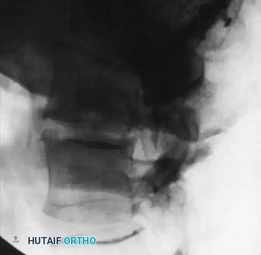
Advanced diagnostic imaging is critical for the precise anatomical mapping of the destructive process. Plain radiographs remain the standard initial study, though radiographic findings typically lag behind clinical symptoms by 2 weeks to 3 months. Early radiographic findings include subtle disc space narrowing and loss of the normal contour of the vertebral endplates.
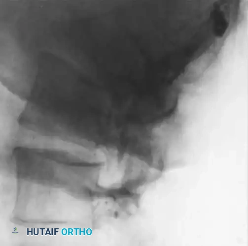
Fig. 40-7A: Early radiographic appearance of spinal osteomyelitis demonstrating minimal disc space narrowing, but a relatively normal endplate and subchondral region.
As the infection progresses, profound disc space narrowing, endplate destruction, and subchondral lytic defects become glaringly evident.
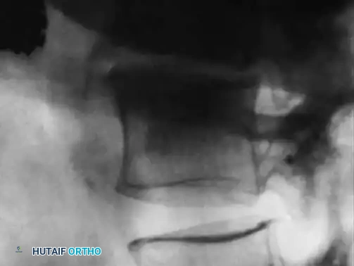
Fig. 40-7B: Progressive infection showing a severe reduction of disc height associated with the destruction of the endplate and the development of subchondral lytic defects.
Late radiographic findings, often taking up to 2 years to fully manifest following successful eradication, include spontaneous bony ankylosis and the formation of bridging osteophytes.
Fig. 40-7C: After successful treatment and eradication of the infection, note the sclerotic vertebra and the formation of a large bridging osteophyte indicating spontaneous fusion.
Magnetic Resonance Imaging (MRI) with and without gadolinium contrast is the undisputed gold standard for preoperative planning, boasting a sensitivity of 96% and an accuracy of 94% for disc space infections. T1-weighted images demonstrate hypointense signals in the infected vertebral bodies, while T2-weighted images reveal hyperintense signals indicative of profound edema. Gadolinium enhancement is indispensable for delineating the exact extent of epidural abscesses and hyperemic inflammatory tissue. Computed Tomography (CT) is utilized as an adjunct to evaluate the structural integrity of the trabecular bone, assess the degree of subchondral osteolysis, and template the size and trajectory of pedicle screws. Digital templating software is employed to measure the exact degree of regional kyphosis, calculate the required dimensions of the structural interbody cage, and determine the optimal length and diameter of the posterior instrumentation.
Patient positioning is dictated by the chosen surgical approach and requires meticulous attention to detail to prevent pressure necrosis and iatrogenic nerve palsies during prolonged procedures. For anterior approaches to the lumbar spine (retroperitoneal), the patient is placed in the true lateral decubitus position, securely taped to the operating table with an axillary roll placed to protect the brachial plexus. The table may be flexed to open the interval between the rib cage and the iliac crest. For posterior instrumentation and decompression, the patient is positioned prone on a radiolucent Jackson spinal table. This specific table frame is crucial as it allows the abdomen to hang completely free, significantly reducing intra-abdominal pressure, which in turn decreases venous engorgement of the epidural plexus of Batson, thereby minimizing intraoperative blood loss during decompression.
Step-by-Step Surgical Approach and Fixation Technique
Image-Guided Percutaneous Biopsy
When blood cultures are negative and empiric therapy is contraindicated or failing, a CT-guided percutaneous biopsy is the standard of care to obtain definitive tissue for culture and histopathology. The patient is positioned prone on the CT gantry, and a preliminary localization scan is performed. A transpedicular or extrapedicular posterolateral approach is meticulously planned to avoid the neural elements, thecal sac, and vital retroperitoneal structures. Under real-time CT fluoroscopic guidance, a heavy-duty Jamshidi needle is advanced through the pedicle or directly into the lateral aspect of the infected disc space or lytic vertebral body. Multiple large-volume core samples are harvested. It is imperative that these samples are immediately dispatched for comprehensive microbiological analysis, including aerobic, anaerobic, mycobacterial, and fungal cultures, as well as permanent histological pathology to definitively rule out masquerading neoplastic processes.
Open Anterior Debridement and Reconstruction (Corpectomy)
For extensive anterior destruction associated with cord compression or severe kyphosis, an open anterior approach is frequently required. In the lumbar spine, a retroperitoneal approach is utilized to avoid contamination of the peritoneal cavity. Once the retroperitoneal space is accessed, the ureter is identified and mobilized anteriorly. The psoas muscle, which is often severely inflamed or harbors a contiguous abscess, is carefully mobilized posteriorly to expose the lateral aspect of the infected vertebral bodies. The segmental vessels traversing the infected levels are meticulously isolated, ligated, and divided to allow for safe mobilization of the great vessels (aorta or inferior vena cava).
The debridement phase must be radical and uncompromising. Using high-speed burrs, rongeurs, and curettes, all necrotic bone, infected disc material, and purulent granulation tissue are aggressively excised. The debridement extends posteriorly to the posterior longitudinal ligament (PLL). If an epidural abscess is present, the PLL is carefully resected to achieve a complete ventral decompression of the thecal sac. The debridement is considered adequate only when healthy, bleeding cancellous bone is encountered at the superior and inferior vertebral endplates. This bleeding bone provides the necessary vascular bed for the incorporation of the structural graft.
Structural Reconstruction of the Anterior Column
Following radical corpectomy, the resultant anterior column defect must be reconstructed to restore sagittal alignment and bear the physiological axial load. The endplates of the adjacent healthy vertebrae are carefully prepared, ensuring that the subchondral bone is preserved to prevent subsequent graft subsidence. A structural graft is then selected. While autologous tricortical iliac crest or fibular strut grafts are historically the gold standard, modern techniques frequently utilize expandable titanium mesh cages or Polyetheretherketone (PEEK) implants packed with local healthy autograft or commercially available allograft. The cage is carefully impacted into the defect under direct visualization and fluoroscopic guidance, ensuring optimal sizing to restore the native disc height and correct the focal kyphosis without over-distracting the neural elements.
Posterior Tension-Band Stabilization and Instrumentation
Because the infected and reconstructed anterior column is mechanically compromised, supplemental posterior pedicle screw instrumentation is almost universally required to provide immediate rigid stabilization. This posterior tension-band prevents the spine from hinging into kyphosis, protects the anterior structural graft from subsidence, and creates a highly stable biomechanical environment conducive to rapid bony arthrodesis. The patient is repositioned prone, and a standard midline posterior approach is performed. Pedicle screws are inserted at least one to two levels above and below the infected segment using standard anatomical landmarks, fluoroscopy, or advanced robotic/navigation assistance. If a dorsal epidural abscess is identified on preoperative MRI, a wide laminectomy is performed simultaneously to decompress the posterior neural elements and evacuate the purulence. Finally, contoured titanium or cobalt-chrome rods are secured to the pedicle screws, and a posterolateral fusion is performed using autogenous bone graft or advanced orthobiologics.
Complications, Incidence Rates, and Salvage Management
Surgical management of spinal infections is fraught with potential complications, driven by the inherently destructive nature of the disease, the friability of infected tissues, and the complexity of the reconstructive procedures. Intraoperative complications include dural tears, which are significantly more common in the setting of epidural infection due to the profound adherence of inflammatory granulation tissue to the delicate thecal sac. Unintended durotomies must be meticulously repaired with primary non-absorbable sutures, supplemented by fibrin glue and fascial onlay grafts, to prevent the catastrophic formation of a postoperative cerebrospinal fluid (CSF) fistula. Vascular injuries, particularly to the aorta, inferior vena cava, or common iliac vessels during anterior approaches, represent life-threatening emergencies requiring immediate packing, mobilization of vascular surgery teams, and primary repair.
Postoperative mechanical complications are a major concern. Graft subsidence, where the anterior structural cage sinks into the adjacent osteoporotic or compromised vertebral bodies, can lead to a loss of sagittal correction and recurrent neural compression. Pseudoarthrosis, or failure of bony fusion, occurs when the biomechanical environment is insufficiently stable or the biological healing capacity is overwhelmed by persistent infection. Hardware failure, including pedicle screw pullout or rod fracture, is often a direct consequence of underlying pseudoarthrosis. The most dreaded biological complication is the persistence or recurrence of the deep spinal infection, which may manifest as chronic draining sinuses, intractable pain, or systemic sepsis.
Salvage management requires a highly individualized and aggressive approach. Persistent infections often necessitate a return to the operating room for radical re-debridement, copious pulsatile lavage, and the potential exchange of instrumentation if biofilms have formed. In cases of severe graft subsidence or hardware failure, the construct must be revised, often requiring extension of the posterior instrumentation to involve additional healthy vertebral levels to increase the biomechanical purchase, combined with aggressive anterior revision and structural allografting.
| Complication | Estimated Incidence | Etiology/Risk Factors | Salvage Management and Prevention |
|---|---|---|---|
| Dural Tear / CSF Leak | 5% - 12% | Adherent epidural granulation tissue, aggressive ventral decompression. | Primary watertight suture repair, fibrin sealant, flat bed rest for 48-72 hours, subarachnoid lumbar drain if refractory. |
| Graft Subsidence | 10% - 20% | Over-distraction of the disc space, excessive resection of subchondral bone, severe osteoporosis. | Optimization of bone density pre-op, utilization of endplate-sparing techniques, revision surgery with extended posterior fixation if symptomatic. |
| Hardware Failure / Pseudoarthrosis | 8% - 15% | Inadequate initial fixation, persistent infection, patient non-compliance with bracing, smoking. | Revision surgery with extension of fusion levels, robust autogenous bone grafting, utilization of orthobiologics, strict smoking cessation. |
| Persistent / Recurrent Infection | 5% - 10% | Inadequate initial surgical debridement, retained necrotic bone, highly virulent organisms (MRSA), immunocompromise. | Radical surgical re-debridement, removal of loose hardware, placement of antibiotic-impregnated PMMA beads, prolonged 12-week IV antibiotic therapy. |
| Major Vascular Injury | < 1% - 2% | Adherent prevertebral inflammatory tissue, aberrant vascular anatomy, aggressive retraction. | Immediate direct pressure packing, emergent vascular surgery consultation for primary suturing or patch angioplasty. |
Phased Post-Operative Rehabilitation Protocols
The postoperative rehabilitation of a patient following complex surgical reconstruction for a spinal infection is a protracted and highly structured process, divided into distinct phases to balance the need for early mobilization with the absolute necessity of protecting the surgical construct and allowing for biological healing. The acute phase (0 to 6 weeks postoperatively) is primarily focused on the eradication of the residual microscopic infection, wound healing, and the prevention of medical complications associated with prolonged bed rest. Intravenous antimicrobial therapy is initiated immediately after deep tissue cultures are obtained intraoperatively and is typically continued for a minimum of 6 to 8 weeks, strictly tailored based on the final microbiological sensitivities.
During this acute phase, mobilization is carefully initiated under the strict supervision of physical therapy. Depending on the rigidity of the internal fixation and the quality of the patient's bone stock, a rigid external orthosis—such as a Thoracolumbosacral Orthosis (TLSO) for thoracolumbar pathology or a rigid cervical collar for cervical infections—may be utilized whenever the patient is out of bed. This orthosis provides supplemental biomechanical support and serves as a crucial kinesthetic reminder to avoid excessive bending, lifting, or twisting (the BLT precautions). Clinical progress is monitored meticulously via serial neurological examinations and weekly assessments of CRP and ESR levels. A steady, uninterrupted decline in CRP is the most reliable biochemical indicator of successful eradication of the infection.
The subacute phase (6 to 12 weeks) marks the transition from intravenous to highly bioavailable oral antibiotics, provided the patient has demonstrated profound clinical improvement and the CRP has normalized. Radiographic follow-up is critical during this period. Standing, weight-bearing plain radiographs are obtained at the 6-week and 12-week marks to assess the maintenance of sagittal alignment, evaluate for any early signs of graft subsidence, and monitor the progressive incorporation of the bone graft. Physical therapy is advanced to include gentle isometric core strengthening and cardiovascular conditioning, strictly avoiding high-impact activities.
The long-term rehabilitation phase (3 to 6 months and beyond) focuses on the restoration of baseline functional capacity and the confirmation of solid bony arthrodesis. Dynamic flexion-extension radiographs or fine-cut CT scans may be utilized at the 6-month mark to definitively confirm the presence of bridging trabecular bone across the interbody space and the posterolateral gutters. Once solid fusion is confirmed radiographically and the patient remains clinically asymptomatic with normal inflammatory markers, the external orthosis is completely weaned, and the patient is gradually cleared to return to full, unrestricted activities of daily living and occupational duties.
Summary of Landmark Literature and Clinical Guidelines
The contemporary management of spinal infections is heavily informed by a robust body of landmark orthopedic and infectious disease literature. The Infectious Diseases Society of America (IDSA) guidelines serve as the foundational framework for the medical management of native vertebral osteomyelitis, strongly recommending a minimum of 6 weeks of targeted parenteral or highly bioavailable oral antimicrobial therapy, with longer durations reserved for cases complicated by extensive epidural abscesses or retained hardware.
From a diagnostic perspective, the seminal work by Modic et al. definitively established the superiority of MRI, demonstrating a sensitivity of 96%, a specificity of 92%, and an accuracy of 94% for identifying disc space infections, thereby cementing MRI as the undisputed gold standard imaging modality. Thelander and Larsson's critical research on the kinetics of inflammatory biomarkers revolutionized postoperative monitoring, demonstrating that CRP returns to normal rapidly following successful eradication of infection, whereas the ESR may remain spuriously elevated for weeks to months. Furthermore, Richards and Emara confirmed that a secondary, unexpected rise in CRP 4 to 6 days postoperatively strongly heralds a recurrent or nosocomial surgical site infection, dictating immediate clinical investigation.
Surgically, the principles of anterior debridement and reconstruction were championed by Eismont and colleagues, who meticulously documented the anatomical predilection for central and anterior cord syndromes in the setting of epidural compression, and advocated for aggressive anterior decompression. The modern paradigm of combining anterior structural support with posterior pedicle screw instrumentation has been validated by numerous contemporary prospective cohorts, which demonstrate significantly lower rates of pseudoarthrosis, graft subsidence, and progressive kyphosis compared to isolated anterior or posterior approaches. Mastery of these evidence-based guidelines and landmark anatomical studies is absolutely essential for any orthopedic surgeon or neurosurgeon tasked with managing these highly complex and potentially devastating clinical entities.
