Candidate: Can you interpret this anteroposterior shoulder X-ray?
Key Takeaway
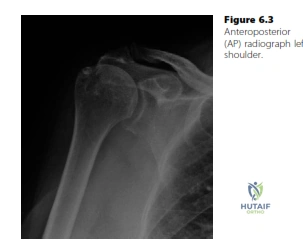
Learn more about Candidate: Can you interpret this anteroposterior shoulder X-ray? and how to manage it. When examining a 76-year-old patient with shoulder pain, a medical candidate performing the assessment might find, on this anteroposterior radiograph, subacromial space narrowing and acromial sclerosis. This indicates severe subacromial impingement and a secondary cuff tear, often confirmed by ultrasound showing torn subscapularis and supraspinatus tendons, requiring management like steroid injections or arthroscopic decompression.
Good afternoon. I have a 76-year-old left-handed fit gentleman presenting with shoulder pain and difficulty with overhead activities. Here is his AP radiograph (Figure 6.3). Please describe the findings.
Candidate: The radiograph shows proximal migration of the humeral head with reduced acromiohumeral distance (less than 7mm). There is no obvious glenohumeral or ACJ arthritis. This pattern is highly suggestive of chronic rotator cuff deficiency.
Candidates often focus solely on the "narrowing" without mentioning the biomechanical implication (proximal migration) or fail to note the absence of arthritis. Some candidates immediately jump to requesting an MRI, failing to recognize that the X-ray findings alone are diagnostic of cuff deficiency.
Start with a structured description: 1. Position/View. 2. Humeral head position (proximal migration indicates cuff deficiency). 3. Acromiohumeral distance (the measurement). 4. Status of the glenohumeral joint (to rule out primary osteoarthritis). 5. The diagnosis: Chronic Rotator Cuff Arthropathy.
You have identified findings suggestive of cuff arthropathy. What is your next step in assessment, and why?
Candidate: I would request an axillary lateral view to assess for static subluxation of the humeral head, which helps identify subscapularis insufficiency, and to rule out any associated structural abnormalities.
Asking for an MRI or Ultrasound before obtaining the full radiographic series (specifically the axillary view). In the context of suspected cuff arthropathy, the axillary view is critical to diagnose anterior subluxation, confirming a massive tear involving the subscapularis.
Demonstrate clinical reasoning: "I would obtain an axillary view to look for anterior subluxation of the humeral head. If present, this confirms an irreparable anterior cuff tear (subscapularis) and shifts my diagnosis toward Rotator Cuff Arthropathy (Hamada stage progression)."
The patient is a 76-year-old artist who wishes to regain function to abduct his arm for painting. Previous steroid injections have failed. What is the surgical management of choice, and what is the biomechanical rationale?
Candidate: I would offer a Reverse Polarity Shoulder Arthroplasty (RSA). This is preferred over standard total shoulder arthroplasty because the reverse design medializes the center of rotation and distalizes the humerus, effectively lengthening the lever arm of the deltoid, allowing it to function as an abductor despite a deficient rotator cuff.
Suggesting an attempted cuff repair or subacromial decompression. In established cuff arthropathy (proximal migration + bony changes), a primary repair is unlikely to hold or provide the functional outcome required for this patient's lifestyle goals.
Key terms: "Reverse Geometry," "Medialization of the Center of Rotation," "Distalization of the Humerus," and "Restoration of Deltoid Tension." Acknowledge that the patient’s functional requirement (painting/abduction) is the driving force for opting for RSA, which is the gold standard for pseudo-paralytic shoulders due to cuff arthropathy.
