Ortho Exam: How to Describe an 84-Year-Old Lady's Shoulder X-ray
Key Takeaway
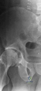
This topic focuses on Ortho Exam: How to Describe an 84-Year-Old Lady's Shoulder X-ray, An 84-year-old lady's left shoulder radiograph reveals severe joint destruction and lost articular anatomy. Her history, which may have the yearold lady describe a painless lump at age 14 and subsequent discharging sinus, indicates a chronic low-grade infection. This condition now results in significant movement restriction, reflecting decades of progressive joint damage.
An 84-year-old female presents after a mechanical fall. You are reviewing her initial shoulder trauma series. On the AP view, you notice the humeral head appears rounded and fixed in internal rotation—the classic "lightbulb sign." Which radiographic view is mandatory to confirm the suspicion of a posterior glenohumeral dislocation?
Candidate: I would order a true axillary lateral view to definitively assess the glenohumeral congruity and confirm the direction of the dislocation.
Stating "I would just get another X-ray" or relying on the Scapular Y view alone. In an elderly patient, a Scapular Y can be notoriously difficult to interpret if the patient cannot abduct sufficiently, and the axillary view is the gold standard for glenoid orientation.
The candidate should state: "I require a true axillary lateral view. While the 'lightbulb sign' on the AP is suggestive of a posterior dislocation, it is not diagnostic. An axillary view allows me to assess the relationship of the humeral head to the glenoid fossa in the axial plane, confirming the posterior displacement, and screening for associated bony pathology like a reverse Hill-Sachs lesion."
Following successful reduction, the patient has chronic weakness and limited elevation. You observe superior migration of the humeral head and subacromial erosion. How do you classify this pathology and what is the underlying mechanism?
Candidate: This is Rotator Cuff Arthropathy. It happens because the cuff is torn, the head moves up, and it grinds against the acromion.
Failing to mention the "force couple" mechanism. Simply saying "it grinds" is too simplistic. Failing to acknowledge the Hamada-Fukuda classification system is a missed opportunity for higher marks.
This is Rotator Cuff Arthropathy (RCA). The mechanism involves the loss of the rotator cuff force couple; specifically, the deltoid muscle becomes unopposed, causing superior migration of the humeral head. This leads to attrition of the subacromial space, acetabularization of the acromion, and eventual collapse of the glenohumeral joint. I would grade this using the Hamada-Fukuda classification, which looks at the acromiohumeral interval and the presence of glenoid erosion.
The patient requires surgery for a complex proximal humerus fracture. Given her age and bone quality, when would you choose a Reverse Total Shoulder Arthroplasty (rTSA) over ORIF?
Candidate: I would choose rTSA if the bone is very osteoporotic or if the head is broken into too many pieces so it won't heal with a plate.
Being too vague. Not mentioning "vascularity risk" or "head-split components." A high-scoring candidate must systematically address the patient, the deformity, and the joint.
I determine the choice based on three pillars: 1. Patient Factors: Older age, severe osteopenia, and physiological demand. 2. Deformity Factors: Complexity of the fracture (e.g., 4-part displacement, head-split component). 3. Joint Prognosis: High risk of Avascular Necrosis (AVN) based on the Hertel criteria (e.g., metaphyseal extension < 8mm, medial hinge disruption). If the fracture pattern predicts failure of ORIF or post-traumatic AVN, rTSA provides a predictable functional outcome by bypassing the need for tuberosity healing in the short term.
