Solve Shoulder & Elbow Cases: When a Patient Presents to Clinic
Key Takeaway
We review everything you need to understand about Solve Shoulder & Elbow Cases: When a Patient Presents to Clinic. When a 72-year-old male with a history of diabetes presents to clinic 8 months post-total shoulder arthroplasty with worsening pain, a CT arthrogram of the left shoulder is the most appropriate initial next step. This imaging helps evaluate for component loosening, which can indicate an underlying infection or mechanical issue, guiding further management despite other negative findings.
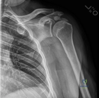
A 72-year-old male presents with a "stiff and painful" left shoulder eight months after an anatomic total shoulder arthroplasty. He has never been pain-free since the index procedure. There are no signs of systemic sepsis. What is your primary differential diagnosis, and why are you concerned about this specific clinical history?
Candidate: The primary differential is periprosthetic joint infection (PJI), specifically with a low-virulence organism like Cutibacterium acnes. Other differentials include aseptic loosening of the glenoid component, subscapularis failure, or adhesive capsulitis. I am concerned because the patient reports never being pain-free since surgery, which is a red flag for occult infection rather than mechanical wear or late-onset aseptic failure.
Focusing heavily on aseptic loosening as the primary diagnosis. Failing to highlight why "never being pain-free" is a critical historical marker for chronic indolent infection. Ignoring the patient's diabetic status as a risk factor.
Systematically structure the differential into "Biological" (Infection) and "Mechanical" (Loosening/Soft Tissue). Highlight that an arthroplasty that is painful from the outset suggests an unrecognized, low-virulence PJI—most commonly C. acnes. Explicitly mention that C. acnes forms biofilms that often manifest as stiffness and occult loosening without systemic inflammatory signs, making the clinical history the most sensitive diagnostic tool.
You have ordered a CT arthrogram and plain radiographs. The labs show normal CRP and ESR is 45 mm/hr. The CT arthrogram confirms glenoid loosening. How do you interpret these findings in the context of a suspected shoulder PJI?
Candidate: The normal inflammatory markers do not rule out PJI, as C. acnes infections frequently present with normal ESR/CRP. The CT arthrogram is the investigation of choice as it avoids the metal artifact of MRI, and the contrast extravasation confirms mechanical loosening. Given the early timing (8 months post-op) and the patient's clinical symptoms, I must treat this as a "probable" or "possible" PJI until proven otherwise by deep tissue cultures.
Assuming that because inflammatory markers are normal (or only slightly elevated), the diagnosis of infection is unlikely. Suggesting that a negative frozen section during biopsy would definitively rule out infection.
State clearly that shoulder PJI diagnosis requires a high index of clinical suspicion. Explain that serum inflammatory markers (ESR/CRP) have low sensitivity for C. acnes. The CT arthrogram is the diagnostic imaging modality of choice to confirm component-bone interface failure. Emphasize that in the absence of obvious systemic signs, surgical exploration for deep tissue cultures (held for 14 days) is the mandatory next step.
You are now in the operating room for the revision. You have taken deep tissue cultures. What is your management strategy for the implants? Explain your reasoning regarding DAIR versus two-stage exchange.
Candidate: I would perform a two-stage revision arthroplasty. DAIR is contraindicated because the patient has chronic symptoms (8 months), meaning a mature biofilm exists. I would explant all components, perform a radical synovectomy, and place an antibiotic-loaded cement spacer. After 14-day culture results and an antibiotic-free holiday, I would proceed to Stage 2, likely converting to a reverse total shoulder arthroplasty (rTSA).
Suggesting DAIR as an option, failing to understand that DAIR is only appropriate for early, acute infections. Forgetting to mention the necessity of antibiotic-loaded spacers or the conversion to rTSA.
Articulate that DAIR is reserved for acute postoperative infections (<4 weeks). Due to the chronic nature, biofilm maturity, and confirmed loosening, a two-stage approach is the standard of care. Explain the need for radical debridement, removal of all cement, and the use of high-dose antibiotic-loaded spacers. Justify the eventual conversion to rTSA based on the anticipated subscapularis compromise and the need for reliable glenoid fixation in the setting of previous infection.
