Part of the Master Guide

Master Diagnosis: Shoulder & Elbow Cases (Reproduced with Permission)

Ortho Exam: How to Describe an 84-Year-Old Lady's Shoulder X-ray

23 Apr 2026 83 min read 102 Views
Shoulder and elbow structured oral examination question1

Key Takeaway

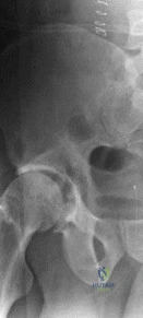
This topic focuses on Ortho Exam: How to Describe an 84-Year-Old Lady's Shoulder X-ray, An 84-year-old lady's left shoulder radiograph reveals severe joint destruction and lost articular anatomy. Her history, which may have the yearold lady describe a painless lump at age 14 and subsequent discharging sinus, indicates a chronic low-grade infection. This condition now results in significant movement restriction, reflecting decades of progressive joint damage.

Comprehensive Exam


00:00

Start Quiz

Question 1

An 84-year-old female presents after a fall onto her outstretched hand. You are asked to describe her shoulder X-ray. Which of the following views is most critical for definitively diagnosing a posterior glenohumeral dislocation when an AP view shows a 'lightbulb' sign?


Explanation

The axillary view is the gold standard for assessing glenohumeral joint congruity and confirming the direction of dislocation (anterior or posterior). While the 'lightbulb' sign on an AP view is suggestive of posterior dislocation, it is not definitive. The Scapular Y view can indicate posterior dislocation if the humeral head is posterior to the glenoid, but it's a sagittal view. The West Point and Stryker notch views are specific for glenoid rim defects or Hill-Sachs lesions, respectively, not primary dislocation diagnosis.

Question 2

An 84-year-old woman presents with long-standing shoulder pain and weakness, with active elevation limited to 60 degrees. Her X-ray shows superior migration of the humeral head, acromial erosion, and significant glenohumeral joint space narrowing. Which of the following is the most likely diagnosis?


Explanation

Rotator cuff arthropathy (RCA) is characterized by chronic, massive rotator cuff tears leading to superior migration of the humeral head, resulting in direct articulation between the humeral head and the acromion. This pathological contact causes secondary degenerative changes, including acromial erosion, glenohumeral joint space narrowing (typically superiorly), and often extensive osteophyte formation. Primary glenohumeral osteoarthritis usually shows concentric or inferior joint space narrowing without significant superior migration. CPPD can cause degenerative changes but not typically with this degree of superior migration. Adhesive capsulitis shows no significant radiographic findings. Seronegative spondyloarthropathies would show erosive changes, often with sacroiliitis, but not the specific pattern of RCA.

Question 3

An 84-year-old osteoporotic lady sustains a proximal humerus fracture after a low-energy fall. Her X-ray shows a fracture involving the surgical neck, greater tuberosity, and lesser tuberosity, with articular displacement. According to the Neer classification, how would you classify this fracture?


Explanation

The Neer classification divides the proximal humerus into four anatomical parts: the humeral head (articular segment), greater tuberosity, lesser tuberosity, and humeral shaft. A fracture involving all three tuberosities and the surgical neck (which separates the head from the shaft) constitutes four distinct displaced segments, thus classifying it as a 4-part fracture. Each displaced segment (>1cm displacement or >45 degrees angulation) counts as a 'part.' A 2-part involves one displaced segment, a 3-part involves two displaced segments (e.g., head + greater tuberosity + shaft). Articular displacement is a characteristic of 4-part fractures but not a primary classification part itself. Valgus-impacted is a specific stable variant, usually 2- or 3-part.

Question 4

When reviewing the shoulder X-ray of an 84-year-old female, which radiographic finding is most indicative of severe osteoporosis, beyond just fracture presence?


Explanation

Trabecular thinning and cortical attenuation (thinning of the outer bone layer) are direct radiographic signs of reduced bone mineral density characteristic of osteoporosis. While osteoporosis predisposes to fractures, the other options are signs of degenerative joint disease (subchondral cysts, joint space narrowing, osteophyte formation) or rotator cuff arthropathy (acromial erosion), not direct indicators of systemic bone density loss.

Question 5

An X-ray of an 84-year-old lady's shoulder shows significant inferomedial glenohumeral joint space narrowing, subchondral sclerosis, and large inferior osteophytes. There is no evidence of superior migration of the humeral head. Which diagnosis is most consistent with these findings?


Explanation

Primary glenohumeral osteoarthritis (GHOA) typically presents with inferomedial joint space narrowing, subchondral sclerosis, and significant osteophyte formation, particularly inferiorly (humeral head and glenoid). Crucially, there is no superior migration of the humeral head, differentiating it from rotator cuff tear arthropathy. CPPD can mimic OA but often shows chondrocalcinosis. Septic arthritis would show rapid joint destruction, effusion, and possibly periarticular osteopenia, less typically prominent osteophytes. Avascular necrosis would show subchondral collapse, crescent sign, and eventual secondary OA.

Question 6

An 84-year-old lady presents with recurrent anterior glenohumeral instability. Which specialized radiographic view is most effective for visualizing a bony Bankart lesion or an anterior glenoid rim fracture?


Explanation

The West Point axillary view is specifically designed to profile the anterior-inferior glenoid rim, making it superior for detecting bony Bankart lesions or anterior glenoid rim fractures, which are common sequelae of anterior glenohumeral dislocation. The Stryker Notch view is for Hill-Sachs lesions (posterolateral humeral head compression fracture). The Scapular Y view assesses dislocation direction. The Grashey view is a true AP. The Apical Oblique view can also show glenoid rim pathology but less specifically than West Point.

Question 7

When describing the shoulder X-ray of an elderly patient, you note an apparent non-union of the acromion. Which specific view would be most crucial to confirm an os acromiale and differentiate it from an acute fracture?


Explanation

The outlet view (or supraspinatus outlet view) is optimal for evaluating the acromial morphology, including the presence of an os acromiale, by projecting the acromion en face. An os acromiale is a developmental failure of fusion of the acromial apophyses. While other views might incidentally show it, the outlet view provides the best profile. The Zanca view is specific for the AC joint. Axillary view is for glenohumeral congruity. Transthoracic is for humeral shaft.

Question 8

A 'Grashey view' is requested for an 84-year-old female's shoulder. What is the primary purpose of this specific projection?


Explanation

The Grashey view is a 'true AP' view of the glenohumeral joint, achieved by internally rotating the patient approximately 30-45 degrees to align the glenoid parallel to the X-ray beam. This eliminates overlap of the humeral head and glenoid, allowing for accurate assessment of joint space and articulation. While rotator cuff integrity cannot be assessed directly, its sequelae (e.g., superior migration) are better appreciated.

Question 9

On an AP internal rotation view of an 84-year-old woman's shoulder, which anatomical landmark is best visualized en face?


Explanation

The AP internal rotation view brings the lesser tuberosity into profile, facing medially. The greater tuberosity is seen medially overlapping the humeral head. Conversely, the AP external rotation view profiles the greater tuberosity laterally.

Question 10

When reviewing an AP external rotation view of an elderly patient's shoulder, which structure is typically seen in profile laterally on the humeral head?


Explanation

The AP external rotation view rotates the humerus externally, bringing the greater tuberosity into profile on the lateral aspect of the humeral head. The lesser tuberosity is then positioned anteriorly and medially, often superimposing on the humeral head.

Question 11

An 84-year-old lady presents with a proximal humerus fracture. Her X-rays reveal a surgical neck fracture with 1.2cm displacement and 50 degrees of angulation. According to the Neer classification, how would you classify this fracture?


Explanation

The Neer classification system defines 'parts' based on significant displacement (>1cm or >45 degrees angulation) of the four major segments: humeral head (articular segment), greater tuberosity, lesser tuberosity, and humeral shaft. In this scenario, the surgical neck fracture separates the humeral head from the shaft. Since there is 1.2cm displacement and 50 degrees angulation, these two segments (head and shaft) are considered significantly displaced relative to each other. With only these two main segments displaced, it is classified as a 2-part surgical neck fracture. A 3-part would involve one tuberosity in addition to the head-shaft displacement, and a 4-part would involve both tuberosities plus the head-shaft displacement. Valgus-impacted fractures are a specific stable variant, usually 1- or 2-part, where the head is impacted into the shaft in valgus.

Question 12

An 84-year-old female sustains a proximal humerus fracture. Her X-ray shows a fracture through the surgical neck and a fracture of the greater tuberosity, with the articular segment displaced in a valgus-impacted pattern. The lesser tuberosity is intact. How would this fracture be classified using the Neer system?


Explanation

This describes a Neer 3-part fracture (humeral head, greater tuberosity, and shaft, with the surgical neck fracture separating the head from the shaft, and the greater tuberosity also fractured). The 'valgus-impacted' descriptor indicates a stable, often comminuted, but well-aligned fracture pattern where the head is driven into the shaft in valgus. It modifies the 3-part classification but doesn't change the number of displaced parts. The four parts are humeral head, greater tuberosity, lesser tuberosity, and shaft. Here, the head, greater tuberosity, and shaft are involved, making it a 3-part.

Question 13

An 84-year-old woman falls and presents with a painful, deformed shoulder. Her X-rays show a comminuted proximal humerus fracture with the humeral head clearly dislocated anteriorly from the glenoid fossa. What is the most appropriate description of this injury?


Explanation

The presence of both a fracture and a dislocation warrants the comprehensive term 'fracture-dislocation.' While it may also be a Neer 4-part fracture, 'fracture-dislocation' specifically captures both components of the injury. Anterior glenohumeral dislocation is only part of the injury. Luxatio erecta is a rare inferior dislocation. Pathologic fracture suggests an underlying bone lesion, which is not stated.

Question 14

For an 84-year-old lady with a comminuted 4-part proximal humerus fracture, which radiographic feature is most indicative of a potential need for reverse total shoulder arthroplasty (rTSA) over open reduction internal fixation (ORIF)?


Explanation

A head split component (fracture extending through the articular surface of the humeral head) or significant articular damage makes anatomical reduction difficult or impossible, often leading to poor outcomes with ORIF. In the elderly, especially with osteoporotic bone, rTSA is often preferred for complex 3- and 4-part fractures, particularly when articular comminution or displacement suggests avascular necrosis risk or inability to achieve stable fixation with ORIF. Valgus-impacted fractures are generally more stable. Intact tuberosities or minimal comminution would favor ORIF. Intact rotator cuff tendons would be important for an anatomic TSA, but rTSA bypasses a non-functional cuff.

Question 15

An 84-year-old female's shoulder X-ray following a fall shows a single, minimally displaced fracture of the greater tuberosity (<5mm displacement). Which of the following is generally the most appropriate initial management based on this X-ray finding?


Explanation

Minimally displaced greater tuberosity fractures (<5mm for non-dominant, <3mm for dominant arm, or as per local protocol) are typically managed non-operatively with initial immobilization (e.g., sling) for comfort, followed by early gentle range of motion exercises to prevent stiffness and facilitate healing. Surgical fixation is generally reserved for significant displacement, often exceeding 5mm, or involvement of the articular surface. Arthroscopy or arthroplasty are not indicated for this simple injury.

Question 16

Which radiographic finding in an 84-year-old's shoulder X-ray, weeks after a surgical neck fracture treated non-operatively, would raise the greatest concern for delayed union or non-union?


Explanation

Persistent fracture line with surrounding lucency (resorption) and lack of bridging callus formation at the fracture site are classic radiographic signs of delayed union or non-union. Early callus formation indicates healing. Absence of new displacement is a good sign. Soft tissue swelling is expected post-injury. Maintained glenohumeral joint space is unrelated to fracture healing.

Question 17

An 84-year-old female presents with persistent shoulder pain months after a proximal humerus fracture. Her X-ray shows increased density of the humeral head, flattening of the articular surface, and subchondral collapse ('crescent sign'). These findings are most suggestive of:


Explanation

The radiographic features described (increased density/sclerosis, flattening, subchondral collapse, 'crescent sign') are pathognomonic for avascular necrosis (AVN) of the humeral head, which is a common complication following displaced proximal humerus fractures, especially 3- and 4-part injuries due to disruption of the blood supply. Rotator cuff arthropathy would show superior migration. GHOA would show joint space narrowing and osteophytes but not necessarily increased density and collapse initially. Septic arthritis causes rapid joint destruction and effusion. CPPD shows chondrocalcinosis and often OA.

Question 18

A fracture of the anatomical neck of the humerus is distinct from a surgical neck fracture radiographically by its location. The anatomical neck lies:


Explanation

The anatomical neck is the groove separating the humeral head (articular surface) from the tuberosities. Fractures here are intra-articular and have a higher risk of avascular necrosis due to disruption of the blood supply entering through the metaphyseal bone. The surgical neck is distal to the tuberosities, a common site for extra-articular fractures.

Question 19

An 84-year-old lady complains of pain localized to the superior aspect of her shoulder, exacerbated by overhead activities. Her X-ray series includes a Zanca view. Which radiographic findings would be most consistent with symptomatic acromioclavicular (AC) joint osteoarthritis?


Explanation

AC joint osteoarthritis, like other degenerative arthropathies, is characterized by joint space narrowing, osteophyte formation, and subchondral sclerosis, specifically affecting the articulation between the distal clavicle and the acromion. A Zanca view is a specialized AP projection with cephalic tilt to optimally visualize the AC joint. Superior migration of the humeral head points to rotator cuff arthropathy. Subacromial spurring is associated with impingement, not directly AC joint OA. Erosions of the distal clavicle can be seen in inflammatory arthritis (e.g., RA, CPPD) or osteolysis, but OA predominantly shows sclerosis and osteophytes. Glenohumeral joint narrowing is GHOA.

Question 20

A 84-year-old female's shoulder X-ray shows glenohumeral joint space narrowing, subchondral sclerosis, and, notably, linear calcifications within the hyaline cartilage. This specific calcification pattern is highly suggestive of:


Explanation

Linear or punctate calcifications within the joint cartilage (hyaline or fibrocartilage) are the hallmark radiographic sign of calcium pyrophosphate deposition disease (CPPD), also known as chondrocalcinosis. CHADD or calcific tendinitis involves calcifications within tendons or bursae, usually amorphous. Gout causes erosions with overhanging edges, often without joint space calcification. Septic arthritis involves rapid joint destruction.

Question 21

In differentiating chronic inflammatory arthritis (e.g., rheumatoid arthritis) from osteoarthritis on a shoulder X-ray of an 84-year-old, which finding would be most characteristic of rheumatoid arthritis?


Explanation

Rheumatoid arthritis in the shoulder is characterized by concentric joint space narrowing (affecting all aspects of the joint equally), marginal erosions (particularly at the bare areas), and often periarticular osteopenia. In later stages, it can also lead to rotator cuff tears and superior migration. Osteoarthritis typically causes asymmetrical joint space narrowing (inferomedial for GHOA), prominent osteophytes, and subchondral sclerosis, usually without true erosions. Superior migration without erosions is more classic for rotator cuff arthropathy. RA is often bilateral.

Question 22

When evaluating a shoulder X-ray, how can an osteophyte be radiographically differentiated from an osteochondroma?


Explanation

The key radiographic feature differentiating an osteochondroma from an osteophyte (or other bone lesion) is the continuity of its cortical and medullary bone with the underlying parent bone. Osteophytes are bony outgrowths at joint margins, associated with degenerative changes, and do not necessarily show this medullary continuity. Osteochondromas can be sessile or pedunculated and originate from the bone surface, often near growth plates, not exclusively at joint margins.

Question 23

A 84-year-old man presents with acute, severe shoulder pain. His X-ray shows soft tissue swelling and a lytic lesion with an overhanging edge near the greater tuberosity. While rare in the shoulder, these findings are most suggestive of:


Explanation

Although less common in the shoulder than in the foot, gout can affect any joint. The characteristic radiographic signs of chronic gout are soft tissue swelling due to tophi, 'punched-out' lytic lesions (erosions) with sclerotic margins, and often a classic 'overhanging edge' (Martel sign). Acute calcific tendinitis presents with amorphous calcifications. OA flare is typically joint space narrowing and osteophytes. Stress fractures are subtle cortical breaks. Septic arthritis leads to rapid joint destruction and effusion.

Question 24

The 'acromiohumeral interval' (AHI) is a key measurement on shoulder X-rays. In an 84-year-old female with long-standing shoulder pain, a significantly reduced AHI (e.g., <6mm) is most indicative of:


Explanation

A severely reduced acromiohumeral interval (distance between the undersurface of the acromion and the superior aspect of the humeral head) is a hallmark radiographic sign of a chronic, massive rotator cuff tear. Without an intact rotator cuff, the unopposed pull of the deltoid muscle causes superior migration of the humeral head. A normal AHI is typically 7-14mm. Impingement can involve spurs, but true superior migration signifies a tear. Adhesive capsulitis has no radiographic findings. GHOA typically causes inferior or concentric narrowing.

Question 25

An 84-year-old lady's shoulder X-ray shows a large, well-circumscribed, amorphous calcification overlying the supraspinatus tendon insertion. She reports acute, severe pain. This presentation is most consistent with:


Explanation

A large, amorphous, well-defined calcification in a tendon, particularly the supraspinatus, in the context of acute severe pain, is characteristic of acute calcific tendinitis, also known as Calcium Hydroxyapatite Deposition Disease (CHADD). The calcifications are typically within the tendon itself, not the joint space, and are not linear like CPPD. Chronic rotator cuff tears may show superior migration but not necessarily such a dense calcification. Loose bodies are intra-articular and often faceted. Septic arthritis is an infection.

Question 26

Radiographically, how can a chronic, massive rotator cuff tear leading to arthropathy be differentiated from an acute traumatic rotator cuff tear on a standard shoulder X-ray series?


Explanation

Chronic, massive rotator cuff tears, especially those leading to arthropathy, are characterized by distinct radiographic signs of superior migration of the humeral head and secondary degenerative changes such as acromial erosion and glenoid cartilage loss (rotator cuff arthropathy). Acute tears, particularly partial ones, often show no definitive changes on plain X-rays, although a large effusion or minor bone avulsions might be seen.

Question 27

On an 84-year-old patient's shoulder X-ray, you observe diffuse, fine, stippled calcifications within the subacromial-subdeltoid bursa. This finding is most likely indicative of:


Explanation

Diffuse, stippled calcifications within the subacromial-subdeltoid bursa are characteristic of chronic inflammatory bursitis, often secondary to chronic impingement or irritation. While calcific tendinitis involves tendon calcification, bursal calcification is a distinct entity. CPPD involves articular cartilage. Synovial osteochondromatosis produces numerous loose bodies. Myositis ossificans is heterotopic bone formation within muscle.

Question 28

An 84-year-old lady presents after falling on an outstretched arm. Her X-ray shows an anterior glenohumeral dislocation. What is a 'bony Bankart lesion' that might be visible on specific views?


Explanation

A bony Bankart lesion is an avulsion fracture of the anterior-inferior glenoid rim, typically occurring during anterior glenohumeral dislocation when the humeral head impacts and avulses a piece of the glenoid. A compression fracture of the posterolateral humeral head is a Hill-Sachs lesion. A posterior glenoid rim fracture is a reverse bony Bankart. A superior labral tear is a SLAP lesion, which is soft tissue. Greater tuberosity fractures are separate.

Question 29

After a suspected posterior shoulder dislocation in an 84-year-old, which radiographic finding on the humeral head is indicative of a 'reverse Hill-Sachs lesion'?


Explanation

A reverse Hill-Sachs lesion (or McLaughlin lesion) is an impression fracture on the anteromedial aspect of the humeral head, occurring when the humeral head impacts against the posterior glenoid rim during a posterior dislocation. A Hill-Sachs lesion (classic) is on the posterolateral aspect from anterior dislocation.

Question 30

An 84-year-old patient presents with their arm fixed in abduction and external rotation, with the humeral head palpable inferiorly. Her X-ray confirms an inferior glenohumeral dislocation. What is the clinical term for this rare type of dislocation?


Explanation

Luxatio erecta is a rare type of inferior glenohumeral dislocation where the humeral head is displaced inferiorly, and the arm is fixed in an abducted and externally rotated position, often above the head. Anterior and posterior dislocations are more common. Subluxation implies partial dislocation. Multidirectional instability is a clinical diagnosis.

Question 31

For an 84-year-old lady with a history of recurrent anterior shoulder dislocations, which specific radiographic finding would indicate chronic instability and potential future surgical intervention?


Explanation

Hill-Sachs lesions (compression fracture of the posterolateral humeral head) and bony Bankart lesions (avulsion fracture of the anterior-inferior glenoid rim) are common bony sequelae of recurrent anterior glenohumeral dislocations. Their presence indicates significant damage from prior dislocations and can contribute to recurrent instability, often necessitating surgical stabilization. Subchondral sclerosis is general OA. Flattening of GT is not specific. Superior migration is RCA. Intact anatomical neck is irrelevant.

Question 32

On an 84-year-old patient's shoulder X-ray, you observe a solitary, well-defined, expansile lytic lesion in the proximal humerus without periosteal reaction. What is the most common benign bone tumor in this age group, and a likely differential for such a lesion (though often asymptomatic)?


Explanation

A solitary, well-defined lytic lesion in an elderly patient, especially if asymptomatic, could be an enchondroma, which is a common benign cartilage tumor. In the proximal humerus, these are often incidental findings. While metastatic lesions (option D) are more common in the elderly as a general rule for bone lesions, the description 'well-defined, expansile, no periosteal reaction' less fits typical aggressive metastasis, which are often poorly defined, lytic or blastic, with or without periosteal reaction. Osteosarcoma and Ewing sarcoma are aggressive malignant tumors typically seen in younger patients and would have aggressive features. Chondrosarcoma is a malignant cartilage tumor, but often has more aggressive features or calcifications. Given the phrasing of 'most common benign bone tumor... and a likely differential,' enchondroma is the best answer.

Question 33

An 84-year-old female's X-ray reveals multiple, ill-defined, sclerotic lesions (blastic appearance) throughout the proximal humerus and scapula. This radiographic pattern, particularly in an elderly patient, is most suggestive of:


Explanation

Multiple, ill-defined sclerotic (blastic) lesions in bone, especially in an elderly patient, are highly characteristic of metastatic disease from primary tumors like prostate cancer (most common blastic mets in men) or breast cancer (can be mixed lytic/blastic in women). Multiple myeloma typically presents with 'punched-out' purely lytic lesions. Osteochondromatosis and enchondromatosis are benign. Paget's disease can cause sclerotic changes, but often with cortical thickening and bone enlargement, usually not diffuse ill-defined lesions.

Question 34

You suspect a metastatic lesion in the proximal humerus of an 84-year-old woman. What radiographic characteristic, beyond a lytic or blastic appearance, would specifically raise concern for pathological fracture risk?


Explanation

A cortical breach or thinning of the cortex greater than 50% of the bone's width significantly compromises structural integrity and indicates a high risk of pathological fracture. This is a critical radiographic sign to look for in patients with suspected metastatic bone disease. Subchondral sclerosis and osteophytes are signs of degenerative joint disease. Maintained joint space is not directly related to fracture risk of a shaft lesion. Well-defined margins are less indicative of fracture risk than cortical involvement.

Question 35

How would you radiographically differentiate septic arthritis of the glenohumeral joint from advanced primary glenohumeral osteoarthritis in an 84-year-old lady?


Explanation

Septic arthritis is characterized by rapid, uniform (concentric) joint space narrowing due to cartilage destruction, along with early subchondral bone erosions and overall periarticular osteopenia. While effusion might be present, it's not always definitively visible on plain X-rays. Osteoarthritis, by contrast, has a slower progression, typically asymmetric joint space narrowing (often inferomedial), prominent osteophytes, subchondral sclerosis, and cysts, without the rapid, destructive pattern of infection.

Question 36

An 84-year-old patient with diabetes presents with chronic, low-grade shoulder pain and a discharging sinus. Her X-ray shows periosteal reaction, cortical thickening, and medullary sclerosis in the proximal humerus. These findings are most suggestive of:


Explanation

Periosteal reaction, cortical thickening, and medullary sclerosis (often with a sequestrum or involucrum, though not specified here) are classic radiographic signs of chronic osteomyelitis. This is particularly relevant in patients with predisposing factors like diabetes and a draining sinus. Osteoarthritis and rotator cuff arthropathy are joint diseases. Calcific tendinitis involves tendon calcification. Stress fractures show subtle lucency or sclerosis without such extensive reactive bone changes.

Question 37

An 84-year-old lady has a history of right shoulder replacement. Her current X-ray shows an anatomic total shoulder arthroplasty (TSA). Which components would you expect to see on the X-ray?


Explanation

An anatomic Total Shoulder Arthroplasty (TSA) aims to replicate normal anatomy. It consists of a humeral component (stem with a modular head, typically metal) and a glenoid component (a polyethylene liner usually cemented into a metallic baseplate that is fixated to the glenoid bone). Option A is hemiarthroplasty. Option D is a reverse TSA. Option E describes a reverse TSA setup with components flipped. Option B is incomplete.

Question 38

On the X-ray of an 84-year-old lady with a Reverse Total Shoulder Arthroplasty (rTSA), what is the key radiographic characteristic differentiating it from an anatomic TSA?


Explanation

In a reverse total shoulder arthroplasty (rTSA), the 'ball' (glenosphere) is fixed to the glenoid bone, and the 'socket' (humeral cup/liner) is on the humeral side, effectively reversing the native anatomy. This allows the deltoid to act as the primary elevator, compensating for a deficient rotator cuff. An anatomic TSA has the ball on the humerus and socket on the glenoid.

Question 39

Which radiographic finding is a common long-term complication specifically associated with Reverse Total Shoulder Arthroplasty (rTSA)?


Explanation

Scapular notching is a common and specific complication of reverse total shoulder arthroplasty, occurring when the medial aspect of the humeral implant impinges on the inferior scapular neck during adduction and internal rotation, leading to bony erosion. Instability of the humeral head in the glenoid is for anatomic TSA. Resorption of the greater tuberosity is more relevant to proximal humerus fractures or hemiarthroplasty. Dislocation of the metal humeral head is for anatomic TSA. Avascular necrosis of the glenoid is rare.

Question 40

You are reviewing a follow-up X-ray for an 84-year-old with an anatomic TSA. What radiographic sign would be most concerning for glenoid component loosening?


Explanation

Progressive lucency (a radiolucent line) greater than 1-2mm at the bone-cement or bone-implant interface around the glenoid component is the most significant radiographic sign of aseptic loosening of the glenoid component in a TSA. Hill-Sachs lesion is related to dislocation. Rotator cuff calcification is calcific tendinitis. Increased AHI suggests rotator cuff tear. Scapular notching is rTSA specific.

Question 41

What is the primary radiographic feature used to diagnose scapular notching in an X-ray of an rTSA patient?


Explanation

Scapular notching is precisely defined as the erosion of the inferior scapular neck by the humeral component of the rTSA, specifically the polyethylene liner impacting the bone during certain movements. It's a progressive, visually distinct radiographic finding. Glenoid subsidence is different, and the other options are not directly related to notching.

Question 42

On an 84-year-old lady's shoulder X-ray, you observe localized areas of increased bone density, particularly beneath the articular cartilage in the glenohumeral joint. What term describes this finding, and what does it typically indicate?


Explanation

Sclerosis refers to increased bone density, often seen as 'whiteness' on an X-ray. Subchondral sclerosis is a classic radiographic sign of degenerative joint disease (osteoarthritis), representing a response to increased stress on the underlying bone. Osteopenia is decreased bone density. Lucency is decreased density. Sequestrum is dead bone in osteomyelitis. Erosion is bone loss, often from inflammatory arthritis.

Question 43

An X-ray of an elderly shoulder shows a cyst-like lesion in the humeral head subchondral bone, with sclerotic margins, but no communication with the joint. How would you best describe this finding, and what is its typical etiology?


Explanation

Geodes, also known as subchondral cysts, are common findings in osteoarthritis. They are fluid-filled cavities that form within the subchondral bone, often with sclerotic margins. While they can be large, they typically do not communicate with the joint space. Avascular necrosis shows collapse and increased density. A simple bone cyst is a different entity. Metastatic lesions would be more irregular. Septic arthritis causes destruction.

Question 44

An X-ray of an 84-year-old patient reveals a hooked or curved acromial morphology. This finding is most relevant to the pathogenesis of:


Explanation

A hooked or curved (Type II or Type III according to Bigliani's classification) acromial morphology is a well-established anatomical risk factor for subacromial impingement syndrome, as it reduces the space for the rotator cuff tendons to glide beneath the acromion. AC joint OA, calcific tendinitis, AVN, and Bankart lesions are not primarily caused by acromial morphology.

Question 45

On an AP shoulder X-ray, you observe calcification in the soft tissues superior to the coracoid process, possibly within the coracoclavicular ligaments. This finding could indicate:


Explanation

Calcification or ossification of the coracoclavicular ligaments (conoid and trapezoid) can occur, sometimes post-traumatically (e.g., after an AC joint injury where the ligaments were stretched or torn) or as an idiopathic process. It's not a normal aging variant. While it might be associated with prior AC dislocation, 'ossification of the coracoclavicular ligaments' directly describes the radiographic finding. Coracoid impingement is more about morphology. Subcoracoid bursitis typically doesn't calcify in this manner.

Question 46

What constitutes an 'adequate' shoulder X-ray series for an initial evaluation of trauma in an 84-year-old lady?


Explanation

An adequate shoulder trauma series typically includes at least three views: an AP view (often a Grashey true AP is preferred), a Scapular Y view (for assessing dislocation direction and scapular body), and an Axillary view (crucial for glenohumeral congruity, anterior/posterior dislocation, and glenoid rim pathology). These three views provide a comprehensive initial assessment.

Question 47

When systematically describing an 84-year-old's shoulder X-ray, using the 'ABC'S' mnemonic (Alignment, Bone, Cartilage, Soft tissues), what does the 'C' (Cartilage) primarily refer to on plain radiographs?


Explanation

On plain radiographs, cartilage itself is radiolucent and not directly visualized. Its integrity and thickness are inferred by the width of the joint space. Joint space narrowing indicates cartilage loss. While cartilage calcifications (CPPD) and erosions (inflammatory arthritis) can be seen, the primary assessment of 'cartilage' with ABC'S on plain film refers to joint space width.

Question 48

When reviewing a shoulder X-ray, you notice a distinct, thin, radiopaque line projected across the humeral head, not conforming to anatomical structures. What is the most likely explanation for this finding?


Explanation

A thin, radiopaque line that does not conform to known anatomical structures or fracture patterns, especially if it appears too sharp or linear, is highly suspicious for a radiographic artifact, such as a clothing seam, zipper, or hair braid. Stress fractures, pathological fractures, and AVN (crescent sign) have specific radiographic appearances related to bone pathology. Vascular calcifications typically follow vessel paths.

Question 49

In the Neer classification of proximal humerus fractures, what are the criteria for considering a fracture fragment 'displaced' and counting it as an additional 'part'?


Explanation

According to the Neer classification, a fracture fragment is considered 'displaced' (and thus counts as an additional 'part' beyond the initial two segments of head and shaft) if there is greater than 1cm of displacement or greater than 45 degrees of angulation. This threshold helps distinguish significantly displaced fractures requiring more aggressive treatment from minimally displaced ones.

Question 50

An 84-year-old female presents with acute shoulder pain following a fall. Her X-ray shows a fracture of the surgical neck and a fracture of the greater tuberosity, with both fragments significantly displaced. The lesser tuberosity and articular head are intact. What is the Neer classification for this injury?


Explanation

In the Neer classification, the four anatomical parts are the humeral head, greater tuberosity, lesser tuberosity, and the shaft. If a surgical neck fracture (separating head from shaft) and a displaced greater tuberosity fracture are present, this involves three displaced segments (head-shaft unit, and the greater tuberosity), making it a 3-part fracture. The lesser tuberosity and articular head are intact, so it's not 4-part or an articular displacement.

Question 51

Which finding on a shoulder X-ray of an 84-year-old is most indicative of early post-traumatic changes following a shoulder dislocation, rather than chronic instability?


Explanation

A small glenohumeral joint effusion (fluid in the joint space) can be an acute sign of trauma, including dislocation. Large Hill-Sachs and bony Bankart lesions suggest more significant or recurrent trauma. Superior migration of the humeral head indicates chronic rotator cuff deficiency. Glenoid osteophytes are a sign of chronic degenerative change.

Question 52

An 84-year-old lady presents with chronic shoulder pain. Her X-ray shows diffuse periarticular osteopenia, concentric joint space narrowing, and marginal erosions in both glenohumeral joints. These findings are highly characteristic of:


Explanation

Bilateral, symmetrical, concentric joint space narrowing, periarticular osteopenia, and marginal erosions are classic radiographic features of rheumatoid arthritis. Primary OA is typically asymmetric, with osteophytes and sclerosis. Rotator cuff arthropathy involves superior migration. Septic arthritis is usually unilateral and rapidly destructive. CPPD involves chondrocalcinosis.

Question 53

What is the primary utility of an 'axillary lateral view' in the shoulder series of an 84-year-old patient with suspected trauma?


Explanation

The axillary lateral view is crucial for evaluating glenohumeral joint congruity and definitively identifying the direction of dislocation (anterior, posterior, or inferior) as it provides a true lateral projection of the articulation. It is also important for detecting glenoid rim fractures. Other views are better suited for AC joint, clavicle fractures, or subacromial space visualization.

Question 54

An 84-year-old patient has a long-standing total shoulder arthroplasty. On a follow-up X-ray, you notice a large, localized area of bone resorption around the distal tip of the humeral stem, without obvious infection. This is most likely indicative of:


Explanation

Localized bone resorption (osteolysis) around a prosthetic component, especially if progressive and without signs of infection, is a hallmark of aseptic loosening. It is often caused by particulate wear debris triggering an inflammatory response that leads to bone loss. Stress shielding refers to bone atrophy due to reduced load transfer, usually more diffuse. Heterotopic ossification is bone formation in soft tissues. Periprosthetic fracture is a distinct event. Component fatigue refers to material failure, not bone resorption.

Question 55

Which radiographic sign is often observed in the late stages of rotator cuff arthropathy, indicating severe joint destruction?


Explanation

Severe superior migration of the humeral head leading to direct articulation with the acromion, causing both acromial and glenoid erosion, is characteristic of late-stage rotator cuff arthropathy. This indicates extensive loss of the rotator cuff's stabilizing function. Inferior osteophytes are typical of primary OA. AC joint widening implies dislocation. Bicipital groove presence is normal. Scapular body fractures are traumatic.

Question 56

On an 84-year-old lady's shoulder X-ray, you notice an anatomical variant where the acromion is not completely fused, appearing as a separate ossicle. This is known as:


Explanation

An os acromiale is a developmental anomaly where one or more of the acromial ossification centers fail to fuse. It can predispose to subacromial impingement and rotator cuff tears. Sprengel's deformity is congenital elevation of the scapula. Osgood-Schlatter is apophysitis of the tibial tubercle. Accessory navicular is an extra bone in the foot. Pelligrini-Stieda lesion is calcification of the MCL at the femoral attachment.

Question 57

An 84-year-old female presents with acute severe pain and limited range of motion after a minor trauma. Her X-ray shows marked periarticular osteopenia, joint space narrowing, and a 'pressure erosion' on the posterior aspect of the humeral head. This combination is highly suggestive of:


Explanation

Neuropathic (Charcot) arthropathy in the shoulder, though rare, can present with marked periarticular osteopenia, joint space narrowing, fragmentation, and 'pressure erosions' or 'resorption' of the humeral head and glenoid, often with associated soft tissue swelling and instability. This occurs due to loss of proprioception and pain sensation. Post-traumatic arthritis would have a clear history and signs of prior trauma. Other options do not typically cause such rapid, severe destruction with pressure erosions.

Question 58

When evaluating a shoulder X-ray, the presence of 'vacuum phenomenon' within the glenohumeral joint space (gas shadows) is typically seen in:


Explanation

The vacuum phenomenon (presence of gas within the joint space) is a common finding in degenerative joint disease (osteoarthritis). It is believed to be due to nitrogen gas released from synovial fluid into areas of negative pressure within the joint, often during traction or joint manipulation. It is generally not seen in inflammatory or septic conditions.

Question 59

Which radiographic finding is most characteristic of chronic superior labrum anterior posterior (SLAP) tear on a plain shoulder X-ray in an 84-year-old, if any at all?


Explanation

SLAP (Superior Labrum Anterior Posterior) tears are soft tissue injuries involving the labrum and biceps anchor. They are not directly visible on plain radiographs. Diagnosis typically requires MRI with or without arthrogram, or direct visualization during arthroscopy. Plain X-rays might show indirect signs of associated pathology (e.g., degenerative changes), but not the SLAP tear itself.

Question 60

An 84-year-old lady presents with chronic, diffuse shoulder pain. Her X-ray shows diffuse osteopenia, narrowing of the glenohumeral joint space, and evidence of mild subchondral erosions without significant osteophyte formation. What systemic condition should be considered in the differential diagnosis?


Explanation

Diffuse osteopenia, joint space narrowing (often concentric), and mild subchondral erosions without prominent osteophytes are characteristic features of inflammatory arthropathies, particularly Rheumatoid Arthritis, which can affect the shoulder. Primary osteoarthritis typically presents with subchondral sclerosis and prominent osteophytes. Gout would show punched-out erosions with overhanging edges. Psoriatic arthritis might show periostitis or 'pencil-in-cup' deformity, but is less common in the shoulder. Polymyalgia Rheumatica is a clinical diagnosis without specific radiographic findings.

Question 61

On an AP X-ray of an 84-year-old patient, you notice small, well-defined, ring-like calcifications within the soft tissues of the shoulder, not clearly associated with tendons or bursae. These findings might be suggestive of:


Explanation

Synovial osteochondromatosis is a metaplastic condition where the synovial membrane forms cartilaginous nodules that can detach and grow, leading to multiple, small, often ring-like or amorphous calcified loose bodies within the joint space or bursae. Acute calcific tendinitis has amorphous calcifications within tendons. Vascular calcifications follow vessel paths. Pneumarthrosis is gas. Foreign bodies are usually solitary.

Question 62

An 84-year-old lady reports increasing pain and stiffness in her shoulder after an anatomical total shoulder arthroplasty performed 5 years ago. Her X-ray shows a stable humeral component, but there is significant widening of the acromiohumeral interval. This widening is most likely due to:


Explanation

In an anatomic total shoulder arthroplasty, a widening of the acromiohumeral interval strongly suggests a rotator cuff tear. The rotator cuff is essential for centralizing the humeral head on the glenoid. If it tears post-arthroplasty, the deltoid's unopposed pull leads to superior migration of the humeral head, increasing the acromiohumeral distance. Polyethylene wear or glenoid loosening would affect joint space or implant stability, not directly widen the AHI to this extent. Subacromial impingement is often associated with a reduced AHI initially, then tears. Periprosthetic infection would typically show aggressive osteolysis.

Question 63

An X-ray of an 84-year-old patient's shoulder demonstrates severe degenerative changes in the glenohumeral joint, including joint space narrowing, subchondral sclerosis, and large osteophytes. Notably, there is also evidence of osteolysis affecting the distal clavicle. This combination of findings should prompt consideration of:


Explanation

Osteolysis of the distal clavicle can occur as an isolated condition, often due to repetitive microtrauma (e.g., weightlifting) or prior AC joint injury. When seen in conjunction with severe glenohumeral osteoarthritis in an elderly patient, it suggests a multifactorial etiology, likely degenerative arthritis affecting multiple shoulder joints, possibly exacerbated by previous trauma or chronic stress. While septic arthritis could cause osteolysis, affecting both GH and AC joints with typical OA signs is less common for infection. SLE is a possibility but osteolysis of the distal clavicle is not a hallmark. RCA would show superior migration, not necessarily distal clavicle osteolysis.

Question 64

On an 84-year-old lady's shoulder X-ray, you observe a well-defined, radiolucent lesion within the humeral head. This lucency, especially if it is expansile and cortically thinned, should raise concern for which type of underlying bone pathology?


Explanation

A well-defined radiolucent lesion ('lucency') in bone indicates an area of decreased bone density, often representing a lytic lesion. In an 84-year-old, this could range from benign entities like an enchondroma (especially if well-defined and expansile) or a simple bone cyst to more concerning pathologies like a lytic metastasis or multiple myeloma. Cortical thinning and expansion would further increase suspicion for a space-occupying lesion. Sclerosis and osteophytes are increased density. Blastic metastasis is increased density. Calcific tendinitis involves calcification, not lucency.

Dr. Mohammed Hutaif
Medically Verified Content by
Prof. Dr. Mohammed Hutaif
Consultant Orthopedic & Spine Surgeon
Chapter Index