Chronic Anterior Glenohumeral Dislocation: A Case Study on Diagnosis & Imaging Challenges
Key Takeaway
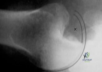
Chronic anterior glenohumeral dislocation diagnosis involves a thorough clinical exam revealing deformity and restricted ROM. Advanced imaging, including CT with 3D reconstruction and MRI, is crucial to assess glenoid bone loss (e.g., bony Bankart) and humeral head defects (e.g., Hill-Sachs lesion), often missed in initial radiographs, guiding appropriate surgical planning.
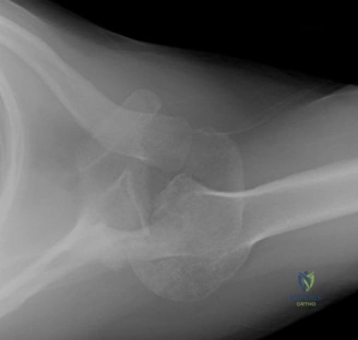
A 68-year-old male presents with a 4-month history of right shoulder pain and stiffness following a fall. He reports a "missed" injury from the initial emergency visit. On examination, there is profound global shoulder stiffness, an anteriorly flattened deltoid, and a firm, palpable fullness in the subcoracoid region. Given this clinical presentation and the provided imaging, what is your primary diagnosis, and what is the crucial clinical sign on physical examination that confirms the mechanical nature of this lesion?
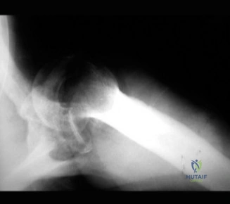
Candidate: The diagnosis is a chronic anterior glenohumeral dislocation. The patient has a missed injury. The physical exam shows a block to range of motion, and the radiographs confirm the humeral head is anterior to the glenoid. The most significant finding is the lack of external rotation.
Candidates often fail to describe the specific physical exam finding with the required terminology. Simply saying "the shoulder is stiff" is insufficient. A weak answer fails to connect the "lack of external rotation" to the "engaging Hill-Sachs lesion" and ignores the importance of the axillary lateral view in the diagnosis.
The diagnosis is a neglected, chronic anterior glenohumeral dislocation. The key clinical sign is an absolute, fixed mechanical block to external rotation. This is pathognomonic for an 'engaging' Hill-Sachs lesion, where the posterolateral humeral head defect is impaled on the anterior glenoid rim. The definitive radiographic diagnostic is the axillary lateral view, which demonstrates the humeral head displaced anterior to the glenoid fossa, confirming the diagnosis that the initial AP views likely obscured.
You are now in the planning phase for this patient. Why is a closed reduction contraindicated at this four-month stage, and what advanced imaging will you request to guide your surgical management?
Candidate: I would not attempt a closed reduction because it has been too long and the tissue is scarred. I would order a CT scan to look at the bone loss before surgery.
Failing to mention the specific risks of closed reduction (fracture, vascular injury) and failing to explain *why* the CT scan is necessary (e.g., assessing the "glenoid track" or quantifying bone loss for arthroplasty planning).
Closed reduction is strictly contraindicated after 3-4 weeks due to dense fibrotic adhesions and potential osteoporosis. Forcing a reduction carries an unacceptably high risk of iatrogenic proximal humerus fracture, axillary artery injury, or brachial plexus traction. I would request a high-resolution CT scan with 3D reconstructions. This is mandatory to quantify the degree of anterior glenoid bone loss and to evaluate the size and 'trackability' of the Hill-Sachs lesion, which is essential to determine if an anatomic or reverse total shoulder arthroplasty is required.
Given the patient is 68, has a 4-month old dislocation, and CT evidence of Goutallier stage 3 fatty infiltration of the rotator cuff, what is your surgical procedure of choice and what are the key technical steps to ensure a successful outcome?
Candidate: I would perform a Reverse Total Shoulder Arthroplasty (RTSA). I would use the deltopectoral approach, release the capsule, remove the humeral head, and then put in the components.
Missing the technical "how-to" points: candidates often neglect the risks to the axillary nerve during capsular release or fail to mention the rationale for *in situ* resection of the humeral head to prevent humerus fracture.
A Reverse Total Shoulder Arthroplasty is indicated due to rotator cuff compromise and massive osseous deficiency. Key technical steps include: 1) A deltopectoral approach with careful identification and protection of the axillary nerve. 2) Extensive circumferential soft tissue release of the fibrotic capsule. 3) In situ resection of the humeral head to avoid iatrogenic fracture of the humeral shaft. 4) Accurate glenoid baseplate placement, often slightly posterior/inferior to account for anterior bone loss. 5) Use of a cemented humeral stem for immediate fixation in osteopenic bone.
