Acad Orthop Surg Challenge: Diagnose Shoulder Pain Case 1
Key Takeaway
Looking for accurate information on Acad Orthop Surg Challenge: Diagnose Shoulder Pain Case 1? A partial-thickness rotator cuff tear is common in older patients, presenting with insidious, diffuse deltoid pain, night pain, and exacerbated by overhead activities. Patients typically maintain full or nearly full active range of motion, experiencing pain but not significant weakness with resisted shoulder flexion, a key differentiator discussed in **acad orthop surg**.
A 76-year-old right-hand-dominant male presents with chronic, progressive lateral shoulder pain. He describes the pain as "diffuse" and notes it is worse at night when rolling onto the shoulder. How do you interpret the mechanism of "night pain" in this clinical context, and what does the patient's specific method of localizing this pain reveal about the underlying pathology?
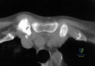
Candidate: Night pain occurs because the patient is lying on the shoulder, causing compression. The patient pointing to the lateral deltoid area with their whole hand suggests a rotator cuff problem rather than a localized joint issue like AC joint arthritis.
Failure to explain the biomechanics of night pain (loss of gravity-assisted inferior traction). A failing candidate also misses the neuroanatomical explanation for the "deltoid sign," failing to link the suprascapular/axillary nerve crossover.
Night pain in this demographic is often due to the loss of gravity-assisted inferior traction on the humerus when supine, leading to superior migration of the humeral head and subsequent compression of the subacromial bursa against the coracoacromial arch. The "Deltoid Sign"—where the patient cups the lateral shoulder with their entire hand—indicates referred pain mediated by the C5/C6 nerve roots. This confirms subacromial pathology, whereas a single-finger localized sign would steer me toward AC joint arthrosis.
Physical examination reveals full passive range of motion. How does this finding influence your differential diagnosis, and what does the presence of "algogenic paresis" (pain-inhibited weakness) vs. "pseudoparalysis" tell you about the integrity of the rotator cuff?
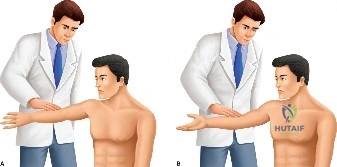
Candidate: Full passive ROM rules out adhesive capsulitis. Algogenic paresis means the patient is weak because it hurts, whereas pseudoparalysis means they cannot physically move the arm due to a massive tear.
Candidates often forget to mention the "rotator cable" or the functional force couple. They also fail to connect the physical finding of full passive motion to the specific exclusion of capsular fibroplasia.
Full passive ROM effectively excludes adhesive capsulitis, which is characterized by fibroplasia and a mechanical block to motion. Algogenic paresis suggests a partial-thickness tear where the rotator cable remains structurally competent, allowing for active elevation. In contrast, pseudoparalysis is a sign of a massive, chronic full-thickness tear where the glenohumeral force couple is disrupted, leading to loss of active motion despite preserved passive mobility. I would verify this by performing a diagnostic injection test to see if strength improves when the pain is addressed.
You have decided to evaluate the patient for surgery. Explain the Ellman Classification system and how it dictates your intraoperative decision-making regarding "in situ" vs. "takedown" repair.
Candidate: Ellman classifies partial tears by location—A for articular, B for bursal, C for interstitial—and depth: Grade I is <25%, Grade II 25-50%, and Grade III >50%. For >50%, we usually repair it.
Candidates often fail to describe the technical difference between *in situ* transtendon repair and a full takedown, and they forget to justify *why* we favor one over the other (e.g., maintaining length-tension relationships).
The Ellman classification categorizes partial tears by location and depth. The "50% rule" is my threshold for repair. If <50%, I perform debridement and subacromial decompression. If >50%, I repair. For an *in situ* transtendon repair, I preserve the intact bursal fibers to maintain the native length-tension relationship and structural integrity. A formal takedown involves completing the tear to perform a standard repair, which offers better visualization but sacrifices the remaining healthy tendon fibers. In a 76-year-old, *in situ* repair is often preferred to maintain the biomechanical construct.
