Advanced Management of Fracture Nonunions: Biomechanics, Bone Grafting, and Surgical Reconstruction
Key Takeaway
Fracture nonunions represent a complex intersection of biological failure and mechanical instability. This comprehensive guide details the evidence-based management of nonunions, emphasizing the critical roles of orthobiologics, bone grafting, and advanced surgical reconstruction. From optimizing host biology to executing precise surgical techniques like exchange nailing and distraction osteogenesis, orthopedic surgeons will find actionable, textbook-level protocols to restore skeletal integrity and function in challenging clinical scenarios.
Comprehensive Introduction and Patho-Epidemiology
The successful management of a fracture nonunion remains one of the most formidable and resource-intensive challenges in orthopedic surgery. A nonunion is classically defined by the U.S. Food and Drug Administration (FDA) as a fracture that is at least nine months old and has not shown any radiographic signs of progression toward healing for three consecutive months. However, in contemporary orthopedic practice, a nonunion is recognized clinically the moment the surgeon determines that the fracture has no further biologic potential to heal without operative intervention. This clinical definition often prompts intervention long before the arbitrary nine-month mark, particularly in fractures with massive bone loss, severe soft tissue stripping, or established infection.
Epidemiologically, nonunions complicate approximately 5% to 10% of all fractures, though this incidence varies drastically depending on the anatomic location, fracture morphology, and host biology. High-energy diaphyseal fractures of the tibia, scaphoid waist fractures, and fractures of the proximal fifth metatarsal base exhibit notoriously high nonunion rates due to their precarious vascular supply. The etiology of nonunion is inherently multifactorial, representing a catastrophic failure of the mechanical environment, the biological milieu, or a synergistic combination of both. The contemporary understanding of fracture healing heavily relies on Giannoudis’s "Diamond Concept," which posits that successful osteogenesis requires the simultaneous presence of osteogenic cells, an osteoconductive scaffold, osteoinductive growth factors, adequate mechanical stability, and a robust vascular supply.
Furthermore, the physiological status of the host cannot be overstated. A compromised host will predictably fail a mechanically sound construct. Systemic factors such as chronic nicotine abuse, uncontrolled diabetes mellitus, peripheral vascular disease, and severe malnutrition profoundly impair the microvascular proliferation required for callus formation. Nicotine, in particular, acts as a potent vasoconstrictor and competitive inhibitor of oxygen binding to hemoglobin via carbon monoxide, leading to cellular hypoxia at the fracture gap. Furthermore, recent literature has highlighted an epidemic of undiagnosed metabolic bone diseases in nonunion populations. Severe Vitamin D deficiency (25-OH Vitamin D < 20 ng/mL) and secondary hyperparathyroidism are rampant and must be corrected to optimize the host before any surgical reconstruction is attempted.
Occult infection is a critical epidemiological factor that must be actively ruled out in every presumed aseptic nonunion. Studies indicate that up to 15% of nonunions without clinical signs of infection (erythema, draining sinus tracts, or systemic sepsis) harbor indolent pathogens, most commonly Staphylococcus epidermidis or Cutibacterium acnes. These organisms form resilient biofilms on necrotic bone and orthopedic implants, rendering systemic antibiotics ineffective and necessitating radical surgical debridement. Therefore, the modern orthopedic surgeon must approach every nonunion as infected until intraoperative cultures and frozen sections definitively prove otherwise.
Detailed Surgical Anatomy and Biomechanics
A profound mastery of osseous vascular anatomy and the biomechanics of fracture healing is the foundation upon which all nonunion surgery is built. The diaphyseal blood supply of long bones is dual in nature, consisting of the intramedullary nutrient artery system and the periosteal capillary network. In a pristine state, the nutrient artery supplies the inner two-thirds of the diaphyseal cortex, while the periosteal vessels supply the outer one-third. However, following high-energy trauma or surgical intervention (such as intramedullary reaming), the intramedullary blood supply is obliterated. The bone must then rely entirely on the periosteal network, which undergoes a transient reversal of flow to supply the full thickness of the cortex. Surgical approaches for nonunion must meticulously respect this altered physiology; excessive periosteal stripping during plate application can devascularize the remaining viable bone, converting a biologically active nonunion into a dead, atrophic segment.
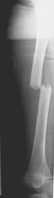
Biomechanically, fracture healing is governed by Perren’s Strain Theory. Strain is defined as the change in gap length divided by the original gap length under physiological loading. For primary bone healing (osteonal remodeling without callus) to occur, absolute stability must be achieved, keeping gap strain below 2%. For secondary bone healing (endochondral ossification with callus formation), relative stability is required, with strain levels between 2% and 10%. If the strain exceeds 10%, the mechanical environment will only support the formation of granulation tissue or fibrous tissue, leading to a hypertrophic nonunion. Hypertrophic nonunions are characterized by abundant, biologically viable callus that fails to bridge the gap due to excessive micromotion. The biological engine is running, but the mechanical clutch is slipping.
Conversely, atrophic nonunions represent a failure of biology rather than pure mechanics. In these scenarios, the fracture gap may experience appropriate strain, but the absence of vascularity, osteoprogenitor cells, or growth factors prevents the initiation of the inflammatory and reparative phases of healing. Radiographically, the bone ends appear osteopenic, sclerotic, or completely resorbed, resembling a "pencil-point" deformity. The biomechanical strategy here must shift: the surgeon cannot rely on the host's innate healing response. Instead, absolute stability must be provided (typically via rigid compression plating) to protect the massive biological augmentation (bone grafting) required to bridge the defect.
The biomechanics of the selected implant also dictate the healing pathway. Intramedullary nails function as load-sharing devices located at the neutral axis of the bone, providing excellent resistance to bending while allowing controlled axial micromotion (dynamization) to stimulate secondary healing. Plates, when applied in compression, act as load-bearing or load-sharing devices that provide absolute stability, neutralizing bending, torsional, and shear forces. External fixators, particularly circular tensioned-wire constructs (Ilizarov), offer unique viscoelastic properties. They provide sufficient stiffness to allow immediate weight-bearing while permitting axial micromotion, which is the fundamental biomechanical prerequisite for distraction osteogenesis and the tension-stress effect.
Exhaustive Indications and Contraindications
The decision-making process in nonunion surgery requires a meticulous matching of the patient's biological capacity, the nonunion classification (Weber and Cech), and the mechanical properties of the proposed construct. Surgical intervention is indicated when non-operative modalities (e.g., prolonged immobilization, biophysical stimulation) have failed or are predictably doomed to fail based on fracture morphology.
Indications and Contraindications Matrix
| Nonunion Type / Host Status | Recommended Surgical Intervention | Absolute Contraindications | Relative Contraindications |
|---|---|---|---|
| Hypertrophic Diaphyseal | Exchange intramedullary nailing; Compression plating. | Active deep infection (for internal hardware). | Severe soft tissue compromise precluding safe approach. |
| Atrophic Diaphyseal | Open debridement, decortication, rigid plating, and copious autografting. | Uncorrected metabolic bone disease; Active infection. | Active smoking; Poor soft tissue envelope. |
| Oligotrophic | Biological augmentation (BMAC, percutaneous graft) +/- mechanical stabilization. | Severe angular deformity requiring open correction. | Inability to comply with weight-bearing restrictions. |
| Infected Nonunion | Stage 1: Radical debridement, hardware removal, antibiotic spacer. Stage 2: Reconstruction. | Single-stage internal fixation with massive grafting. | Cierny-Mader Class C host (amputation may be indicated). |
| Segmental Defect (>4 cm) | Masquelet technique (induced membrane) OR Ilizarov bone transport. | Inadequate vascular runoff to support a viable limb. | Severe psychiatric illness precluding frame management. |
| Periarticular Nonunion | Rigid dual-plating with structural autograft/allograft; Arthrodesis if joint destroyed. | Active joint sepsis (requires staged approach). | Advanced age with low demand (consider primary arthroplasty). |
Absolute contraindications to complex limb salvage and nonunion reconstruction primarily revolve around the physiological status of the host. A Cierny-Mader Class C host—a patient whose medical comorbidities or systemic illness makes the operative intervention more life-threatening than the disease itself—should not be subjected to multiple, prolonged reconstructive surgeries. In such cases, functional amputation or chronic suppression may be the most appropriate and humane course of action. Furthermore, the presence of active, purulent infection absolutely precludes the definitive use of massive structural allografts or permanent internal fixation devices until the pathogen has been eradicated through radical debridement and targeted antimicrobial therapy.
Relative contraindications include active, unapologetic smoking and uncorrected endocrinopathies. Elective nonunion takedown should be delayed until the patient has achieved absolute smoking cessation for a minimum of four to six weeks, confirmed by negative serum or urine cotinine levels. Proceeding with complex bone grafting in an active smoker drastically increases the risk of graft resorption, persistent nonunion, and deep surgical site infection, often converting a salvageable limb into an amputation candidate.
Pre-Operative Planning, Templating, and Patient Positioning
Meticulous preoperative planning is the most critical determinant of success in nonunion surgery. The evaluation begins with a comprehensive metabolic workup. A complete metabolic panel, including serum calcium, phosphorus, alkaline phosphatase, 25-OH Vitamin D, parathyroid hormone (PTH), and thyroid-stimulating hormone (TSH), must be obtained. Vitamin D levels must be aggressively repleted to a minimum of 30 ng/mL (preferably >40 ng/mL) prior to surgery. Furthermore, inflammatory markers, including Erythrocyte Sedimentation Rate (ESR) and C-Reactive Protein (CRP), are mandatory to screen for occult osteomyelitis. If inflammatory markers are elevated, a preoperative aspiration of the nonunion site under fluoroscopic or ultrasound guidance should be attempted to identify the offending pathogen and tailor perioperative antibiotic prophylaxis.
Imaging modalities must be exhaustively utilized to define the mechanical and biological problem. Standard orthogonal radiographs of the entire bone, including the joints above and below, are the baseline requirement to assess deformity (varus/valgus, apex anterior/posterior, translation) and existing hardware integrity. However, plain radiographs are notoriously inaccurate for determining the true extent of bridging trabeculae. A thin-slice Computed Tomography (CT) scan with multiplanar reconstruction and metal artifact reduction sequence (MARS) is the gold standard. The CT scan accurately delineates the volume of the osseous defect, the presence of necrotic sequestrum, and the precise anatomy of articular extensions. In cases where the biological activity of the nonunion is equivocal (e.g., differentiating oligotrophic from atrophic), nuclear scintigraphy utilizing Technetium-99m combined with Indium-111 labeled white blood cells can provide definitive functional imaging.
Digital templating is mandatory. The surgeon must anticipate the required implant length, diameter, and screw trajectory. If an exchange nailing is planned, the surgeon must template for a nail that is at least 1.5 to 2.0 mm larger in diameter than the existing hardware to ensure adequate mechanical stiffness and sufficient reaming volume. If plating is planned, the plate must be long enough to allow a minimum of four bicortical screws (eight cortices) of fixation on each side of the nonunion, though six cortices may suffice in healthy metaphyseal bone. The volume of the defect must be calculated to determine the appropriate bone graft harvest site. Small defects (<10-15 cc) can be managed with local autograft or anterior iliac crest bone graft (ICBG). Massive defects (>20 cc) require posterior ICBG, Reamer-Irrigator-Aspirator (RIA) harvest from the femur, or the use of massive structural allografts supplemented with orthobiologics.
Patient positioning in the operating room must facilitate simultaneous access to the nonunion site, the bone graft harvest site, and orthogonal fluoroscopy. For femoral and tibial nonunions, the patient is typically positioned supine on a radiolucent flat table with a bump under the ipsilateral hip. This allows unhindered access to the anterior and posterior iliac crests, as well as the entire lower extremity. The use of a sterile tourniquet is recommended for tibial and periarticular nonunions to minimize blood loss during the meticulous debridement phase, though it must be deflated prior to final closure to ensure adequate hemostasis and assess the viability of the bleeding bone ends (the "paprika sign").
Step-by-Step Surgical Approach and Fixation Technique
The surgical execution of a nonunion takedown requires a systematic, uncompromising approach. The overriding principles are the eradication of necrotic tissue, the restoration of mechanical alignment and stability, and the optimization of the biological environment.
Protocol for Occult Infection Rule-Out
Regardless of the preoperative suspicion of infection, every nonunion must be treated as potentially infected. Upon exposing the nonunion site, before the administration of systemic prophylactic antibiotics (if the patient is hemodynamically stable), the surgeon must obtain a minimum of five distinct deep tissue samples from the pseudarthrosis and the bone-implant interface. These samples are sent for aerobic, anaerobic, mycobacterial, and fungal cultures. Furthermore, a sample of the pseudarthrosis tissue must be sent for immediate frozen section analysis. The presence of greater than five polymorphonuclear leukocytes (PMNs) per high-power field (HPF) is highly suggestive of acute infection, which drastically alters the surgical algorithm, often necessitating a staged approach rather than definitive internal fixation.
Surgical Management of Hypertrophic Nonunions: Exchange Nailing
Hypertrophic nonunions possess a robust biological drive but lack mechanical stability. The surgical goal is to alter the mechanical environment without disrupting the established vascularity.
1. Hardware Removal: The existing intramedullary nail and locking screws are removed.
2. Closed Reaming: A ball-tipped guide wire is passed across the nonunion site under fluoroscopic guidance. Sequential reaming is performed, over-reaming the canal by 1.5 to 2.0 mm larger than the previously extracted nail. This creates a larger working diameter for a stiffer implant and deposits autologous bone graft (reamings) directly into the nonunion site.
3. Nail Insertion and Locking: A larger diameter, stiffer intramedullary nail is inserted. Depending on the fracture pattern and rotational stability, the nail is statically or dynamically locked.
4. Biological Preservation: Crucially, the nonunion site is not opened. An open approach would strip the periosteum, devascularize the hypertrophic callus, and potentially convert a hypertrophic nonunion into an atrophic one.
Surgical Management of Atrophic Nonunions: Open Takedown and Plating
Atrophic nonunions require a complete biological overhaul and absolute mechanical stability.
1. Extensile Exposure: An extensile surgical approach is utilized. The periosteum is carefully elevated, preserving soft tissue attachments to the surrounding musculature.
2. Excisional Debridement: The fibrous pseudarthrosis is radically excised. Sclerotic, avascular bone ends are resected using a high-speed burr or oscillating saw until healthy, punctate bleeding bone is encountered (the "paprika sign"). The medullary canals of both the proximal and distal segments must be opened and reamed to restore intramedullary vascular continuity.
3. Judet Decortication: Osteoperiosteal decortication (shingling) is performed using a sharp osteotome for 2 to 3 cm proximal and distal to the nonunion. This elevates thin slivers of cortical bone attached to the periosteum, massively increasing the vascular surface area for graft incorporation.
4. Rigid Fixation: A heavy-duty construct, typically a broad dynamic compression plate (DCP) or locking compression plate (LCP), is applied. If the fracture pattern allows, absolute stability is achieved via axial compression.
5. Massive Bone Grafting: The defect and the decorticated area are packed tightly with autologous bone graft (ICBG or RIA). In cases of large volume requirements, the autograft may be expanded with allograft chips and augmented with osteoinductive agents such as recombinant human Bone Morphogenetic Protein-2 (rhBMP-2) applied on a collagen sponge carrier.
Surgical Management of Segmental Defects: The Masquelet Technique
For massive bone defects (>4 cm) resulting from trauma or radical debridement of an infected nonunion, the Masquelet (induced membrane) technique is highly effective.
1. Stage 1: Radical debridement of all necrotic bone and soft tissue. A polymethylmethacrylate (PMMA) cement spacer (often antibiotic-impregnated) is placed into the defect. The limb is stabilized with an external fixator or appropriate internal hardware. Over the next 6 to 8 weeks, a highly vascularized, biologically active pseudosynovial membrane forms around the cement spacer.
2. Stage 2: The surgical site is reopened. The induced membrane is carefully incised, and the PMMA spacer is extracted. The biological chamber created by the membrane is then densely packed with cancellous autograft (typically RIA) combined with allograft. The membrane is sutured closed over the graft, providing a vascularized envelope that prevents graft resorption and promotes rapid consolidation.
Complications, Incidence Rates, and Salvage Management
Despite meticulous planning and flawless surgical execution, the treatment of fracture nonunions is fraught with complications. The compromised biological state of the limb, combined with the extensive nature of the required surgeries, places the patient at significant risk for adverse outcomes.
Common Complications and Salvage Strategies
| Complication | Estimated Incidence | Etiology / Risk Factors | Salvage Management Strategy |
|---|---|---|---|
| Persistent Nonunion | 10% - 20% | Inadequate mechanical stability; Insufficient graft volume; Persistent host optimization failure (smoking); Unrecognized occult infection. | Re-evaluation of mechanics and biology. Revision internal fixation with massive autografting (RIA) and orthobiologics (BMP-2). |
| Deep Surgical Site Infection | 5% - 15% | Prolonged operative time; Poor soft tissue envelope; Inadequate initial debridement of infected nonunion; Hematoma formation. | Radical surgical debridement, hardware retention (if stable) or removal (if loose), targeted IV antibiotics, placement of antibiotic PMMA beads. |
| Hardware Failure | 5% - 10% | Fatigue failure of plates/screws due to persistent nonunion (race between healing and metal fatigue); Inadequate construct stiffness. | Hardware removal, optimization of the mechanical construct (e.g., dual plating, larger diameter nail), and biological augmentation. |
| Donor Site Morbidity (ICBG) | 10% - 25% | Lateral femoral cutaneous nerve injury; Hematoma; Chronic pelvic pain; Iliac wing fracture. | Transition to RIA for large graft volumes; Local wound care; Neuromodulators for nerve pain. |
| Limb Length Discrepancy (LLD) | 15% - 30% | Resection of atrophic/necrotic bone ends without compensatory distraction; Malunion. | Shoe lifts for LLD < 2 cm. Distraction osteogenesis (Ilizarov) or contralateral epiphysiodesis/shortening for LLD > 2-3 cm. |
The most devastating complication is the development of a recalcitrant, infected nonunion with massive bone loss that is refractory to multiple reconstructive attempts. In these catastrophic scenarios, the surgeon must have a frank, multidisciplinary discussion with the patient regarding limb salvage versus amputation. Salvage of a multiply-operated, stiff, insensate, and painful limb is often a disservice to the patient. A well-executed below-knee or above-knee amputation, followed by early prosthetic fitting and aggressive rehabilitation, frequently provides a superior functional outcome and a faster return to society than years of relentless, failed limb salvage procedures.
For massive defects where amputation is refused or contraindicated, a free vascularized fibular graft (FVFG) remains a viable microsurgical salvage option. By anastomosing the peroneal vessels of the fibular graft to the recipient vessels in the traumatized limb, the surgeon transfers a biologically live, structurally sound bone segment that bypasses the hostile local environment of the nonunion bed.
Phased Post-Operative Rehabilitation Protocols
The postoperative rehabilitation of a reconstructed nonunion is a delicate, protracted process that requires constant communication between the orthopedic surgeon, the physical therapist, and the patient. The protocol must be highly individualized, dictated primarily by the mechanical stability of the fixation construct and the biological quality of the host bone.
Phase 1: Protection and Early Mobilization (Weeks 0 to 6)
The primary goals of the initial phase are wound healing, mitigation of edema, prevention of deep vein thrombosis (DVT), and maintenance of adjacent joint mobility.
* Weight-Bearing: The weight-bearing status is strictly dependent on the implant. For diaphyseal nonunions treated with an exchange intramedullary nail, immediate weight-bearing as tolerated is often encouraged, as axial loading promotes dynamization and stimulates secondary bone healing. Conversely, for atrophic nonunions treated with decortication, massive structural grafting, and compression plating, strict non-weight-bearing or touch-down weight-bearing (toe-touch) is mandatory to prevent catastrophic hardware failure before biological incorporation occurs.
* Joint Mobility: Early, aggressive passive and active-assisted range of motion (ROM) of the joints proximal and distal to the nonunion is critical to prevent arthrofibrosis, a common sequela of prolonged limb dysfunction.
* Medical Management: DVT prophylaxis (e.g., low molecular weight heparin or direct oral anticoagulants) is maintained per institutional protocols. Strict adherence to smoking cessation is monitored.
Phase 2: Progressive Loading and Biophysical Stimulation (Weeks 6 to 12)
As the inflammatory and early reparative phases of healing progress, the focus shifts to progressive mechanical loading to stimulate osteoblastic activity via Wolff's Law.
* Radiographic Monitoring: Orthogonal radiographs are obtained at 6, 8, and 12 weeks. The surgeon looks for signs of graft incorporation, blurring of the fracture lines, and the absence of hardware failure (e.g., broken screws, plate pullout).
* Weight-Bearing: For plated constructs, partial weight-bearing (e.g., 25% to 50% of body weight) is initiated once early radiographic callus or graft incorporation is visualized.
* Biophysical Stimulation: Adjuvant non-invasive therapies are frequently deployed. Pulsed Electromagnetic Fields (PEMF) induce a time-varying magnetic field that generates an electrical current, upregulating BMP expression and osteoblast proliferation. Low-Intensity Pulsed Ultrasound (LIPUS) can also be utilized to stimulate angiogenesis and chondrocyte proliferation in the fracture gap.
Phase 3: Strengthening and Return to Function (Months 3 to 6+)
Once definitive radiographic union is achieved—defined as bridging trabeculae across at least three of four cortices on orthogonal radiographs—the patient transitions to functional restoration.
* Strengthening: Progressive resistive exercises are initiated to rebuild the severely atrophied musculature. Isotonic and isokinetic strengthening optimizes the mechanical environment surrounding the newly healed bone.
* Gait Training: Normalization of gait mechanics is emphasized, weaning the patient off assistive devices (crutches, canes) as tolerated.
* Return to Activity: Return to heavy manual labor or high-impact sports is delayed until the bone has undergone significant remodeling, often taking 9 to 12 months from the time of the definitive reconstruction.
Summary of Landmark Literature and Clinical Guidelines
The modern management of fracture nonunions is built upon a foundation of landmark scientific discoveries and rigorous clinical trials. A comprehensive understanding of this literature is essential for evidence-based orthopedic practice.
- The Diamond Concept (Giannoudis et al., 2007): This seminal paper revolutionized the conceptual framework of fracture healing. Giannoudis established that successful bone repair requires the spatial and temporal orchestration of osteogenic cells, an osteoconductive scaffold, osteoinductive mediators, and mechanical stability, all underpinned by adequate vascularity. This concept forms the basis of all modern biological augmentation strategies in nonunion surgery.
- Weber and Cech Classification (1976): Based on extensive clinical and radiographic observations, Weber and Cech classified nonunions into biologically active (hypertrophic, oligotrophic) and biologically inactive (atrophic) categories. This classification remains the gold standard because it directly dictates the surgical algorithm: mechanical stabilization for active nonunions, and biological augmentation combined with stabilization for inactive nonunions.
- Discovery of Bone Morphogenetic Proteins (Marshall Urist, 1965): Urist’s discovery that demineralized bone matrix could induce ectopic bone formation via BMPs laid the groundwork for the field of orthobiologics. The subsequent development of recombinant human BMPs (rhBMP-2 and rhBMP-7) provided surgeons with potent, off-the-shelf osteoinductive agents to treat recalcitrant atrophic nonunions.
- The LEAP Study (Lower Extremity Assessment Project): This massive, multicenter prospective study evaluated the outcomes of limb salvage versus amputation for severe lower extremity trauma. It definitively highlighted the catastrophic impact of smoking and poor host biology on the success rates of limb reconstruction, establishing stringent criteria for patient optimization prior to undertaking complex nonunion surgery.
- Principles of Distraction Osteogenesis (Gavriil Ilizarov, 1989): Ilizarov’s discovery of the tension-stress effect—that gradual, controlled distraction of living tissue stimulates the regeneration of bone and soft tissue—revolutionized the treatment of massive segmental defects and infected nonunions. The Ilizarov method remains the ultimate salvage tool for defects that exceed the capacity of traditional bone grafting techniques.
- The SPRINT Trial (Bhandari et al., 2008): While primarily focused on acute tibial shaft fractures, the Study to Prospectively Evaluate Reamed Intramedullary Nails in Patients with Tibial Fractures (SPRINT) provided level I evidence regarding the biological impact of reaming. The trial demonstrated a trend toward lower nonunion rates with reamed nails in closed fractures, supporting the principle that reamings act as an autologous bone graft, a concept heavily utilized in the exchange nailing of hypertrophic nonunions.
This academic synthesis is based on established protocols from Hutaifortho's Operative Orthopaedics and has been medically reviewed by Prof. Dr. Mohammed Hutaif, Consultant Orthopedic & Spine Surgeon. It is designed to assist orthopedic residents, fellows, and practicing surgeons in surgical preparation and board reviews (AAOS, FRCS, Arab Board).
This academic resource was prepared and medically reviewed by Prof. Dr. Mohammed Hutaif, Consultant Orthopedic & Spine Surgeon. It is formulated specifically for medical students, orthopedic residents, and surgeons preparing for high-stakes board examinations (AAOS, FRCS Tr & Orth, Arab Board).
