Benign Bone Tumors: Operative Management & Surgical Techniques
Key Takeaway
This comprehensive guide details the operative management of benign bone tumors and tumor-like lesions. Designed for orthopedic surgeons and residents, it covers indications, biomechanics, and step-by-step surgical techniques for unicameral bone cysts, aneurysmal bone cysts, osteoid osteomas, and cartilaginous lesions. Evidence-based protocols for extended curettage, radiofrequency ablation, and postoperative rehabilitation are thoroughly examined to optimize patient outcomes and minimize recurrence.
Comprehensive Introduction and Patho-Epidemiology
The operative management of benign bone tumors and tumor-like conditions represents a complex intersection of musculoskeletal oncology, advanced biomechanics, and precise surgical execution. While these osseous lesions lack the capacity for distant metastatic dissemination, their localized biological behavior can be profoundly destructive. The capacity of these tumors to induce severe local tissue degradation, extensive cortical expansion, and catastrophic pathologic fractures necessitates highly specific, evidence-based surgical interventions. Historically, the orthopedic approach to benign osseous lesions relied heavily on aggressive open en bloc resections or, conversely, simple intralesional curettage. These historical modalities often carried unacceptable morbidity profiles, including massive structural defects, or yielded prohibitive local recurrence rates. Over the past three decades, the surgical paradigm has definitively shifted toward joint-sparing, minimally invasive, and adjuvant-enhanced techniques that prioritize both oncologic eradication and immediate biomechanical restoration.
To master the surgical management of benign bone tumors, the orthopedic surgeon must first possess a profound understanding of their patho-epidemiology and biological staging. The Enneking staging system for benign musculoskeletal tumors remains the foundational framework for surgical decision-making. Stage 1 (latent) lesions, such as asymptomatic non-ossifying fibromas or static enchondromas, are characterized by a well-defined margin, a thick reactive sclerotic rim, and no progressive growth; these typically require only observation. Stage 2 (active) lesions, such as unicameral bone cysts (UBCs) or osteoid osteomas, exhibit progressive growth, cause mild to moderate symptoms, and expand the host bone while remaining contained within natural anatomical barriers. Stage 3 (aggressive) lesions, most notably aneurysmal bone cysts (ABCs) and giant cell tumors (GCTs) of bone, demonstrate rapid, locally destructive growth, cortical breakthrough, and extension into adjacent soft tissue compartments. The Enneking stage directly dictates the required surgical margin, ranging from simple percutaneous intervention to extended intralesional curettage with aggressive adjuvants, or even wide marginal resection.
The epidemiology and molecular pathophysiology of these lesions further guide surgical strategy. Unicameral bone cysts predominantly affect the metaphyses of long bones in skeletally immature patients, driven by an underlying venous outflow obstruction that leads to increased intraosseous pressure and subsequent osteoclastic resorption. Conversely, aneurysmal bone cysts are driven by an upregulation of the USP6 oncogene, resulting in highly vascular, destructive, multiloculated blood-filled cavities. Fibrous dysplasia, driven by a somatic activating mutation in the GNAS1 gene, results in the replacement of normal lamellar bone with mechanically inferior woven bone and fibrous stroma, leading to the classic "shepherd's crook" deformity. Osteoid osteomas secrete high levels of prostaglandins (specifically PGE2), which mediate the intense, nocturnal pain characteristic of the disease and the dense reactive sclerosis surrounding the highly vascularized nidus.
The modern orthopedic armamentarium has evolved to address these specific pathophysiological mechanisms. The integration of high-speed burring, chemical adjuvants (such as phenol and hydrogen peroxide), thermal adjuvants (liquid nitrogen cryotherapy and polymethylmethacrylate cementation), percutaneous sclerotherapy, and radiofrequency ablation (RFA) has revolutionized operative management. This definitive chapter delineates the surgical indications, biomechanical considerations, step-by-step operative approaches, and phased postoperative protocols for the most frequently encountered benign bone tumors, synthesizing decades of peer-reviewed literature into highly actionable, master-level surgical strategies.
Detailed Surgical Anatomy and Biomechanics
A profound comprehension of osseous biomechanics is the cornerstone of surgical planning for benign bone tumors. The presence of a lytic lesion within the medullary canal or cortex fundamentally alters the mechanical properties of the host bone, creating significant stress risers. The structural integrity of a long bone is largely dependent on its cross-sectional geometry, specifically the polar moment of inertia, which dictates torsional rigidity. Because the polar moment of inertia is proportional to the fourth power of the radius ($J /propto r^4$), even minor reductions in cortical thickness or small eccentric cortical defects can exponentially decrease the bone's resistance to torsional forces. Consequently, benign lesions that cause endosteal scalloping or eccentric cortical expansion dramatically increase the susceptibility to spiral pathologic fractures under normal physiologic loads.
The creation of a surgical cortical window for intralesional curettage further compromises local biomechanics. When a closed cylindrical tube (the diaphyseal cortex) is converted into an "open section" via a cortical window, its torsional strength is reduced by up to 70-90%, depending on the size of the defect. The stress concentration factor ($K_t$) at the corners of a rectangular cortical window is exceptionally high, predisposing the bone to iatrogenic fracture propagation. To mitigate this, orthopedic oncologists must design cortical windows that are oval or elliptical, with gently rounded corners, thereby dispersing stress lines. Furthermore, the length of the cortical window should not exceed the absolute minimum required for complete visualization and access to the tumor margins, and its width should ideally remain less than 30% of the bone's total circumference to preserve basic axial load-bearing capacity.
Anatomic location heavily influences the biomechanical failure mode and subsequent reconstructive strategy. In the proximal femur, lesions such as fibrous dysplasia or large solitary bone cysts are subjected to massive compressive forces on the medial calcar and extreme tensile forces on the lateral cortex. The dysplastic woven bone of fibrous dysplasia lacks the anisotropic lamellar orientation required to resist these bending moments, inevitably leading to progressive varus bowing (the shepherd's crook deformity) and eventual tension-sided failure. Surgical reconstruction in this anatomical zone cannot rely on standard plates and screws, which are prone to pull-out failure in mechanically inferior bone. Instead, intramedullary load-sharing devices, such as cephalomedullary nails, are mandatory to bypass the dysplastic segment and transfer forces directly to the healthy distal diaphysis.
Conversely, in the hand and phalanges, where enchondromas frequently present, the biomechanical priority shifts toward preserving the delicate extensor mechanism and preventing joint contractures. The phalangeal cortices are extraordinarily thin, and cystic expansion can reduce them to an eggshell-like consistency. Surgical access must navigate between the central slip and lateral bands without violating the articular cartilage. Reconstruction of these small, high-stress cavities often necessitates the use of injectable, in situ-curing calcium phosphate or hydroxyapatite cements. These synthetic bone substitutes offer immediate, high compressive strength (often exceeding that of normal cancellous bone), which neutralizes the stress riser effect and permits the immediate active range of motion essential for preventing catastrophic flexor or extensor tendon adhesions.
Exhaustive Indications and Contraindications
The decision to proceed with operative intervention for a benign bone tumor requires a nuanced risk-benefit analysis, balancing the natural history of the lesion against the inherent risks of surgery. Prophylactic intervention is strictly indicated when the risk of pathologic fracture outweighs the morbidity of the procedure. The Mirels' scoring system remains a highly validated, objective tool for predicting fracture risk in long bone lesions. It evaluates four parameters: site (upper limb, lower limb, trochanteric region), pain (mild, moderate, functional), lesion type (blastic, mixed, lytic), and size relative to bone diameter (<1/3, 1/3–2/3, >2/3). A Mirels' score of 8 or higher is generally accepted as an absolute indication for prophylactic internal fixation, as the fracture risk exceeds 15%, while a score of 9 carries a fracture risk approaching 30%.
For cystic lesions such as Unicameral Bone Cysts (UBCs), indications for intervention include a high risk of pathologic fracture (defined biomechanically as cortical thickness < 2 mm or cyst diameter encompassing > 85% of the bone cross-section), active cysts abutting the physis causing impending growth disturbance, or recurrent pain indicating micro-fracture. A critical relative contraindication for immediate cyst curettage is the presence of an acute, displaced pathologic fracture. In these scenarios, the preferred strategy is to allow the fracture to heal over 6 to 8 weeks; the influx of the fracture hematoma occasionally induces spontaneous cyst consolidation, rendering surgical curettage unnecessary. If the cyst persists after fracture union, definitive intervention can then be pursued.
Aneurysmal Bone Cysts (ABCs) and Giant Cell Tumors (GCTs) present a more aggressive clinical profile, making observation alone a contraindication. Because these lesions rapidly destroy bone and expand into soft tissues, surgical intervention is almost universally indicated upon diagnosis to prevent catastrophic mechanical failure and alleviate severe pain. In the spine, osteoblastomas and aggressive ABCs frequently cause painful scoliosis or neurological compromise due to epidural extension. Here, the indication for surgery is absolute, often requiring urgent decompression, marginal en bloc resection, and complex multi-level spinal instrumentation.
Radiofrequency ablation (RFA) has become the gold standard for osteoid osteomas, indicated for intractable nocturnal pain refractory to NSAIDs. However, specific contraindications to RFA must be rigorously respected. RFA is absolutely contraindicated for lesions located within 1.0 to 1.5 cm of critical neurovascular structures (such as the spinal cord, nerve roots, or major peripheral nerves) due to the unpredictable sphere of thermal necrosis, which can cause irreversible neural injury. In these anatomically precarious locations, computer-navigated open burr-down resection or en bloc excision remains the safest and most definitive surgical option.
| Tumor Type | Primary Surgical Indications | Relative/Absolute Contraindications | Preferred Surgical Modality |
|---|---|---|---|
| Unicameral Bone Cyst (UBC) | Mirels score $/ge$ 8, cortical thickness < 2mm, persistent pain, physeal threat. | Acute unhealed pathologic fracture (allow healing first). | Percutaneous injection (steroid/BMA) or open curettage + grafting. |
| Aneurysmal Bone Cyst (ABC) | Progressive expansion, impending fracture, severe pain, neurologic deficit (spine). | Observation is contraindicated due to aggressive natural history. | Extended intralesional curettage + high-speed burr + adjuvants + PMMA/graft. |
| Osteoid Osteoma | Intractable nocturnal pain, NSAID failure, painful scoliosis, joint synovitis. | Nidus < 1 cm from spinal cord or major nerve (Absolute contraindication for RFA). | CT-guided Radiofrequency Ablation (RFA). Open resection if near nerves. |
| Enchondroma | Symptomatic pain, impending fracture, radiographic signs of malignant transformation. | Asymptomatic, static lesions (Stage 1 latent) should be observed. | Intralesional curettage + calcium phosphate cement or autograft. |
| Fibrous Dysplasia | Severe bowing (shepherd's crook), impending fracture, symptomatic nonunion. | Use of plates/screws (high failure rate in dysplastic bone). | Prophylactic intramedullary nailing + structural cortical allografting. |
Pre-Operative Planning, Templating, and Patient Positioning
Meticulous pre-operative planning is the most critical phase in the operative management of benign bone tumors. The orthopedic surgeon must synthesize clinical data, advanced imaging, and histopathological confirmation prior to making any incision. High-quality orthogonal radiographs of the entire involved bone are mandatory to assess the overall mechanical axis, identify skip lesions, and evaluate cortical integrity. Magnetic Resonance Imaging (MRI) with and without intravenous gadolinium contrast is essential to delineate the intraosseous extent of the tumor, evaluate for soft tissue extension, and map the proximity of the lesion to critical neurovascular bundles and adjacent physes. For osteoid-producing lesions like osteoid osteoma and osteoblastoma, thin-slice (1 mm) computed tomography (CT) is the imaging modality of choice to precisely localize the central radiolucent nidus within the surrounding dense reactive sclerosis.
If a pre-operative biopsy is required—particularly to rule out malignant mimics such as telangiectatic osteosarcoma in the case of an ABC, or low-grade chondrosarcoma in the case of an enchondroma—the biopsy must be performed with strict adherence to oncologic principles. The biopsy tract must be oriented longitudinally and placed directly in line with the planned definitive surgical incision. Transverse incisions are strictly forbidden, as they contaminate multiple anatomical compartments and complicate future resections. Meticulous hemostasis must be achieved during the biopsy to prevent a post-operative hematoma from seeding tumor cells into adjacent, uncontaminated tissue planes. In modern practice, image-guided core needle biopsy has largely replaced open incisional biopsy, significantly reducing the risk of tract contamination.
Digital templating is an absolute requirement when prophylactic stabilization or deformity correction is planned. For lesions requiring intramedullary nailing, such as fibrous dysplasia of the femur, the surgeon must template the entire length of the bone to select an implant that achieves adequate purchase in healthy host bone both proximal and distal to the lesion. If a shepherd's crook deformity is present, the surgeon must pre-operatively calculate the angles for corrective closing wedge osteotomies required to restore the mechanical axis and permit the passage of a rigid cephalomedullary nail. The templating process must also account for the volume of the osseous defect created by curettage to ensure adequate availability of structural allograft, cancellous autograft, or synthetic bone cement.
Patient positioning and operating room setup must be optimized to facilitate unhindered surgical access, fluoroscopic imaging, and the potential need for extensile approaches. The patient is typically positioned on a fully radiolucent operative table. A pneumatic tourniquet should be applied for all extremity lesions to ensure a bloodless surgical field, which is absolutely critical for identifying the subtle visual distinctions between normal reactive bone and residual tumor tissue during curettage. For highly vascular lesions like ABCs, the tourniquet also prevents massive, life-threatening intraoperative hemorrhage. The fluoroscopy unit (C-arm) must be positioned to allow seamless transition between anteroposterior and lateral views without compromising the sterile field. For complex pelvic or spinal lesions, intraoperative CT-based computer navigation systems (e.g., O-arm with StealthStation) should be integrated into the surgical setup to guide precise burring and hardware placement.
Step-by-Step Surgical Approach and Fixation Technique
Intralesional Curettage and Adjuvant Therapy (ABCs, GCTs, Chondroblastomas)
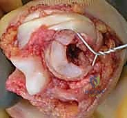
The gold standard for aggressive benign lesions (Enneking Stage 3) is extended intralesional curettage supplemented with chemical and thermal adjuvants. The procedure begins with an extensile longitudinal incision, carefully excising any prior biopsy tracts. The underlying fascia is incised in line with the skin, and the tumor is approached via standard internervous and intermuscular planes. Once the affected bone is exposed, a precise, oval-shaped cortical window is created using a small drill bit and sharp osteotomes. The window must be large enough to allow direct visualization of the entire tumor cavity, including the most proximal and distal recesses.
Meticulous curettage is then performed using a series of straight, angled, and reverse-angled ring curettes. The surgeon must systematically scrape the cavity walls until all gross, macroscopic tumor tissue (often described as hemorrhagic, spongy, or fleshy) is evacuated. Following gross removal, the critical phase of extended curettage begins. A high-speed spherical burr (e.g., Midas Rex) is introduced into the cavity. The burr is used to systematically abrade the entire endosteal surface, breaking down all microscopic bony septations and extending the resection margin 1 to 2 millimeters into normal, healthy reactive bone. Continuous cold saline irrigation is mandatory during burring to clear debris and prevent premature thermal necrosis of the healthy bone.
Once the cavity is visually clear of tumor, adjuvant therapy is applied to eradicate microscopic disease and reduce recurrence rates from >30% to <10%. Phenol (an 89% solution) is commonly used; it acts as a potent chemical cautery agent, denaturing proteins and inducing cell death. It is carefully applied to the cavity walls using cotton-tipped applicators for three cycles of two minutes each, taking extreme care to protect surrounding soft tissues from chemical burns. The phenol is then immediately neutralized with copious 70% isopropyl alcohol lavage, followed by normal saline irrigation. Alternatively, liquid nitrogen (cryotherapy) or an argon beam coagulator can be utilized to achieve a 2- to 3-millimeter zone of thermal necrosis. The resultant defect is then meticulously dried and packed with polymethylmethacrylate (PMMA) bone cement. PMMA not only provides immediate, rigid structural support but also generates an exothermic reaction (reaching temperatures up to 80°C) that serves as a final thermal adjuvant.
Percutaneous Techniques and Radiofrequency Ablation (UBCs, Osteoid Osteoma)
For Unicameral Bone Cysts (UBCs) requiring intervention, percutaneous injection is the first-line surgical modality. The patient is positioned supine on a radiolucent table under general anesthesia. Under live biplanar fluoroscopy, two large-bore Jamshidi trocars are advanced percutaneously into the cyst cavity—one positioned at the proximal pole and the other at the distal pole. The stylets are removed, and the characteristic clear, straw-colored serous fluid is aspirated. A cystogram is then performed by injecting radiopaque contrast material to confirm that the needles are within the cyst and to rule out loculations that might prevent the therapeutic agent from reaching the entire cavity. The cyst is then aggressively flushed with sterile normal saline to mechanically disrupt the fibrous cyst lining. Finally, 40 to 80 mg of methylprednisolone acetate or autologous bone marrow aspirate (harvested concurrently from the anterior iliac crest) is injected through the distal needle while the proximal needle acts as a vent.
For Osteoid Osteomas, CT-guided Radiofrequency Ablation (RFA) has superseded open resection. The procedure is performed in the CT suite under deep sedation or general anesthesia, as the thermal ablation is intensely painful. A localizing grid is placed on the skin, and a thin-slice CT scan identifies the exact coordinates of the hyperemic nidus. A rigid biopsy cannula is advanced through the cortex and positioned precisely in the geometric center of the nidus. A biopsy is often taken prior to ablation. The RFA probe (typically a 17-gauge electrode with a 5 mm to 10 mm active tip) is then introduced through the cannula. The generator is activated, heating the tissue to precisely 90°C for exactly 6 minutes. This temperature profile ensures complete coagulative necrosis of the nidus and a 1 cm surrounding margin, effectively destroying the prostaglandin-secreting cells.
Prophylactic Internal Fixation and Structural Grafting (Fibrous Dysplasia)
Surgical intervention for Fibrous Dysplasia necessitates a fundamentally different biomechanical approach, as the dysplastic woven bone is inherently weak and prone to progressive deformity. When addressing a symptomatic lesion in the proximal femur, the patient is positioned on a fracture table. If a severe shepherd's crook deformity precludes the passage of an intramedullary device, the surgeon must first perform one or more closing-wedge valgus osteotomies at the apex of the deformity.
Once the mechanical axis is restored, the medullary canal is reamed sequentially. Reaming must be performed with caution, as the dysplastic bone can be highly vascular and prone to massive intraoperative hemorrhage. A rigid, titanium cephalomedullary nail is then inserted, bypassing the entire dysplastic segment. The proximal interlocking screws must achieve purchase in the dense bone of the femoral head, while the distal locking screws must anchor securely in healthy diaphyseal cortical bone. Because cancellous autograft is rapidly resorbed and incorporated into the dysplastic process, any significant structural defects or osteotomy sites must be grafted with rigid cortical allograft struts (e.g., fibular allograft), which provide durable mechanical support and resist osteoclastic resorption.
En Bloc Resection and Marginal Excision (Osteochondroma)
Osteochondromas (exostoses) require meticulous marginal excision to prevent recurrence and eliminate mechanical irritation. The surgical approach is dictated by the lesion's anatomic location (e.g., distal femur, proximal tibia). The critical oncologic principle is the complete, unviolated excision of the hyaline cartilage cap and its overlying perichondrium, as this is the site of active proliferation and potential malignant transformation.
The surgeon dissects the overlying soft tissues, muscles, and neurovascular structures bluntly, sweeping them away from the exostosis. The bursa that frequently overlies the cartilage cap is excised en bloc with the tumor. The surgeon must absolutely avoid violating or fragmenting the bluish-white cartilage cap. Once the bony stalk is isolated, curved Hohmann retractors are placed circumferentially to protect surrounding structures. A broad osteotome or an oscillating saw is used to resect the stalk completely flush with the surrounding host bone cortex. The specimen is sent for histopathological analysis to confirm the benign nature of the cap (typically < 1.5 cm thick in adults; caps > 2 cm raise high suspicion for secondary peripheral chondrosarcoma). The cortical defect is smoothed with a rasp, and bone wax may be applied to minimize postoperative medullary bleeding.
Complications, Incidence Rates, and Salvage Management
Despite the utilization of advanced surgical techniques and adjuvants, the operative management of benign bone tumors is associated with a distinct profile of complications. The most persistent and challenging complication is local recurrence, which is highly dependent on the tumor's biological aggressiveness and the adequacy of the initial surgical margin. Aneurysmal bone cysts and giant cell tumors treated with simple curettage alone historically exhibited recurrence rates exceeding 30-50%. The advent of high-speed burring and chemical/thermal adjuvants has successfully driven this incidence down to approximately 5-10%. When recurrence does occur, it typically manifests within the first 24 months post-operatively. Salvage management for recurrent aggressive benign lesions involves a repeat, more aggressive extended curettage, often utilizing a different adjuvant modality (e.g., switching from phenol to liquid nitrogen), or, in refractory cases, progressing to wide marginal en bloc resection and complex endoprosthetic reconstruction.
Pathologic fracture following intralesional curettage is another significant complication, particularly when the cortical window is excessively large or when the defect is inadequately reconstructed. The use of PMMA bone cement significantly reduces this risk by providing immediate compressive strength; however, the bone-cement interface can become a stress riser over time, potentially leading to periprosthetic fractures. If a post-operative fracture occurs, it is managed based on standard traumatology principles, typically requiring open reduction and internal fixation (ORIF) with rigid plating, often supplemented with additional structural bone grafting to address the void.
Physeal arrest and subsequent growth deformity are profound concerns when operating on skeletally immature patients, particularly during the treatment of metaphyseal lesions like UBCs or chondroblastomas (which classically cross the physis into the epiphysis). Iatrogenic injury to the physis during curettage or thermal damage from adjuvants can lead to premature partial or complete physeal closure, resulting in limb length discrepancies or angular deformities. Salvage strategies for physeal arrest include epiphysiodesis of the contralateral limb to equalize length, or complex corrective osteotomies and spatial frame applications (e.g., Ilizarov or Taylor Spatial Frame) for angular corrections.
| Complication | Incidence Rate (%) | Associated Tumor / Procedure | Salvage Management Strategy |
|---|---|---|---|
| Local Recurrence | 5% - 15% (with adjuvants) | ABCs, GCTs, Osteoblastoma | Repeat extended curettage with alternative adjuvants, or wide en bloc resection. |
| Post-Op Pathologic Fracture | 3% - 8% | Large UBCs, Fibrous Dysplasia | Rigid internal fixation (ORIF or IM Nailing) + structural allografting. |
| Physeal Arrest / Deformity | 2% - 5% | UBC injections, Chondroblastoma | Contralateral epiphysiodesis, corrective osteotomy, or external fixation lengthening. |
| Thermal Nerve Injury | < 1% | RFA for Osteoid Osteoma | Often irreversible. Prevention is key (avoid RFA if nidus is <1cm from nerve). |
| Malignant Transformation | < 1% (Solitary) up to 30% (Syndromic) | Enchondroma (Ollier's), Osteochondroma | Immediate wide oncologic resection and staging for secondary chondrosarcoma. |
Phased Post-Operative Rehabilitation Protocols
The post-operative rehabilitation protocol following the surgical management of benign bone tumors is highly individualized. It is dictated entirely by the anatomic location of the lesion, the size of the residual cortical defect, and the specific reconstructive modality employed (e.g., PMMA cement vs. cancellous autograft vs. structural allograft). The overarching goal of rehabilitation is to safely restore the patient to their pre-morbid functional baseline while strictly protecting the surgical construct from premature mechanical failure.
Phase I: Immediate Post-Operative Protection (Weeks 0 - 6)
For lower extremity lesions where the cortical defect or window exceeds 30% of the bone's diameter, or where prophylactic intramedullary nailing was performed for fibrous dysplasia, strict protective weight-bearing is mandatory. Patients are restricted to touch-down weight-bearing (TDWB) or partial weight-bearing (PWB) using bilateral crutches or a walker. This protects the bone from extreme bending and torsional forces while early callus formation begins. Conversely, for upper extremity lesions—particularly enchondromas of the hand reconstructed with high-compressive-strength synthetic cements—immediate active range of motion (AROM) is aggressively encouraged. Prolonged immobilization of the digits inevitably leads to devastating adhesive capsulitis, flexor tendon adhesions, and permanent stiffness. In the upper extremity, the mechanical strength of the cement allows for early motion, though heavy lifting and gripping are restricted.
Phase II: Progressive Loading and Mobility (Weeks 6 - 12)
At the 6-week mark, orthogonal radiographs are obtained to assess the incorporation of bone grafts, the stability of synthetic cements, and the early bridging of any osteotomy sites. If radiographic evidence of healing is satisfactory and the patient is pain-free upon palpation of the surgical site, weight-bearing status is progressively advanced. Lower extremity patients transition from crutches to a single cane, and eventually to full weight-bearing (FWB) as tolerated. Physical therapy focuses on restoring normal gait mechanics, normalizing joint kinematics, and initiating closed-kinetic-chain strengthening exercises to rebuild the atrophied periarticular musculature.
Phase III: Advanced Strengthening and Return to Activity (Months 3 - 6+)
Once full, painless weight-bearing is achieved and radiographic consolidation is confirmed, the rehabilitation protocol shifts toward high-level functional restoration. Isokinetic strengthening, proprioceptive training, and sport-specific drills are introduced. Return to high-impact activities, contact sports, or heavy manual labor is generally restricted until at least 6 months post-operatively, and only after complete radiographic incorporation of the graft or complete cortical remodeling around a cement mantle is visualized.
Radiographic surveillance is a critical, ongoing component of the post-operative protocol. Baseline orthogonal radiographs are obtained immediately in the Post-Anesthesia Care Unit (PACU).
