Master the Principles of Biopsy: Your Guide to Accurate Tissue Diagnosis
Key Takeaway
Here are the crucial details you must know about Master the Principles of Biopsy: Your Guide to Accurate Tissue Diagnosis. The principles of biopsy for suspected sarcoma involve performing it at a specialised sarcoma centre with histopathology support. It must be planned with the oncology surgeon, obtain representative tissue (often from the periphery), avoid violating surrounding compartments, and have the biopsy tract marked by tattoo to aid definitive surgical excision.
Introduction and Epidemiology
Musculoskeletal oncology represents a highly specialized and technically demanding domain within orthopedic surgery, where diagnostic accuracy and procedural precision directly dictate patient survival and limb salvage potential. In this context, the biopsy is not merely a diagnostic adjunct; it is the final, definitive staging procedure in the management of bone and soft tissue sarcomas. Errors in biopsy technique, anatomical placement, or execution can lead to catastrophic oncological outcomes, including unnecessary amputation, local recurrence, and compromised overall survival.
Sarcomas encompass a heterogeneous group of mesenchymal malignancies that are broadly classified into soft tissue sarcomas and primary bone sarcomas. Epidemiologically, primary bone sarcomas—most notably osteosarcoma, Ewing sarcoma, and chondrosarcoma—are exceptionally rare, accounting for less than 0.2% of all malignant neoplasms. They often present with a bimodal age distribution, peaking in the second decade of life and again in the sixth decade (often secondary to Paget's disease or prior irradiation). Soft tissue sarcomas are slightly more common but still represent only approximately 1% of adult malignancies.
Sarcoma Classification and Multidisciplinary Approach
The cornerstone of modern orthopedic oncology is the multidisciplinary team (MDT) approach. All patients presenting with a suspected sarcoma, an indeterminate bone lesion, or an isolated musculoskeletal metastasis must be referred to a dedicated musculoskeletal tumor unit prior to any surgical intervention, including biopsy. The MDT typically consists of fellowship-trained orthopedic oncology surgeons, musculoskeletal radiologists, specialized soft-tissue and bone histopathologists, medical oncologists, and radiation oncologists.
Appropriate early referral to a sarcoma unit is critical. Careful diagnostic workup and coordinated management play a fundamental role in achieving optimal oncological and functional outcomes. The biopsy must be planned as a collaborative effort between the operating oncology surgeon and the interventional radiologist. Comprehensive discussion and planning from the time of initial clinical presentation through to postoperative care, adjuvant therapy, and long-term surveillance are mandatory.
Surgical Anatomy and Biomechanics
A profound understanding of compartmental anatomy is an absolute prerequisite for performing a musculoskeletal biopsy. The fundamental oncological principle is that a biopsy tract contaminates the anatomical compartment it traverses with malignant cells. Therefore, the biopsy tract must be considered a contaminated zone that will require en bloc excision during the definitive tumor resection.
Compartmental Barriers and Tumor Spread
Anatomical compartments are defined by natural barriers to tumor extension, which include major fascial septa, periosteum, articular cartilage, and cortical bone. Tumors constrained within these boundaries are considered intracompartmental (Enneking Stage A), whereas those that breach these barriers are classified as extracompartmental (Enneking Stage B).

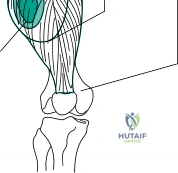
When planning a biopsy, the surgeon must ensure that the needle or surgical incision does not violate adjacent, uninvolved compartments. For example, a lesion located in the vastus intermedius (anterior compartment of the thigh) must be biopsied directly through the anterior compartment. Passing a needle through the lateral intermuscular septum from the posterior or medial compartment would contaminate a previously clean space. This iatrogenic contamination can definitively convert a potential limb-sparing resection into a mandatory amputation to achieve negative margins.

Biomechanical Considerations of Cortical Windows
In open biopsies of bone, creating a cortical window introduces a significant stress riser, altering the polar moment of inertia of the diaphysis or metaphysis. The biomechanical integrity of the bone is acutely compromised, drastically increasing the risk of a pathological fracture under physiological loads. A fracture through a primary bone sarcoma disseminates tumor cells into the surrounding soft tissues (extracompartmental spread) and fracture hematoma, worsening the prognosis and complicating local control.
Therefore, cortical windows must be meticulously planned. They should be circular or oval to minimize stress concentration factors at sharp corners, and they must be limited to the minimum geometric size necessary for adequate tissue sampling and visualization.

Indications and Contraindications
The primary indication for a biopsy in musculoskeletal oncology is to obtain a definitive histological diagnosis to guide subsequent neoadjuvant therapy, radiation, or surgical resection. Biopsy modalities include Fine Needle Aspiration (FNA), Core Needle Biopsy (CNB), Open Incisional Biopsy, and Excisional Biopsy.
Indications for Needle Biopsy
Core needle biopsy is currently the gold standard for the initial histological diagnosis of both bone and soft tissue sarcomas. It provides structural tissue architecture, which is critical for grading and subtyping sarcomas. This is a distinct advantage over FNA, which only provides cytological detail and is generally insufficient for primary sarcoma diagnosis. Indications for CNB include any suspected primary bone or soft tissue malignancy where tissue architecture is required for immunohistochemistry, flow cytometry, and molecular cytogenetics (e.g., identifying the t(11;22) translocation in Ewing sarcoma).

Contraindications for Needle Biopsy
Absolute contraindications include lesions situated in close proximity to major neurovascular structures where the required trajectory puts these structures at unacceptable risk of injury. Relative contraindications include severe coagulopathy or bleeding diathesis, which could lead to a massive post-procedural hematoma. A hematoma is oncologically disastrous as it acts as a fluid vehicle for tumor cell dissemination throughout the fascial planes, effectively expanding the required resection volume.

Operative vs Non Operative Biopsy Indications
| Biopsy Modality | Primary Indications | Contraindications | Key Surgical Considerations |
|---|---|---|---|
| Core Needle Biopsy (CNB) | Suspected primary sarcoma; Metastatic disease; Soft tissue masses >5cm. | Inaccessible lesions; High risk of neurovascular injury; Bleeding diathesis. | Gold standard. Requires multiple passes (14G or 16G). Tract must be excisable. |
| Fine Needle Aspiration (FNA) | Confirming local recurrence; Documenting metastatic spread to lymph nodes. | Primary diagnosis of sarcoma (insufficient architecture for grading). | High non-diagnostic rate for primary mesenchymal tumors. Cytology only. |
| Open Incisional Biopsy | Non-diagnostic CNB; Need for large tissue volume (e.g., suspected lymphoma or atypical infection). | Poorly planned incision that compromises future limb salvage; Transverse incisions. | Requires strict hemostasis. Longitudinal incisions only. Defect must be plugged. |
| Excisional Biopsy | Small (<3cm) superficial soft tissue masses; Characteristic benign bone lesions (e.g., osteoid osteoma). | Suspected primary sarcoma >5cm; Deep subfascial lesions. | May result in positive margins requiring wide re-excision if histology returns malignant. |
Pre Operative Planning and Patient Positioning
Preoperative planning is arguably the most critical phase of the biopsy process. A biopsy must never be performed before complete local and systemic staging is obtained, as the biopsy artifact (edema, hemorrhage) can severely degrade the quality of subsequent imaging.
Staging and Imaging Modalities
Local staging requires a contrast-enhanced MRI (T1, T2 fat-suppressed, and post-gadolinium sequences) of the entire anatomical compartment to define the tumor's exact extent, its relationship to neurovascular bundles, and the presence of skip metastases. Plain radiographs in orthogonal planes are essential for evaluating bone matrix production (osteoid, chondroid) and periosteal reactions (e.g., Codman's triangle, sunburst appearance). Systemic staging typically involves a high-resolution CT of the chest (to rule out pulmonary metastases) and a whole-body Technetium-99m bone scan or FDG PET-CT.

Needle Biopsy Templating
The needle entry point and tract require meticulous thought. The trajectory must be planned by the orthopedic oncology surgeon who will perform the definitive tumor resection. The fundamental rule is that the biopsy tract will need to be excised en bloc with the tumor if malignancy is confirmed. Therefore, the biopsy tract must lie directly in line with the planned extensile surgical incision, avoiding skin flaps and uninvolved muscle bellies.
Patient Positioning
Patient positioning must facilitate both the biopsy and the future definitive resection. The patient should be positioned exactly as they would be for the definitive surgery. For example, a lesion in the posterior thigh should be biopsied with the patient in the prone position. Ensure that gravity and positioning do not distort the soft tissue planes, which could lead to inadvertent contamination of adjacent compartments once the patient is repositioned or ambulates.

Detailed Surgical Approach and Technique
The surgical technique for any biopsy must adhere to strict oncological principles. The procedure should ideally be performed at a specialized sarcoma center with appropriate intraoperative histopathology support (frozen section).
Principles of Core Needle Biopsy
Core needle biopsy is typically performed under ultrasound or CT guidance to ensure accuracy and avoid necrotic zones.
1. The entry site is chosen based on the surgeon's preoperative templating.
2. Local anesthetic is infiltrated into the skin and subcutaneous tissues, avoiding deep injection that could distort fascial planes or hydro-dissect tumor cells into adjacent tissues.
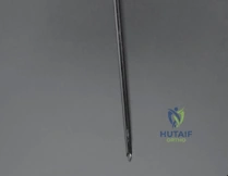
3. A coaxial needle system is preferred. The outer cannula remains in place while the inner cutting needle is advanced, allowing multiple core samples to be taken through a single fascial puncture, minimizing tract seeding.
4. Samples must include representative tissue. The central portion of a large high-grade sarcoma is often necrotic and non-diagnostic. The needle should target the viable, enhancing periphery or the pseudo-capsule.
5. The biopsy tract must be marked with a permanent tattoo (e.g., India ink) at the skin entry site to ensure it is easily identified and excised during definitive surgery.
Principles of Open Incisional Biopsy
When an open biopsy is indicated (typically after a non-diagnostic CNB), the execution must be flawless.
1. Incision: Always use a longitudinal incision. Transverse incisions contaminate multiple longitudinal fascial compartments and are exceedingly difficult, if not impossible, to excise en bloc during limb salvage surgery.
2. Dissection: Dissection must be direct, minimizing the raising of subcutaneous tissue flaps. Do not follow internervous or intermuscular planes, as this contaminates the areolar spaces between muscle bellies. Instead, split the muscle longitudinally directly over the tumor.
3. Tourniquet Use: A tourniquet may be used to maintain a bloodless field, but the limb must not be exsanguinated with an Esmarch bandage, as this mechanical compression can squeeze tumor cells into the systemic venous circulation. Simply elevate the limb for 3-5 minutes before inflation. The tourniquet must be deflated prior to closure to ensure absolute hemostasis.
4. Bone Window: If the tumor is intraosseous, a small, oval cortical window is created using a high-speed burr or drill. Avoid sharp corners to mitigate stress risers.
5. Tissue Sampling: Obtain tissue from the viable periphery. Send samples for fresh frozen section to confirm that diagnostic tissue (not just necrotic debris) has been obtained, as well as for permanent formalin fixation, flow cytometry, and microbiology cultures.
6. Defect Management: The cortical defect must be plugged to prevent postoperative hematoma and tumor spillage. Polymethylmethacrylate (PMMA) bone cement or bone wax is tightly packed into the window.
7. Closure: Meticulous, multi-layered closure is mandatory to prevent hematoma and wound dehiscence. Drains should be avoided if possible. If a drain is absolutely necessary to prevent a massive hematoma, it must exit directly in line with the surgical incision, close to the wound, so the drain tract can be easily excised later.


Excision of Benign Bone Tumors and Cyst Curettage
For radiographically benign lesions (e.g., Unicameral Bone Cysts, Aneurysmal Bone Cysts, Enchondromas), an excisional biopsy or intralesional curettage may serve as both the diagnostic and definitive therapeutic procedure.
Bone Cyst Curettage Technique:
1. An adequately sized cortical window is created to allow complete visualization of the cyst cavity.
2. Meticulous intralesional curettage is performed using various sized angled curettes to scrape the reactive bone until normal, healthy, punctate bleeding cancellous bone is encountered.
3. Adjuvant Therapy: To reduce local recurrence rates (especially in aggressive benign lesions like Giant Cell Tumor of Bone or ABCs), chemical or physical adjuvants are utilized. These include high-speed burring to extend the margin by 1-2 mm, application of phenol (followed by alcohol neutralization), hydrogen peroxide, or liquid nitrogen (cryotherapy).
4. Reconstruction: The resulting cavitary defect is then reconstructed. This can be achieved with autologous bone graft, allograft, synthetic bone substitutes, or PMMA bone cement. Cement provides immediate structural stability and its exothermic reaction acts as an additional thermal adjuvant against microscopic residual disease.


Malignant Tumor Principles
If the biopsy confirms a malignant primary bone tumor, definitive management involves wide local excision. The oncological goal is complete removal of the tumor covered by a continuous shell of healthy, normal tissue (wide margin). The previous biopsy tract, the entire involved bone segment, and the surrounding reactive zone (pseudocapsule) are excised en bloc. Reconstructive options following wide excision include modular endoprostheses (megaprostheses), osteoarticular allografts, vascularized fibular autografts, or rotationplasty, depending on patient age, tumor location, and expected functional demands.


Complications and Management
The complications associated with musculoskeletal biopsies are not merely technical failures; they are often oncological disasters. The classic study by Mankin et al. demonstrated that biopsies performed at non-specialized centers resulted in a significantly higher rate of diagnostic errors, wound complications, and inappropriate alterations in the definitive surgical plan.
Consent and Risks
Informed consent must explicitly detail the unique risks of the procedure. Patients must be warned that neurovascular injury and infection are primary risks. Furthermore, they must understand the possibility of tumor seeding along the biopsy tract. Patients should also be counseled that a second needle biopsy or a subsequent open biopsy may be necessary if the initial specimen is inadequate, necrotic, or non-diagnostic.

Common Complications and Salvage Strategies
| Complication | Estimated Incidence | Pathophysiology / Consequence | Prevention and Salvage Strategies |
|---|---|---|---|
| Inadequate Diagnostic Tissue | 10-15% (CNB) | Sampling of necrotic center instead of viable periphery. Prevents initiation of neoadjuvant therapy. | Prevention: Image guidance targeting the enhancing periphery. Fresh frozen section during open biopsy. Salvage: Repeat CNB or proceed to open incisional biopsy. |
| Post-Biopsy Hematoma | 2-5% | Inadequate hemostasis. Blood acts as a vehicle, spreading malignant cells throughout the compartment. | Prevention: Deflate tourniquet before closure. Plug cortical windows with PMMA. Strict layered closure. Salvage: May require expansion of the definitive wide excision margins to include the entire hematoma bed. |
| Pathologic Fracture | 1-3% | Stress riser from an oversized or sharply angled cortical window. Contaminates adjacent compartments. | Prevention: Small, circular/oval windows. Protected weight-bearing post-op. Salvage: Immobilization. Do not perform internal fixation (rods/plates), as this disseminates tumor. May necessitate amputation. |
| Biopsy Tract Contamination | Variable | Transverse incision or traversing uninvolved compartments. Prevents limb salvage. | Prevention: Strict longitudinal incisions. Plan tract with the resecting surgeon. Salvage: Amputation or highly morbid radical resection. |
| Infection | <2% | Introduction of skin flora into necrotic tumor mass. Delays neoadjuvant chemotherapy. | Prevention: Strict aseptic technique. Prophylactic antibiotics for open procedures. Salvage: Aggressive debridement, targeted IV antibiotics. |

Post Operative Rehabilitation Protocols
Postoperative rehabilitation following a biopsy is dictated by the structural integrity of the bone and the anticipated oncological treatment plan.
For soft tissue core needle biopsies, patients can typically resume normal activities within 24-48 hours, monitoring closely for signs of hematoma or expanding mass.
For open bone biopsies or extensive curettage, the rehabilitation protocol must prioritize the prevention of a pathologic fracture.
1. Weight-Bearing Restrictions: If a significant cortical window was created, the patient must be placed on strict non-weight-bearing or touch-down weight-bearing precautions using crutches or a walker until definitive resection or adequate bone healing occurs.
2. Immobilization: Depending on the location and size of the defect, a brace or splint may be applied to protect against torsional forces.
3. Wound Care: Close monitoring of the wound is essential, especially if the patient is scheduled to begin neoadjuvant chemotherapy or radiation therapy shortly after the biopsy. These systemic treatments significantly impair fibroblast proliferation and wound healing. Sutures or staples should often be left in place longer than the standard 14 days (up to 21-28 days) to prevent
Clinical & Radiographic Imaging

