Clinical Case Study: Distal Femoral Osteosarcoma Diagnosis & Management
Key Takeaway
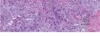
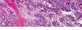
Diagnosing adolescent distal femoral osteosarcoma involves evaluating progressive bone pain, followed by comprehensive imaging. Radiographs show aggressive lytic/sclerotic lesions; MRI details local tumor extent and neurovascular involvement. CT chest screens for metastases. A CT-guided core needle biopsy provides the definitive histopathological diagnosis, essential for tailoring effective orthopedic oncology treatment.
A 16-year-old male presents with a 4-month history of progressive knee pain. The pain is worse at night and poorly responsive to NSAIDs. On examination, there is a firm, fixed mass over the distal femur with venous engorgement. Looking at the initial plain radiograph below, what is your primary differential diagnosis, and how would you structure your immediate investigation?
Candidate: The radiographic appearance—a metaphyseal lesion with cortical destruction, "sunburst" periosteal reaction, and a Codman's triangle—is highly suggestive of a high-grade malignant bone tumor, most likely osteosarcoma. I would exclude Ewing sarcoma, osteomyelitis, and aneurysmal bone cyst. I would order an MRI of the entire femur, systemic staging with a CT chest and PET-CT, and bloods including ALP and LDH.
Candidates often jump immediately to biopsy or offer a long list of benign differentials without acknowledging the aggressive nature of the lesion. Failing to mention that the biopsy must be deferred until after staging imaging is a critical failure, as poor biopsy placement can compromise future limb-salvage options.
The candidate must define the lesion as "aggressive" based on the Codman’s triangle and sunburst reaction. They must articulate a "Stepwise Staging Protocol": 1) Local staging (MRI of the whole bone for skip lesions), 2) Systemic staging (CT chest for pulmonary mets, PET-CT for osseous mets), 3) Laboratory workup (ALP/LDH as prognostic markers), and 4) Meticulous biopsy planning, emphasizing that the biopsy must be performed by the treating oncological surgeon to ensure the tract is exciseable.
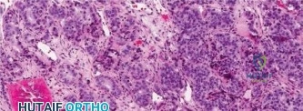
The biopsy confirms high-grade osteosarcoma. You are now discussing the surgical plan. The patient is anxious about the prospect of an amputation versus limb salvage. What are the specific criteria you use to determine if this patient is a candidate for limb salvage, and how do you address the biopsy tract during the definitive procedure?
Candidate: Limb salvage is indicated if we can achieve wide oncologic margins, preserve the neurovascular bundle, and provide adequate soft tissue coverage. Regarding the biopsy tract, it must be excised en bloc with the primary specimen. I would use an elliptical incision encompassing the tract to ensure no local seeding remains.
Failing to mention the "functional" aspect: a salvage limb that is non-functional or requires multiple future surgeries may be inferior to a high-quality amputation/rotationplasty. Also, ignoring the risk of infection in large endoprosthetic reconstructions is a common oversight.
A sophisticated candidate frames this around "Oncologic vs. Functional outcome." 1) Criteria: Neurovascular integrity (as seen on MRI), ability to achieve 2cm+ bony margins, and skin/soft tissue envelope viability. 2) Biopsy: The tract is "contaminated tissue" and must be excised with a 1cm margin of surrounding skin and subcutaneous tissue. 3) Reconstruction: Mention the role of the gastrocnemius flap to manage dead space and provide vascularized cover for the megaprosthesis, as this directly reduces infection risk.
The patient has undergone resection and reconstruction with a rotating-hinge distal femoral replacement. Post-operatively, he is concerned about his long-term activity levels and the durability of the implant. How do you manage his expectations, and what does the "Huvos Grade" represent in his follow-up care?
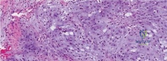
Candidate: I would advise against high-impact activities to preserve implant longevity. The Huvos grade refers to the percentage of tumor necrosis observed in the resected specimen after neoadjuvant chemotherapy. Grade III or IV, which is >90% necrosis, indicates a good response and better prognosis.
Focusing only on the implant and ignoring the systemic nature of the disease. Failing to state that Huvos grading is the single most powerful prognostic indicator is a major omission for an FRCS-level answer.
State that the rotating-hinge design is intended to reduce stress at the bone-implant interface, but emphasize that the primary risk remains aseptic loosening and infection. Regarding Huvos: It is a 4-grade scale. Grade I/II (<90% necrosis) is a poor prognostic factor, while Grade III/IV (>90%) is a good prognostic factor. This confirms the efficacy of the MAP (Methotrexate, Adriamycin, Cisplatin) protocol and guides the intensity of adjuvant chemotherapy.
