Adamantinoma of the Distal Tibia: A Detailed Orthopedic Case Study on Diagnosis
Key Takeaway
Diagnosing adamantinoma, a rare bone tumor often affecting the distal tibia, involves a comprehensive approach. It typically presents with chronic, progressive pain and swelling, sometimes with acute exacerbation from a pathological fracture. Clinical examination reveals a firm osseous mass. Radiographs are crucial, often showing an eccentric, expansile lytic lesion with a 'soap bubble' appearance and cortical thinning.
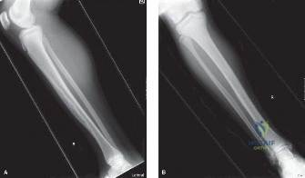
A 32-year-old construction supervisor presents with an 18-month history of insidious distal leg pain, recently exacerbated by a minor misstep. On examination, there is a firm, non-mobile, bony-hard mass at the anteromedial distal tibia. Anteroposterior and lateral radiographs are provided below.
Describe the radiographic findings and provide your differential diagnosis.
Candidate: The radiograph shows an eccentric, expansile lytic lesion in the distal tibial metadiaphysis. It has a "soap bubble" appearance with internal trabeculation and cortical thinning. There is a pathological fracture through the anterior cortex. My differential includes Adamantinoma, fibrous dysplasia, and an aneurysmal bone cyst.
Candidates often fail to highlight the specific tibial predilection, which is the most critical clue. They may list generic differentials like "giant cell tumor" without addressing the fact that GCT is epiphysio-centric, whereas this lesion is clearly diaphyseal/metaphyseal. They also often fail to mention the sclerotic rim, which points toward a more indolent process.
A high-scoring answer describes the lesion using structured terminology: location (diaphyseal/metaphyseal), margins (sclerotic rim indicating slow growth), matrix (lytic/soap-bubble), and cortical status (expansile with breach/pathological fracture). Acknowledging the "classic" tibial presentation for Adamantinoma in this age group (20-40) is essential. Must differentiate from Fibrous Dysplasia (ground glass) and Osteofibrous Dysplasia (younger age, purely cortical, no medullary expansion).
The biopsy confirms an Adamantinoma. You are planning the surgical management. What are the key oncologic principles you must adhere to, and how do you handle the biopsy tract?
Candidate: The goal is wide en-bloc resection because Adamantinoma is resistant to chemo and radiotherapy. I must perform a wide resection of the tumor with a cuff of normal tissue. Crucially, I must include the entire previous biopsy tract in the resection specimen to prevent local recurrence.
Missing the requirement to excise the biopsy tract. Some candidates suggest curettage and bone grafting, which is inappropriate for a malignant lesion and carries a very high recurrence rate. They may also suggest adjuvant therapy, failing to acknowledge that Adamantinoma is notoriously radio- and chemo-resistant.
The candidate defines the surgical goal as R0 wide resection for local control. They explicitly state the necessity of resecting the biopsy site as part of the surgical approach. They classify the tumor (MSTS Stage IIB, G1T2M0) and justify the choice of wide excision over limb salvage amputation based on the absence of distant metastasis and the feasibility of reconstructing the tibial defect.
You have resected the segment. What are your considerations for reconstructive options, and what are the specific risks associated with using a structural intercalary allograft?
Candidate: I would use a structural intercalary allograft augmented with an intramedullary nail and plates. The risks include non-union at the host-graft junction, deep infection, graft fracture, and the prolonged time required for incorporation.
Failing to mention the biological augmentation (e.g., autograft) required to facilitate union. Also failing to mention the necessity of structural stability (IM nail + plate) to prevent graft fatigue and fracture during the incorporation phase.
The candidate discusses the "Allograft-Host Junction" as the primary site of failure. They mention augmentation with autogenous bone graft to increase osteoinductive potential. They correctly identify that allograft incorporation is slow and requires long-term protection, explaining why a construct combining an intramedullary device (for axial strength) and a plate (for rotational stability) is the Gold Standard for this defect.
