Principles of Orthopedic Oncology: Biopsy, Limb Salvage, and Reconstruction
Key Takeaway
Orthopedic oncology demands a rigorous, multidisciplinary approach to maximize patient survival while optimizing limb function. This comprehensive guide details the critical principles of musculoskeletal tumor management, including biopsy techniques, neoadjuvant protocols, and advanced limb salvage reconstructions. From giant cell tumor curettage to complex periacetabular resections and expandable endoprostheses, mastering these evidence-based surgical strategies is essential for orthopedic residents, fellows, and practicing consultants managing primary sarcomas and metastatic bone disease.
Comprehensive Introduction and Patho-Epidemiology
The surgical management of musculoskeletal tumors has undergone a profound paradigm shift over the last four decades, evolving from a discipline characterized by radical, function-sacrificing amputations to one defined by sophisticated limb salvage techniques. Historically, radical amputation was the unyielding standard of care for primary bone sarcomas, yet overall survival remained abysmal due to the inevitable progression of micrometastatic disease. Today, the integration of neoadjuvant and adjuvant chemotherapy, high-resolution cross-sectional imaging (MRI, PET-CT), and modular endoprosthetic design has established limb salvage surgery as the gold standard for over 85% of patients. The foundational literature—spanning William Enneking’s revolutionary staging systems to modern cooperative group trials (e.g., Rizzoli, COSS, EURAMOS)—dictates a strict adherence to oncologic principles to prevent local recurrence while maximizing biomechanical function.
Understanding the patho-epidemiology of these lesions is paramount for the treating orthopedic surgeon. Primary bone sarcomas exhibit a distinct bimodal age distribution. Osteosarcoma and Ewing's sarcoma predominantly afflict adolescents and young adults during periods of rapid skeletal growth, typically localizing to the metaphyses and diaphyses of long bones, respectively. Conversely, chondrosarcoma and secondary osteosarcomas (arising from Paget's disease or prior irradiation) typically present in the fifth to seventh decades of life. The molecular characterization of these tumors has become an integral component of diagnosis and targeted therapy. For instance, the identification of the t(11;22)(q24;q12) translocation resulting in the EWSR1-FLI1 fusion protein is pathognomonic for Ewing's sarcoma, while MDM2 and CDK4 amplification define parosteal and low-grade central osteosarcomas.
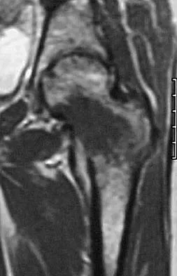
Benign aggressive lesions, such as Giant Cell Tumor (GCT) of bone, present a different patho-epidemiological challenge. While histologically benign and lacking the metastatic inevitability of high-grade sarcomas, GCTs are locally destructive, osteolytic lesions that typically occur in the epiphyses of skeletally mature young adults. The pathophysiology is driven by the neoplastic stromal cells expressing Receptor Activator of Nuclear Factor Kappa-B Ligand (RANKL), which aggressively recruits and activates multinucleated osteoclast-like giant cells. This understanding has led to the utilization of targeted monoclonal antibodies, such as Denosumab, as neoadjuvant therapy to downstage massive, joint-threatening lesions prior to extended curettage.
Metastatic bone disease represents the most common malignancy of bone, with the skeleton serving as the third most frequent site for metastatic dissemination following the lung and liver. Carcinomas of the breast, prostate, lung, thyroid, and kidney exhibit a profound osteotropism. The pathophysiology of skeletal metastases involves a complex interplay between tumor cells and the bone microenvironment—often referred to as the "vicious cycle" of bone metastasis—where tumor-secreted factors stimulate osteoclastic bone resorption, which in turn releases bone-derived growth factors (e.g., TGF-beta) that further stimulate tumor proliferation. The management of metastatic disease is inherently palliative, focusing on the prevention of catastrophic pathologic fractures, pain mitigation, and the restoration of ambulatory function in patients with limited life expectancies.
Detailed Surgical Anatomy and Biomechanics
The foundational principle of orthopedic oncology is the concept of compartmental anatomy. The musculoskeletal system is divided into discrete anatomic compartments bounded by robust fascial septa, tendinous insertions, and bone. These natural barriers dictate the local propagation of sarcomas. Tumor growth follows the path of least resistance; thus, robust structures such as major fascial planes, articular cartilage, and the periosteum temporarily constrain tumor extension, creating a "reactive zone" or pseudocapsule. Conversely, neurovascular bundles, lymphatic channels, and intermuscular planes serve as conduits for rapid longitudinal spread. An intimate understanding of these compartments is mandatory; a tumor confined within the anterior compartment of the thigh requires a vastly different resection strategy than one that has breached the vastus intermedius and invaded the posterior compartment via the linea aspera.
Articular anatomy presents unique barriers and pathways for tumor dissemination. Articular cartilage is highly resistant to tumor invasion due to its avascular nature and the presence of anti-angiogenic factors. Consequently, primary bone sarcomas rarely breach the joint space directly through the chondral surface. Instead, intra-articular contamination typically occurs via the capsular reflections or along the cruciate ligaments. In the knee, a distal femoral osteosarcoma may track along the posterior cruciate ligament into the joint space, necessitating an extra-articular total knee resection to achieve a wide margin. Furthermore, the anatomic proximity of critical neurovascular structures dictates resectability. In the proximal tibia, the trifurcation of the popliteal artery and the intimate relationship of the common peroneal nerve to the fibular head demand meticulous dissection to preserve distal limb viability during wide resection.
The biomechanics of limb salvage reconstruction are profoundly complex, requiring the restoration of immediate structural stability and long-term joint kinematics in the setting of massive bone and soft tissue loss. Modular endoprostheses (megaprostheses) rely on intramedullary stems, which transfer immense bending and torsional moments to the remaining host bone. This non-physiologic load transfer frequently results in stress shielding and subsequent aseptic loosening. To mitigate this, modern implants utilize extracortical porous-coated collars (e.g., hydroxyapatite or titanium foam) to promote osseointegration and extracortical bone bridging, thereby sharing the mechanical load and reducing the stress at the stem-tip interface. The kinematics of the reconstructed joint are also altered; a rotating-hinge knee mechanism is universally employed in distal femoral replacements to provide varus-valgus stability while allowing for a degree of axial rotation, reducing the torsional stresses transmitted to the intramedullary stems.
Pelvic biomechanics following internal hemipelvectomy present the most formidable reconstructive challenge. The pelvis acts as the critical load-transferring ring between the axial skeleton and the lower appendicular skeleton. Enneking Type II (periacetabular) resections utterly disrupt this ring, eliminating the hip joint. Reconstructive options, such as saddle prostheses or custom 3D-printed triflange components, attempt to restore the mechanical axis but face immense shear forces, leading to high rates of hardware failure, superior migration, and catastrophic pelvic dissociation. Alternatively, a flail hip (pseudarthrosis) reconstruction deliberately abandons rigid mechanical continuity. The proximal femur is allowed to migrate superiorly and scar into the gluteal musculature. While this severely alters gait biomechanics—resulting in a profound Trendelenburg lurch and significant leg length discrepancy—it eliminates the risk of hardware failure and provides a durable, albeit mechanically compromised, functional outcome.
Exhaustive Indications and Contraindications
The decision algorithm in orthopedic oncology requires a delicate balance between achieving absolute oncologic clearance and preserving a functional, durable extremity. The primary indication for limb salvage surgery in the setting of a primary bone sarcoma is the ability to achieve a wide surgical margin—defined as a continuous cuff of normal, healthy tissue surrounding the tumor and its reactive pseudocapsule—while maintaining a limb that is more functional than an amputation and a prosthesis. This paradigm assumes that the patient is medically optimized to undergo a prolonged, complex reconstructive procedure and the subsequent rigorous rehabilitation protocol.
Biopsy indications are absolute for any suspected primary bone tumor or solitary skeletal metastasis without a known primary. Core needle biopsy (CNB) is indicated as the initial diagnostic modality, providing sufficient tissue architecture for histologic grading, immunohistochemistry, and molecular cytogenetics. Open incisional biopsy is strictly indicated only when multiple CNB attempts are non-diagnostic, or when sufficient tissue volume is required for specific molecular sequencing protocols that cannot be achieved via percutaneous methods. Prophylactic stabilization of metastatic lesions is indicated based on Mirels' criteria; a score of 9 or greater, or a lesion involving more than 50% of the cortical diameter, dictates immediate surgical intervention to prevent pathologic fracture.
Contraindications to limb salvage are rigid and must be respected to prevent catastrophic local recurrence and patient mortality. Absolute contraindications include major neurovascular bundle encasement that cannot be bypassed or reconstructed with interposition grafting, extensive soft tissue contamination from a poorly placed, non-longitudinal prior biopsy, and the presence of uncontrolled fulminant infection in the tumor bed. Relative contraindications involve patient factors such as extreme skeletal immaturity. In very young children (e.g., under 8 years of age) with distal femoral osteosarcomas, the anticipated leg length discrepancy from a massive resection often exceeds 10-12 cm. While expandable endoprostheses are an option, rotationplasty (Van Nes procedure) is often strongly indicated as a highly durable, biologic alternative that avoids the complications of multiple lengthening surgeries.
| Clinical Scenario | Indications for Limb Salvage / Intervention | Contraindications (Absolute and Relative) |
|---|---|---|
| Primary High-Grade Sarcoma | Ability to achieve wide margins; Intact major neurovascular bundle; Responsive to neoadjuvant chemotherapy. | Absolute: NV bundle encasement; Massive biopsy contamination; Active tumor bed infection. Relative: Extreme skeletal immaturity (consider rotationplasty). |
| Benign Aggressive Lesions (GCT) | Joint-threatening lesions; Impending pathologic fracture; Campanacci Grade II/III. | Relative: Massive extra-osseous extension precluding joint preservation (may require en bloc resection rather than curettage). |
| Metastatic Bone Disease | Mirels' Score $/ge$ 9; Intractable pain failing radiotherapy; Actual pathologic fracture. | Absolute: Patient medically unfit for surgery; Life expectancy $< 6$ weeks. Relative: Highly radiosensitive tumors (e.g., myeloma) without impending fracture. |
| Pelvic Tumors (Periacetabular) | Localized disease; Patient acceptance of extensive rehab and potential functional deficits. | Absolute: Bilateral sacral nerve root involvement; Massive visceral invasion precluding clear margins. |
Pre-Operative Planning, Templating, and Patient Positioning
Meticulous pre-operative planning is the crucible in which successful orthopedic oncology outcomes are forged. The planning phase begins with exhaustive high-resolution imaging. A whole-bone MRI of the affected extremity is absolutely mandatory to assess the intra-osseous extent of the tumor, delineate the extra-osseous soft tissue mass, map the relationship to major neurovascular bundles, and critically, to rule out "skip metastases"—discrete tumor foci within the same bone but physically separated from the primary lesion. High-resolution CT of the chest is required for pulmonary staging, while PET-CT evaluates for systemic osseous or visceral metastases. The biopsy tract, which must be clearly marked and documented during the initial diagnostic phase, is mapped on the MRI to ensure it can be excised en bloc with the definitive resection specimen.
Digital templating and advanced 3D modeling have revolutionized the precision of tumor resection. Surgeons utilize specialized software to overlay the MRI tumor volume onto the CT bone models. For primary sarcomas, a planned resection margin of 2 to 3 cm of normal bone beyond the MRI-defined intramedullary extent is standard. Custom, patient-specific 3D-printed cutting guides can be manufactured to ensure the osteotomy matches the pre-operative plan with sub-millimeter accuracy. This is particularly critical in joint-preserving intercalary resections and complex pelvic internal hemipelvectomies, where standard anatomic landmarks are distorted by the tumor mass. The exact dimensions of the required modular endoprosthesis or massive allograft are calculated, ensuring that backup components of varying sizes are immediately available in the operating theater.
Patient positioning and operating room setup must anticipate the need for extensile exposures and potential intra-operative complications. For lower extremity resections, the patient is typically positioned supine on a radiolucent table to facilitate unimpeded fluoroscopic access. A sterile tourniquet is applied proximally; however, it is a strict oncologic rule that the limb must never be exsanguinated with an Esmarch bandage, as the compressive forces can embolize tumor cells into the systemic circulation. The limb is merely elevated for 3 to 5 minutes prior to tourniquet inflation. For complex pelvic resections (Type II or III), the patient is placed in a "floppy" lateral decubitus position. This allows the surgeon to dynamically roll the patient, providing simultaneous access to the anterior ilioinguinal approach for visceral and vascular mobilization, and the posterior gluteal approach for sciatic nerve isolation and iliac osteotomies.
Pre-operative optimization also involves vascular mapping and intervention. For highly hypervascular metastases, particularly those arising from renal cell carcinoma or thyroid carcinoma, pre-operative transarterial embolization is mandatory. This procedure, performed by interventional radiology within 24 to 48 hours prior to the surgical resection, dramatically reduces intra-operative blood loss, preventing catastrophic hemorrhage that can obscure surgical planes and compromise both margin status and patient survival. Furthermore, coordination with plastic surgery for simultaneous soft tissue coverage (e.g., medial gastrocnemius flaps or free latissimus dorsi flaps) is finalized during this planning phase.
Step-by-Step Surgical Approach and Fixation Technique
Principles of Oncologic Biopsy
The biopsy is the final, and arguably the most critical, staging procedure. The surgeon must proceed under the assumption that the lesion is a high-grade sarcoma. The incision must be strictly longitudinal; transverse incisions contaminate multiple extensor and flexor compartments, rendering subsequent limb salvage impossible. The approach must traverse the minimum number of anatomic compartments, passing directly through the muscle belly rather than exploiting intermuscular planes, thereby preventing hematoma from tracking along fascial boundaries. Meticulous hemostasis is mandatory. A post-biopsy hematoma is considered fully contaminated with tumor cells and exponentially expands the required resection volume. The tourniquet must be deflated prior to closure to ensure absolute hemostasis. If the cortical bone must be breached, a round burr is used to create a circular window, avoiding square windows that create stress risers. The defect is plugged with PMMA to prevent tumor spillage. Drains are strictly contraindicated; if unavoidable, they must exit directly in line with the surgical incision.
Wide Resection of Primary Sarcomas
Taking the distal femur as the quintessential model, the procedure begins with an extensile utilitarian incision, incorporating the previous longitudinal biopsy tract in an elliptical fashion. Skin flaps are raised, maintaining a thick layer of subcutaneous fat to preserve vascularity. The superficial femoral artery and vein are identified in the subsartorial canal and meticulously traced distally into the popliteal fossa, separating them from the tumor pseudocapsule. The sciatic nerve and its bifurcation are isolated and protected. The vastus medialis, lateralis, and intermedius are transected at a safe distance from the tumor mass, leaving a robust cuff of normal muscle overlying the lesion. The femur is osteotomized at the pre-calculated level (2-3 cm proximal to the MRI margin). The entire specimen, including the distal femur, the tumor, the pseudocapsule, and the biopsy tract, is removed en bloc. The margins are immediately assessed via frozen section by the musculoskeletal pathologist.
Endoprosthetic and Allograft Reconstruction
Following wide resection, skeletal reconstruction commences. For modular endoprostheses, the host bone medullary canal is reamed. If a cemented stem is chosen, pulsatile lavage and meticulous drying are performed before retrograde injection of PMMA cement. The prosthesis is impacted, ensuring rotational alignment matches the linea aspera. Extracortical porous collars are positioned intimately against the host bone osteotomy site to promote bridging. Soft tissue reconstruction is paramount; the extensor mechanism must be reattached. In proximal tibia replacements, the patellar tendon is sutured to the porous-coated proximal tibial component using heavy non-absorbable tape, and a medial gastrocnemius rotational flap is universally mobilized to cover the prosthesis, providing a robust, vascularized envelope that prevents catastrophic deep infection and aids in extensor mechanism healing.
Extended Curettage for Benign Aggressive Lesions
For Giant Cell Tumors, the goal is total eradication of microscopic disease while preserving the native subchondral bone and articular cartilage. A massive cortical window is created to allow unhindered visual access to the entire tumor cavity. Intralesional curettage is performed aggressively with hand curettes until normal-appearing cancellous bone is reached. Subsequently, a high-speed burr is utilized to systematically break down the bony ridges within the cavity, extending the margin by 2 to 3 millimeters in all directions. Chemical or thermal adjuvants are then applied to induce necrosis of any remaining microscopic cells. Phenol (89%) can be painted onto the cavity walls and neutralized with alcohol, or liquid nitrogen cryotherapy can be utilized. Finally, the defect is packed with PMMA bone cement. The exothermic reaction of the curing PMMA provides an additional thermal adjuvant effect, while its immediate structural stability allows for early weight-bearing.
Pelvic Internal Hemipelvectomy
A Type II periacetabular resection demands unparalleled anatomic command. Utilizing a modified Gibson approach combined with an anterior ilioinguinal exposure, the external iliac vessels and femoral nerve are mobilized anteriorly. Posteriorly, the sciatic nerve is identified at the greater sciatic notch and traced distally. The gluteal musculature is elevated off the ilium, or resected en bloc if involved. Osteotomies are performed through the ilium superior to the acetabulum, and through the superior and inferior pubic rami. The entire periacetabular segment is delivered. Reconstruction, if chosen, involves the implantation of a custom 3D-printed triflange component, which is secured to the remaining ilium, sacrum, and pubis with locking screws. The hip joint is then reconstructed with a constrained or dual-mobility total hip arthroplasty to prevent dislocation in the setting of massive abductor deficiency.
Complications, Incidence Rates, and Salvage Management
The complication profile in orthopedic oncology is exceptionally high, a consequence of massive surgical dissections combined with the profound immunosuppressive and wound-healing detriments of neoadjuvant chemotherapy and radiation. The Henderson Classification is the universally accepted system for categorizing the failure of limb salvage reconstructions, dividing complications into five distinct types: Soft Tissue Failure (Type 1), Aseptic Loosening (Type 2), Structural Failure (Type 3), Infection (Type 4), and Tumor Progression (Type 5).
Infection (Type 4 failure) remains the most devastating non-oncologic complication, with incidence rates ranging from 10% to 15% in primary megaprostheses, and exceeding 30% in pelvic reconstructions. The pathophysiology is driven by the vast dead space, the presence of massive foreign bodies, and the compromised vascularity of irradiated tissues. Management of deep periprosthetic joint infection (PJI) in oncology mirrors standard arthroplasty but is significantly more complex. It typically requires a two-stage revision: complete explantation of the megaprosthesis, placement of a customized antibiotic-loaded PMMA spacer, and 6 to 8 weeks of targeted intravenous antibiotics. If soft tissue coverage is inadequate, free tissue transfer (e.g., rectus abdominis or latissimus dorsi flaps) is mandatory prior to reimplantation. In modern practice, silver-coated endoprostheses or iodine-supported titanium implants are increasingly utilized in high-risk patients to deter biofilm formation.
Aseptic loosening (Type 2) and structural failure (Type 3, including periprosthetic fracture or implant breakage) occur due to the massive biomechanical mismatch between rigid metallic stems and osteopenic host bone. Incidence increases linearly with time, making it a primary concern for young, long-term sarcoma survivors. Salvage management involves revision to longer, thicker stems, often utilizing cemented fixation if the host bone is extensively compromised, or employing compressive osseointegration devices (e.g., Compress® implants) that utilize high-tension spring washers to stimulate continuous bone hypertrophy at the implant-host interface.
Local recurrence (Type 5) represents an oncologic failure and carries a grave prognosis, significantly increasing the risk of subsequent pulmonary metastasis. The incidence of local recurrence following wide resection of high-grade sarcomas is approximately 5% to 10%. Salvage management is highly individualized. If the recurrence is localized and a secondary wide excision can be achieved without compromising major neurovascular structures, re-resection and revision reconstruction may be attempted. However, in cases of massive, multifocal recurrence, or recurrence involving the neurovascular bundle, salvage amputation (e.g., hip disarticulation or forequarter amputation) is the definitive, life-saving intervention.
| Henderson Classification | Description of Failure | Estimated Incidence | Salvage Management Strategy |
|---|---|---|---|
| Type 1 | Soft Tissue Failure (Flap necrosis, tendon rupture, instability) | 5 - 10% | Debridement, local/free flap coverage, constrained liners, allograft tendon reconstruction. |
| Type 2 | Aseptic Loosening (Bone-cement or bone-implant interface failure) | 15 - 20% (at 10 years) | Revision arthroplasty, longer stems, compressive osseointegration devices. |
| Type 3 | Structural Failure (Implant fracture, periprosthetic fracture) | 5 - 8% | Implant exchange, open reduction internal fixation (ORIF) with cortical strut allografts. |
| Type 4 | Infection (Deep periprosthetic joint infection) | 10 - 15% | Two-stage revision, antibiotic spacers, aggressive flap coverage, suppressive antibiotics. |
| Type 5 | Tumor Progression (Local recurrence) | 5 - 10% | Re-resection with wide margins if feasible; Salvage amputation (often required). |
Phased Post-Operative Rehabilitation Protocols
Rehabilitation following orthopedic oncology resections is highly individualized, dictated by the specific anatomic site, the type of reconstruction, and the overarching timeline of the patient's adjuvant chemotherapy or radiation therapy. The immediate post-operative phase (Weeks 0-4) prioritizes wound healing above all functional goals. Chemotherapy agents (e.g., Methotrexate, Doxorubicin) and localized radiation severely inhibit fibroblast proliferation and collagen cross-linking. Consequently, the surgical incision is exceptionally fragile. Sutures or staples must remain in place for a minimum of 3 to 4 weeks, and aggressive range-of-motion exercises that place tension on the incision or rotational flaps are strictly prohibited until primary healing is clinically confirmed.
Weight-bearing protocols are strictly governed by the reconstructive modality. Patients with cemented modular endoprostheses are typically permitted immediate weight-bearing as tolerated, as the PMMA cement provides immediate maximal structural stability. Conversely, reconstructions utilizing massive structural allografts or Allograft-Prosthetic Composites (APCs) require prolonged protection. These biologic reconstructions rely on creeping substitution—a process where host osteoclasts resorb the dead allograft bone while host osteoblasts lay down new bone at the junction. This process is painfully slow, particularly under the suppressive effects of chemotherapy. Patients with intercalary allografts are restricted to toe-touch or partial weight-bearing for 6 to 9 months, until robust radiographic union at the host-graft junctions is definitively observed.
The intermediate phase (Months 1-6) focuses on the restoration of joint kinematics and muscular strength. For distal femoral replacements, achieving 90 degrees of knee flexion is a critical milestone, though active extension is often delayed to protect the patellar tendon reattachment. Physical therapy must be carefully calibrated to the patient's systemic tolerance; chemotherapy-induced fatigue, anemia, and peripheral neuropathy (particularly from Cisplatin) frequently hinder rehabilitation efforts. The use of continuous passive motion (CPM) machines and aquatic therapy (once wounds are completely sealed) are excellent adjuncts.
Long-term functional evaluation and oncologic surveillance form the final, lifelong phase of care. Function is objectively quantified using the Musculoskeletal Tumor Society (MSTS) scoring system, a validated 30-point scale assessing pain, range of motion, strength, joint stability, emotional acceptance, and walking ability. Simultaneously, an intense oncologic surveillance protocol is initiated to detect local recurrence or pulmonary metastases at the earliest, most treatable stages. For high-grade sarcomas, this mandates a physical examination, local cross-sectional imaging (MRI), and non-contrast high-resolution chest CT every 3 months for the first 2 years, every 6 months for years 3 through 5, and annually thereafter for the patient's lifetime.
Summary of Landmark Literature and Clinical Guidelines
The practice of orthopedic oncology is deeply rooted in a robust framework of landmark literature and cooperative group trials that have systematically dismantled historical dogmas. The foundational pillar of the discipline remains William Enneking’s 1980 publication on the surgical staging of musculoskeletal sarcomas. Enneking’s classification system, based on histologic grade (G), anatomic setting/compartment (T), and the presence of metastasis (M), established the universal language by which surgeons define surgical margins (Intralesional, Marginal, Wide, Radical) and predict the probability of local recurrence. This framework remains the absolute standard for surgical planning globally.
The evolution of chemotherapy from experimental to essential was driven by massive cooperative group trials. The Cooperative Osteosarcoma Study Group (COSS) and the Rizzoli Orthopaedic Institute trials in the 1980s and 1990s definitively proved that multi-agent neoadjuvant chemotherapy (comprising High-Dose Methotrexate, Doxorubicin, and Cisplatin—the MAP protocol) increased long-term survival in osteosarcoma from a dismal 20% with surgery alone to over 65%. Furthermore, the histologic evaluation of tumor necrosis in the resected specimen, quantified by the Huvos grading system, was established as the single most powerful prognostic indicator for overall survival, directly guiding the intensity of postoperative adjuvant therapy. The more recent EURAMOS-1 trial, the largest international osteosarcoma trial ever conducted, further refined these protocols, though it highlighted the ongoing challenge of improving outcomes for poor responders (Huvos Grade I/II).
In the realm of benign aggressive tumors, Campanacci’s seminal work on Giant Cell Tumor of bone defined the radiographic staging system (Grades I, II, and III) that dictates surgical intervention. His extensive clinical series established that extended intralesional curettage with high-speed burring and the use of local adjuvants (phenol or liquid nitrogen) could achieve local control rates comparable to en bloc resection, thereby saving countless native joints. More recently, the introduction of Denosumab, a RANKL inhibitor, has been validated in numerous clinical trials as a powerful tool to ossify the margins of massive GCTs, making previously unresectable tumors amenable to joint-preserving curettage.
Finally, the surgical management of metastatic bone disease is universally guided by the Mirels' Criteria, published by Hilton Mirels in 1989. By quantifying the risk of pathologic fracture based on four variables—site, pain, lesion nature (lytic vs. blastic), and size—Mirels provided an objective, reproducible scoring system that transformed prophylactic fixation from a subjective guess to an evidence-based mandate. A score of 9 or greater demands prophylactic stabilization, drastically reducing the morbidity associated with completed pathologic fractures and cementing the role of the orthopedic surgeon in the palliative care of the advanced cancer patient.
