Partial Selective Fasciectomy for Dupuytren's Contracture: Surgical Masterclass
Key Takeaway
Partial selective fasciectomy remains the gold standard surgical intervention for advanced Dupuytren's contracture. This technique involves the meticulous excision of pathological fascial cords while preserving the overlying skin and neurovascular structures. Proper execution requires a thorough understanding of palmar fascial anatomy, precise surgical dissection, and rigorous postoperative rehabilitation to optimize functional outcomes, minimize flare reactions, and prevent recurrence in the affected digits.
Comprehensive Introduction and Patho-Epidemiology
Partial selective fasciectomy remains the gold standard and the most universally performed surgical intervention for the definitive management of advanced Dupuytren’s disease. Historically, surgical treatment paradigms favored radical fasciectomy, a highly aggressive procedure that sought to excise all palmar and digital fascia—both macroscopically diseased and seemingly normal—in an attempt to eradicate the disease. However, longitudinal outcomes demonstrated an unacceptably high complication profile, including devastating rates of hematoma, skin flap necrosis, profound postoperative stiffness, and complex regional pain syndrome (CRPS), without a commensurate reduction in long-term recurrence rates. Consequently, the modern orthopedic consensus has shifted definitively toward partial (selective) fasciectomy. This technique meticulously targets and excises only the macroscopically diseased, hypertrophied fascial cords responsible for the biomechanical contracture, deliberately preserving the overlying dermis, the delicate neurovascular bundles, and the uninvolved fascial structures. This targeted approach strikes an optimal, evidence-based balance between minimizing surgical morbidity and maximizing long-term functional restoration.
To fully appreciate the rationale behind partial selective fasciectomy, the orthopedic surgeon must possess a granular understanding of the patho-epidemiology driving Dupuytren’s diathesis. The condition is fundamentally a progressive fibroproliferative disorder of the palmar aponeurosis. At the cellular level, the pathogenesis is characterized by the aberrant proliferation of fibroblasts and their subsequent phenotypic transformation into contractile myofibroblasts. This differentiation is heavily mediated by localized overexpression of transforming growth factor-beta (TGF-β), basic fibroblast growth factor (bFGF), and platelet-derived growth factor (PDGF). The resulting myofibroblasts express alpha-smooth muscle actin (α-SMA), aligning along the axis of tension and generating the relentless mechanical force that ultimately results in irreversible flexion contractures of the metacarpophalangeal (MCP) and proximal interphalangeal (PIP) joints. The disease progresses through three distinct histological phases: the proliferative phase (characterized by high cellularity and nodule formation), the involutional phase (where myofibroblasts align and begin to contract), and the residual phase (dominated by acellular, dense collagenous cords).
Epidemiologically, Dupuytren’s disease demonstrates a strong genetic predisposition, primarily affecting individuals of Northern European descent, leading to its historical moniker, "Viking disease." The inheritance pattern is generally considered autosomal dominant with variable penetrance. The concept of "Dupuytren’s diathesis," originally popularized by Hueston, identifies a subset of patients with a particularly aggressive, early-onset phenotype. Indicators of a strong diathesis include bilateral hand involvement, ectopic fibromatosis (such as plantar fibromatosis or Ledderhose disease, penile fibromatosis or Peyronie’s disease, and dorsal PIP joint knuckle pads or Garrod’s pads), a strong family history, and onset before the age of 50. Patients exhibiting these diathesis factors are statistically predisposed to higher recurrence rates following any intervention, necessitating meticulous surgical execution and rigorous postoperative surveillance.
Furthermore, environmental and systemic factors play a synergistic role in the phenotypic expression and acceleration of the disease. Chronic microvascular ischemia, often exacerbated by smoking, diabetes mellitus, and chronic alcohol consumption, has been implicated in upregulating the local fibrogenic response. Additionally, there is a well-documented association between epilepsy and Dupuytren’s disease, historically attributed to the use of phenobarbital and phenytoin, which are thought to stimulate fibroblast proliferation. Understanding these complex patho-epidemiological factors is essential for the operating surgeon, as they directly inform preoperative patient counseling, the anticipation of surgical complexity, and the realistic prognostication of long-term outcomes and recurrence risks.
Detailed Surgical Anatomy and Biomechanics
A profound, three-dimensional understanding of the normal and pathological anatomy of the palmar and digital fascia is the absolute cornerstone of safe and effective fasciectomy. The normal palmar aponeurosis is a complex, multi-layered fascial network designed to stabilize the palmar skin during grip and protect the underlying neurovascular and tendinous structures. It consists of longitudinal pretendinous bands, the superficial transverse palmar ligament, and deeper structures including the natatory ligaments in the web spaces. Within the digits, the fascial architecture becomes even more intricate, comprising the spiral bands, the lateral digital sheets, and the stabilizing ligaments of Grayson (volar to the neurovascular bundle) and Cleland (dorsal to the neurovascular bundle). In Dupuytren’s disease, it is critical to recognize that the pathology does not generate de novo structures; rather, it causes the hypertrophy, contracture, and pathological transformation of these pre-existing normal fascial bands into rigid, unyielding "cords."
The biomechanical consequences of these contracting cords dictate the specific clinical deformity observed. The pretendinous cord, arising from the pretendinous band of the palmar aponeurosis, is primarily responsible for isolated metacarpophalangeal (MCP) joint contractures. Because this cord attaches to the base of the proximal phalanx or the distal palmar crease skin, its contraction pulls the proximal phalanx into flexion. The central cord, which is essentially a distal continuation of the pretendinous cord into the digit, courses volarly to the neurovascular bundles and inserts onto the middle phalanx, generating PIP joint contractures. The natatory cord, derived from the natatory ligaments, spans the web spaces and is responsible for adduction contractures, severely restricting the patient's ability to abduct the digits. The lateral cord, intimately associated with the lateral digital sheet, can contribute to both PIP and distal interphalangeal (DIP) joint contractures, often tethering the skin laterally.
Of all the pathological structures encountered during selective fasciectomy, the spiral cord is unequivocally the most surgically treacherous. The spiral cord is not a single anatomical entity but an amalgamation of four distinct structures: the pretendinous band, the spiral band, the lateral digital sheet, and Grayson’s ligament. As this composite structure undergoes myofibroblastic contraction, it essentially "bowstrings" across the PIP joint. Crucially, because the spiral band passes deep to the neurovascular bundle before joining the lateral digital sheet, its contraction fundamentally alters the anatomical position of the digital nerve and artery. The contracting spiral cord displaces the neurovascular bundle proximally, centrally toward the midline of the digit, and superficially toward the dermis. This displacement places the nerve in extreme peril, often positioning it directly beneath the skin incision at the level of the proximal phalanx and the distal palmar crease.
The biomechanics of chronic PIP joint contractures warrant special attention due to their propensity to cause secondary joint pathology. Unlike the MCP joint, which has a cam-shaped metacarpal head and collateral ligaments that are maximally taut in flexion (making it relatively forgiving to chronic flexion), the PIP joint is highly susceptible to secondary capsular and ligamentous contractures. A chronic PIP joint contracture extending beyond 60 degrees generates immense, sustained tension on the extensor mechanism, specifically the central slip inserting on the base of the middle phalanx. Over time, this constant mechanical disadvantage leads to the attenuation and elongation of the central slip. If the central slip becomes functionally incompetent, the lateral bands may subluxate volarly past the axis of rotation of the PIP joint. If this biomechanical failure is not recognized and managed appropriately during the postoperative period, the patient is at high risk of developing a secondary, iatrogenic boutonnière deformity, profoundly compromising the functional outcome of the fasciectomy.
Exhaustive Indications and Contraindications
The decision to proceed with operative intervention in Dupuytren’s disease must be predicated on a careful synthesis of objective clinical measurements, functional impairment, and the patient's overall physiological status. The mere presence of palmar nodules or non-contracting fascial bands is explicitly not an indication for surgery, as these may remain indolent for decades without causing functional deficits. The classic, rapid-screening clinical threshold is a positive Hueston's "tabletop test," wherein the patient is unable to lay the affected palm and digits entirely flat against a hard surface. While highly sensitive for detecting contractures that have progressed beyond the compensatory capacity of the hand, this test must be supplemented with precise, goniometric measurements of each affected joint to establish a baseline and guide surgical planning.
Specific, universally accepted surgical thresholds include an MCP joint contracture of 30 degrees or greater, and any measurable PIP joint contracture exceeding 15 to 20 degrees. The lower threshold for the PIP joint is dictated by its distinct biomechanical vulnerability; PIP contractures are notoriously recalcitrant, tend to progress rapidly, and secondary capsuloligamentous contractures develop swiftly, rendering delayed surgical correction significantly more complex and less predictable. Severe web space contractures that impede functional grasp or the ability to span objects (e.g., holding a large glass or typing) also constitute a strong indication for release. Additionally, in rare instances, profound, intractable pain associated with rapidly growing palmar nodules may warrant intervention, though pain is an atypical primary presenting symptom of Dupuytren's disease and alternative diagnoses should be rigorously excluded.
Contraindications to partial selective fasciectomy must be respected to prevent catastrophic postoperative outcomes. Absolute contraindications include active local or systemic infection, severe vascular insufficiency of the upper extremity (which precludes safe tourniquet use and compromises flap viability), and active Complex Regional Pain Syndrome (CRPS) in the affected limb, as surgical trauma will invariably exacerbate the sympathetic dystrophy. Relative contraindications encompass severe medical comorbidities that contraindicate regional or general anesthesia, and profound cognitive or psychiatric impairment that would prevent strict adherence to the demanding postoperative splinting and rehabilitation protocols. Without compliant postoperative therapy, surgical intervention is virtually guaranteed to fail, often leaving the patient with a stiffer, more contracted hand than their preoperative baseline.
Clinical Indications and Contraindications Matrix
| Category | Specific Criteria / Conditions | Clinical Rationale & Surgical Implications |
|---|---|---|
| Primary Indications | MCP Joint Contracture ≥ 30° | Functional impairment in grasping large objects; highly correctable with standard fasciectomy. |
| PIP Joint Contracture ≥ 15° - 20° | High risk of rapid progression and secondary joint/capsular stiffness; early intervention is critical. | |
| Positive Tabletop Test | Objective evidence of functional loss; inability to place hand flat limits daily activities. | |
| Severe Web Space Contracture | Restricts digital abduction; requires meticulous release of the natatory cords. | |
| Absolute Contraindications | Active Complex Regional Pain Syndrome (CRPS) | Surgical trauma will trigger a severe flare, resulting in devastating, irreversible stiffness. |
| Severe Peripheral Vascular Disease | High risk of digital ischemia and skin flap necrosis post-excision; poor wound healing potential. | |
| Active Local Infection | Risk of deep space hand infection, tenosynovitis, and systemic dissemination. | |
| Relative Contraindications | Inability to Comply with Rehab | Postoperative splinting and therapy are mandatory; non-compliance guarantees recurrent contracture. |
| Mild, Non-progressive Nodules | Surgical morbidity outweighs benefits; high risk of triggering disease acceleration (flare). | |
| Severe Medical Comorbidities | High anesthetic risk; consider minimally invasive options (e.g., percutaneous needle aponeurotomy). |
Pre-Operative Planning, Templating, and Patient Positioning
Meticulous preoperative planning is the linchpin of a successful partial selective fasciectomy. The physical examination must transcend simple goniometry to include a rigorous assessment of the hand's vascular and neurological status. A modified Allen’s test is mandatory to confirm the patency of the superficial palmar arch and the digital arteries, particularly in patients with severe, long-standing contractures where chronic vascular tethering may have occurred. Two-point discrimination should be documented for every digit, as pre-existing subclinical neuropathies are common, and establishing a baseline is critical for medicolegal protection and postoperative assessment. The skin integrity, particularly the presence of deep palmar pits, maceration in the web spaces, or severe dermal adherence to the underlying cords, must be evaluated, as these factors dictate the choice of incision and the potential need for full-thickness skin grafting (dermofasciectomy).
Incision planning is a highly strategic exercise aimed at maximizing surgical exposure of the pathological cords while strictly avoiding the creation of linear scars that cross flexion creases, which would inevitably lead to secondary iatrogenic scar contractures. The two primary workhorse incisions are the Bruner (zigzag) incision and the longitudinal incision designed for subsequent conversion into multiple Z-plasties. The Bruner incision places the apices of the flaps at the mid-axial lines of the digits, avoiding the flexion creases entirely. However, for severe contractures where significant skin lengthening is anticipated upon digital extension, the longitudinal incision with planned Z-plasties is often superior. The longitudinal approach allows for rapid, direct exposure of the cord and the neurovascular bundles. Once the contracture is released, the longitudinal wound is converted into a series of Z-plasties (typically 60-degree angles), which not only breaks up the linear scar but effectively recruits adjacent transverse skin laxity to lengthen the volar skin envelope.
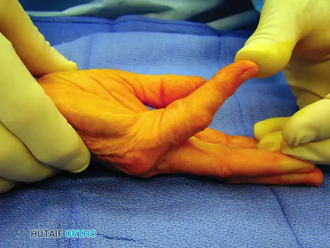
Figure 75-9 A: Preoperative presentation demonstrating a 60-degree metacarpophalangeal joint contracture.
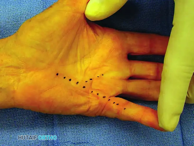
Figure 75-9 B: Dotted lines indicating the underlying pathological cords to be released at surgery.
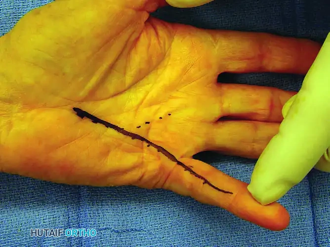
Figure 75-9 C: Solid line outlining the intended longitudinal incision.
For complex, multi-digit involvement, the templating of incisions becomes an exercise in advanced spatial geometry. The surgeon must plan incisions that allow access to the pretendinous, central, and natatory cords simultaneously without creating narrow, ischemic skin bridges between adjacent surgical fields. Marking the anticipated path of the cords and the planned incisions with a sterile surgical pen prior to exsanguination is mandatory, as the anatomical landmarks shift once the tourniquet is inflated and the hand is manipulated.
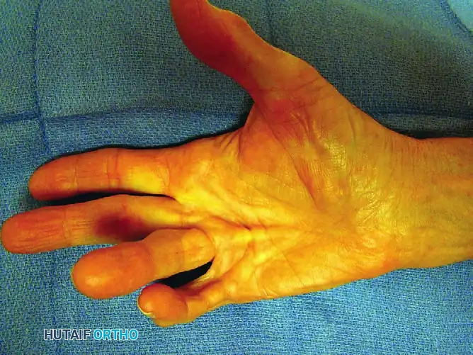
Figure 75-10 A: Complex disease involving the thumb, middle, ring, and small fingers.
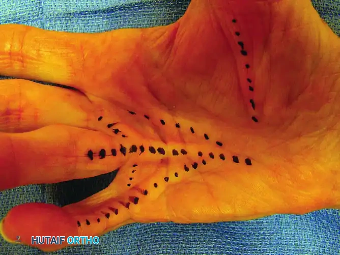
Figure 75-10 B: The cords to be released are dotted with a skin-marking pen.
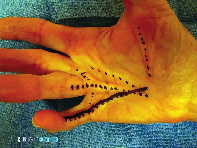
Figure 75-10 C: Planned incisions are solidly outlined before tourniquet inflation.
Patient positioning and anesthetic management are standardized to optimize the surgical environment. The procedure is ideally performed under a regional brachial plexus block (axillary or supraclavicular), which provides excellent intraoperative anesthesia and profound postoperative analgesia, facilitating early mobilization and mitigating the hyper-sympathetic response associated with pain. The patient is positioned supine with the operative arm extended on a radiolucent hand table. A well-padded pneumatic tourniquet is applied to the proximal brachium. Following strict elevation and exsanguination with an Esmarch bandage, the tourniquet is inflated to 250 mm Hg, or precisely 100 mm Hg above the patient's systolic blood pressure. The use of surgical loupe magnification (minimum 2.5x, ideally 3.5x to 4.5x) is non-negotiable, as the differentiation between a glistening digital nerve and a tense, white fascial band is frequently measured in millimeters.
Step-by-Step Surgical Approach and Fixation Technique
The surgical execution of a partial selective fasciectomy demands meticulous tissue handling and an unwavering respect for the altered microanatomy. Following the planned skin incisions, flap elevation is the critical first step. The skin flaps must be elevated at a level just deep to the dermis to preserve the delicate subdermal vascular plexus. In Dupuytren’s disease, the pathological myofibroblasts frequently invade the overlying dermis, creating severe adherence. Sharp dissection with a No. 15 scalpel blade, maintaining constant, gentle traction on the skin edge via skin hooks, is required to separate the dermis from the underlying cord. Blunt dissection or aggressive use of scissors in this subdermal plane will invariably traumatize the vascular plexus, leading to catastrophic full-thickness flap necrosis.
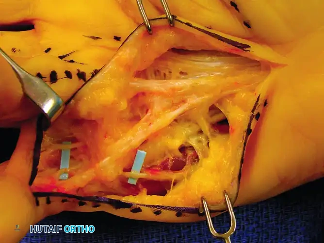
Once the flaps are elevated and secured with temporary retention sutures or lead hands, the dissection of the diseased fascia commences. The cardinal rule of Dupuytren’s surgery is to proceed from proximal to distal, moving from areas of normal, predictable anatomy into the distorted pathological zones. The proximal origin of the pretendinous cord is identified in the mid-palm, sharply transected, and elevated. Grasping this proximal stump with a Kocher clamp provides essential longitudinal traction, dynamically delineating the distal extensions of the cord and pulling the fascial structures away from the deeper neurovascular elements.
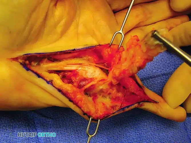
Figure 75-9 D: Disease dissection from proximal to distal, clearly exposing the digital nerves. Note the Kocher clamp on the proximal end of the pretendinous cord providing traction.
As the dissection proceeds distally toward the distal palmar crease and the web spaces, the identification and protection of the neurovascular bundles become paramount. The surgeon must identify the digital nerve and artery in the proximal palm before they enter the diseased tissue. Once identified, the neurovascular bundle is meticulously traced distally, liberating it from the encasing fascial bands. When a spiral cord is present, the surgeon must remain acutely aware that the nerve may be displaced superficially and centrally. A catastrophic pitfall is blindly dividing tight fascial bands in the web space or at the base of the proximal phalanx without direct, continuous visualization of the nerve; the superficially displaced nerve is highly susceptible to transection in this zone.
Figure 75-10 D: The palmar and digital cords are exposed through the longitudinal incision.
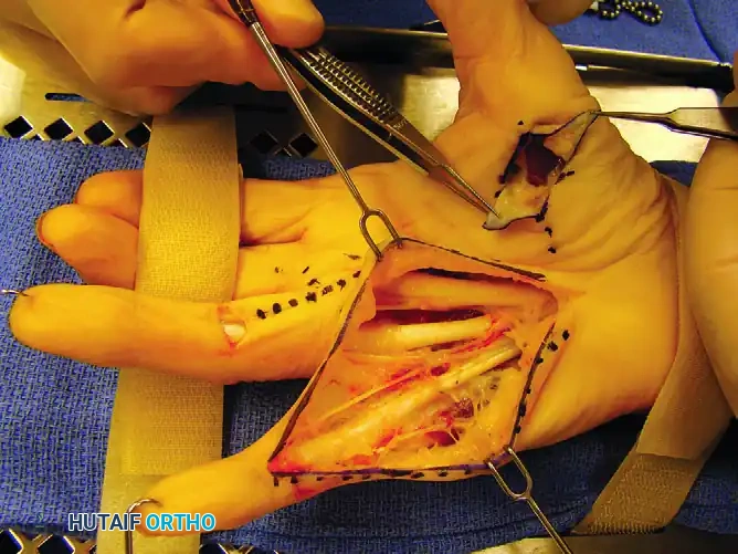
Figure 75-10 E: Exposure after cord excisions. Note the additional transverse incision required to excise the ring finger central cord and the additional thumb cord incision.
Following the complete excision of the pretendinous, central, spiral, and natatory cords, the surgeon must reassess the joints. If a PIP joint contracture persists despite complete fascial clearance, the surgeon faces a critical decision regarding joint release. Stepwise release may involve incising the check-rein ligaments or the accessory collateral ligaments. However, aggressive volar plate release is generally discouraged, as it significantly increases the risk of postoperative joint instability and profound stiffness, often yielding a worse functional outcome than accepting a mild residual flexion contracture.
The "fixation" and closure phase of the operation is as critical as the excision. Prior to closure, the tourniquet is deflated, and meticulous hemostasis is achieved using bipolar electrocautery. Hematoma formation is the nemesis of fasciectomy; it acts as a mechanical barrier to wound healing, a rich nidus for infection, and a potent trigger for a severe fibroproliferative flare reaction. If a longitudinal incision was utilized, it is now converted into Z-plasties. The transposition and fixation of these Z-plasty flaps utilizing fine, non-absorbable monofilament sutures (e.g., 5-0 or 6-0 nylon) serve to lengthen the volar skin envelope, accommodating the newly extended digit without placing the skin under ischemic tension.
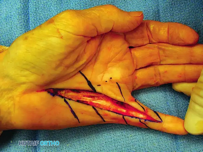
Figure 75-9 E: Planned Z-plasty skin flaps drawn over the longitudinal incision.
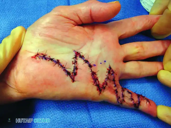
Figure 75-9 F: Subsequent closure of the Z-plasties.
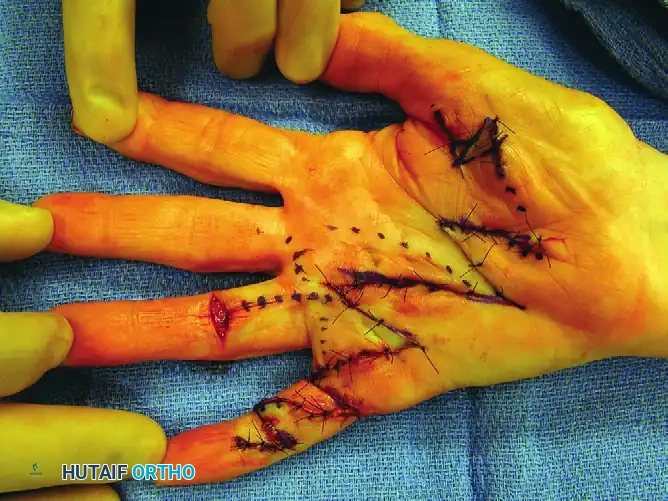
Figure 75-10 F: After meticulous hemostasis, the complex Z-plasty wounds are closed.
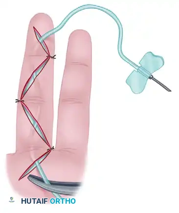
To further combat hematoma formation, active drainage fixation is highly recommended. A closed-suction drainage system can be ingeniously constructed using 21-gauge sterile butterfly catheters. Multiple fenestrations are cut into the catheter tubing, which is then placed deep to the skin flaps.
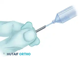
Figure 75-11 A: Multiple tubing holes are made with scissors, and the 21-gauge catheter is placed under the skin flaps.
A highly effective clinical pearl to mitigate the postoperative "flare reaction" involves the instillation of corticosteroids. Prior to activating the suction, 15 to 20 mg of a long-acting corticosteroid, such as betamethasone, can be injected directly into the wound bed via the butterfly catheter. This potent local anti-inflammatory profoundly reduces postoperative edema, stiffness, and pain.
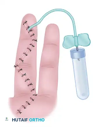
Figure 75-11 B: After wound closure, a 25-gauge needle can be used to instill 15 to 20 mg of betamethasone (Celestone) through the 21-gauge catheter needle.
Finally, the
Clinical & Radiographic Imaging Archive