Myositis Ossificans: A Challenging Orthopedic Oncology Case & Diagnostic Pitfalls
Key Takeaway
Myositis ossificans, a benign ossifying lesion, is often mistaken for soft tissue sarcoma. Differentiating it relies on a detailed history of trauma, clinical presentation, and critical imaging findings like the "zone phenomenon" on CT scans – a mature peripheral ossification with a less dense center. MRI helps characterize soft tissue components, guiding accurate diagnosis and preventing unnecessary interventions.
A 28-year-old rugby player presents with a 3-month history of a persistent, firm, and increasingly painful anterior thigh mass following a blunt injury. Physical exam reveals a firm, fixed, 6cm mass deep in the quadriceps with knee flexion limited to 90 degrees. How do you approach the initial assessment of this patient to reach a diagnosis?
Candidate: I would take a thorough history focusing on the timing of the injury and the nature of the mass. I would perform a physical exam checking for neurovascular status and range of motion. I would then order plain radiographs and an MRI, and likely plan for a biopsy to rule out malignancy.
The candidate suggests a "biopsy" too early. In the context of Myositis Ossificans, an early biopsy is often a "trap." The histological findings in the active phase are highly cellular and can be misdiagnosed as osteosarcoma, leading to unnecessary and potentially catastrophic radical surgery. Furthermore, mentioning MRI before radiographs or failing to emphasize the temporal evolution of the symptoms is a major weakness.
I would approach this by correlating the history of trauma with the "zone phenomenon" seen in imaging. My primary goal is to distinguish this from a malignancy. I would obtain serial plain radiographs—looking for the classic peripheral maturation (centrifugal ossification)—and a CT scan to confirm the radiolucent cleft between the lesion and the femoral cortex. I would explicitly avoid early biopsy due to the risk of histological misdiagnosis. I would classify this as Myositis Ossificans Traumatica and manage it conservatively until radiographic maturity is achieved.
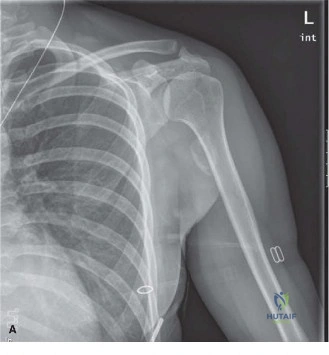
The patient's condition has plateaued, and he remains symptomatic with a 90-degree flexion block. Radiographs now show a well-defined rim of mature bone. Look at these images. What do they tell you about the pathology and the timing for intervention?

Candidate: Figure 1 shows early amorphous calcification. Figure 2 shows the zonal phenomenon, confirming it's Myositis Ossificans. I would operate now to improve the patient's knee flexion.
The candidate fails to appreciate the danger of "early" surgery. Even at 3 months, the lesion may not be metabolically mature. Operating on an immature, active lesion will almost certainly result in recurrence. A good candidate must define how to objectively prove maturity (e.g., normalized ALP, cessation of pain, serial imaging, or a negative three-phase bone scan).
Figure 2 demonstrates the classic zonal phenomenon: a mature, ossified periphery with a central, immature cellular zone. While this confirms the diagnosis, the decision to operate requires proof of metabolic maturity. I would look for normalized serum alkaline phosphatase, resolution of local pain, and a cold/quiescent bone scan. Operating before these signs are met is associated with high rates of recurrence. Only once maturity is confirmed would I proceed with surgical excision, ensuring I use postoperative prophylaxis such as Indomethacin or single-fraction low-dose radiation.
