Case Study: Diagnosing Distal Fibula Osteoid Osteoma in a Young Athlete
Key Takeaway
Osteoid osteoma diagnosis in young athletes often begins with characteristic symptoms like chronic, nocturnal, NSAID-responsive localized pain. Physical examination reveals point tenderness. Definitive diagnosis relies on imaging, with X-rays showing cortical thickening and CT scans precisely locating the radiolucent nidus, often surrounded by reactive sclerosis, as seen in the distal fibula case.
A 16-year-old male presents with a nine-month history of progressive right ankle pain. He is a high-level soccer player. He complains of nocturnal pain that is relieved by ibuprofen. On examination, he has point tenderness over the distal lateral fibula. What is your differential diagnosis, and which clinical features specifically point towards the most likely pathology?
Candidate: The primary differential is an osteoid osteoma. Other considerations would be a stress fracture, osteoblastoma, or subacute osteomyelitis (Brodie abscess). The key features here are the age of the patient, the classic history of nocturnal pain, and the dramatic relief of symptoms with NSAIDs, which is highly characteristic of an osteoid osteoma.
Failing to mention the "classic triad" or specific pain mechanism. Poor candidates often jump straight to "it's a tumor" without differentiating it from common athletic injuries like stress fractures, which are the most common initial misdiagnosis in this cohort.
Structure your answer by acknowledging the differential (Osteoid Osteoma vs. Stress Fracture vs. Osteoblastoma vs. Infection). Explicitly highlight the "biochemical pain" profile: nocturnal exacerbation due to high levels of Prostaglandin E2 in the nidus, and the "NSAID-responsiveness" which functions as a diagnostic test. Mention the demographic (male, 5-25 years) and the specific physical findings (localized cortical tenderness).
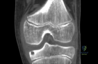
You obtain the following imaging. Describe the findings. What are the potential pitfalls in interpreting the MRI compared to the CT?
Candidate: The CT scan shows a central radiolucent nidus within the lateral cortex of the fibula, surrounded by significant reactive sclerosis. The MRI usually shows extensive marrow edema, which can be misleading and cause a clinician to suspect a malignancy or infection, whereas the CT is the gold standard for defining the nidus.
Calling the edema seen on MRI "inflammation" or "infection" without mentioning that this is the "flare phenomenon" typical of osteoid osteoma. Failing to identify the CT as the definitive modality for surgical planning.
Identify the CT findings: "Target sign" (central mineralization within the nidus) and cortical sclerosis. When discussing the MRI, emphasize that the extensive periosteal and marrow edema is non-specific and can lead to a "mimicry of malignancy" (e.g., Ewing sarcoma). State clearly that the CT is required for precise localization to guide the ablation probe and assess proximity to neurovascular structures.
The lesion is in the distal fibula, only 8mm from the skin. You are planning Radiofrequency Ablation (RFA). What specific technical challenge does this pose, and how do you mitigate it?
Candidate: The main risk is thermal injury to the skin and the superficial peroneal nerve because the lesion is too close to the surface. I would use hydrodissection with sterile fluid, like D5W, to create a buffer zone and physically separate the skin and nerves from the ablation site.
Suggesting saline for hydrodissection without clarifying it's ionic; saline conducts electricity and can interfere with the RFA or cause conductive thermal injury. Failing to mention why the proximity to the skin/nerves is a contraindication to "blind" RFA.
Identify the risk of thermal necrosis of the skin and iatrogenic nerve injury. Specify the use of hydrodissection with a non-ionic solution (e.g., 5% Dextrose in Water) to create a safe thermal buffer. Mention that this is a prerequisite for safe RFA when the nidus is <1.0 cm from the skin. Also, suggest continuous temperature monitoring and skin surface cooling as supplementary safety measures.
