Chondromyxoid Fibroma: Comprehensive Orthopedic Case Study & Diagnostic Imaging Analysis
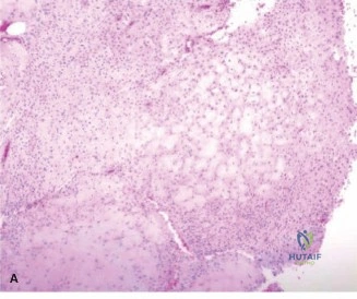
Key Takeaway
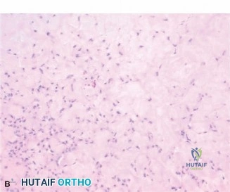
Chondromyxoid fibroma diagnosis involves clinical assessment and advanced imaging. Radiographs reveal eccentric lytic lesions with sclerotic rims. CT refines cortical details. MRI shows characteristic T2-hyperintense, lobulated lesions with internal septations. Definitive diagnosis relies on histopathological confirmation via biopsy, crucial for differentiation from other bone tumors, such as low-grade chondrosarcoma.
A 28-year-old male presents with a 6-month history of worsening anteromedial proximal tibial pain and nocturnal symptoms. On examination, there is a palpable, non-pulsatile, 4cm mass in the proximal tibial metaphysis. Plain radiographs are shown below. Describe the findings and provide your differential diagnosis.
Candidate: The radiograph reveals an eccentric, geographic lytic lesion in the proximal tibial metaphysis. It has well-defined sclerotic margins, internal septations giving a "soap bubble" appearance, and cortical thinning without an aggressive periosteal reaction or soft tissue mass. My differential would include Chondromyxoid Fibroma (CMF), Giant Cell Tumor (GCT), Aneurysmal Bone Cyst (ABC), and potentially a Non-Ossifying Fibroma (NOF) or chondroblastoma.
Candidates often fail to describe the lesion in anatomical terms (e.g., ignoring the metaphyseal location) or neglect to comment on the host-bone response (the sclerotic rim). Failing to mention "no aggressive periosteal reaction" is a missed opportunity to demonstrate oncological safety/reasoning.
Describe using the 'LOCATION' framework: Location (Metaphyseal), Orientation (Eccentric), Cortical status (Intact but thinned), Transition zone (Narrow/Geographic), and Matrix (Soap bubble/septations). Contrast the findings with GCT (usually no sclerotic rim) and Chondroblastoma (epiphyseal), arriving at a logical priority list with CMF at the top.
The MRI and CT findings are shown below. The patient is symptomatic with a thin anteromedial cortex. What is your surgical plan, and how do you address the high recurrence rate associated with this pathology?
Candidate: I would plan for an extended curettage. This involves a cortical window, mechanical removal with a high-speed burr to eliminate the scalloped ridges of the tumor, and the use of chemical or thermal adjuvants, such as hydrogen peroxide or PMMA, to reduce recurrence. I would then reconstruct the defect with a mix of allograft and cement for structural support.
Forgetting to mention the mechanical "burring" of the bony walls. Simple curettage is universally considered insufficient for CMF due to the complex lobular anatomy, and candidates who omit "extended" curettage are seen as clinically outdated.
Articulate the "Extended Curettage" protocol clearly: 1. Wide cortical window. 2. Mechanical debridement via high-speed burr (the most crucial step). 3. Adjuvant therapy (explain the benefit of PMMA's exothermic reaction and hydrogen peroxide's oxidative effect). 4. Reconstruction strategy (emphasizing the subchondral protection with cancellous bone to prevent thermal damage to the cartilage).
Post-operatively, the patient was allowed early weight-bearing. Explain why, and interpret the significance of the radiograph below.
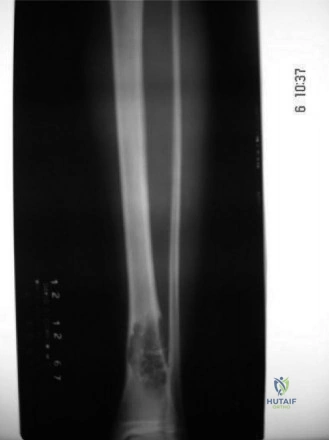
Candidate: The radiograph shows the cement mantle filling the defect. PMMA provides immediate structural stability, unlike autograft or allograft alone, which require incorporation. This allows for early mobilization and weight-bearing. The cement also acts as a thermal adjuvant to the tumor bed.
Failing to mention the "subchondral bone plate" protection. A candidate who ignores the risk of articular cartilage necrosis from the PMMA's exothermic reaction demonstrates a lack of deep surgical anatomical knowledge.
Confirm the patient's rehab benefits from the "Cement Mantel" structural properties. Crucially, add: "I ensured a 5mm layer of cancellous bone was used as a thermal barrier between the subchondral plate and the cement to protect the joint cartilage from exothermic damage, a vital step in high-quality orthopaedic oncology."
