Part of the Master Guide

Master Orthopedic Spine Cases: Sharpen Your Diagnostic Skills

Orthopedic Spine Review | Dr Hutaif Spine Surgery Revie -...

23 Apr 2026 58 min read 151 Views
Orthopedic Spine MCQs: Don't just pmid view abstract, master the topic!

Key Takeaway

Looking for accurate information on ORTHOPEDIC MCQS ONLINE 015Spine? Spinal epidural abscesses require careful management. Risk factors for nonsurgical treatment failure include IV drug abuse, diabetes, age over 65, CRP levels exceeding 115, WBC levels above 12.5, and _Staphylococcus aureus_ infection. Surgical intervention becomes necessary if a patient's neurologic status worsens. For further research, refer to pmid view abstract links provided in studies.

Orthopedic Spine Review | Dr Hutaif Spine Surgery Revie -...

Comprehensive 100-Question Exam


00:00

Start Quiz

Question 1

An 82-year-old male with severe chronic obstructive pulmonary disease and heart failure presents after a mechanical fall from a standing height. He reports significant neck pain. Neurologic examination is unremarkable.

Imaging demonstrates a displaced Type II odontoid fracture. What is the most appropriate management for this patient?


Explanation

In elderly patients (typically >80 years old) with significant medical comorbidities, nonoperative management with a rigid cervical collar is the treatment of choice for Type II odontoid fractures. While the rate of nonunion is high, stable fibrous nonunions are generally well-tolerated. Halo vest immobilization is contraindicated in this demographic due to an unacceptably high rate of morbidity and mortality, primarily from respiratory complications. Surgery is high-risk in the setting of severe comorbidities.

Question 2

A 65-year-old male presents with progressive clumsiness in his hands, difficulty buttoning his shirt, and a wide-based gait. Physical exam reveals a positive Hoffmann's sign bilaterally.

MRI of the cervical spine demonstrates multilevel degenerative spondylosis with cord compression. Which of the following MRI findings is considered the strongest independent predictor of a poor neurologic recovery following surgical decompression?


Explanation

In cervical spondylotic myelopathy (CSM), T1 hypointensity within the spinal cord indicates cystic necrosis, myelomalacia, or gliosis, and is a strong predictor of irreversible spinal cord injury and poor post-surgical outcome. T2 hyperintensity alone can represent reversible edema or inflammation and is a less reliable prognosticator of permanent deficit than T1 hypointensity.

Question 3

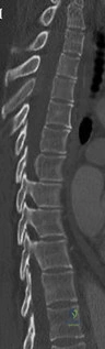
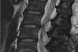
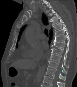
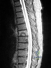
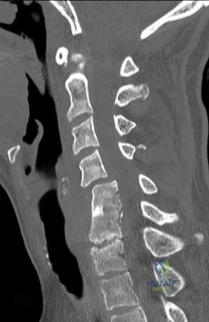
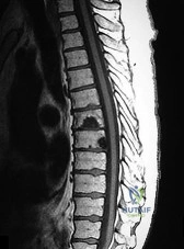
A 55-year-old diabetic male presents with severe mid-thoracic back pain, subjective fevers, and progressive bilateral leg weakness over the past 24 hours. His CRP is 120 mg/L.

An urgent MRI demonstrates a ventral spinal epidural abscess at T8-T10. Which of the following constitutes an absolute indication for urgent surgical decompression?


Explanation

The presence of a progressive neurologic deficit is an absolute indication for emergent surgical decompression in the setting of a spinal epidural abscess. While medical management (IV antibiotics) can be attempted in neurologically intact patients or those with prohibitive surgical risk, any sign of cord or nerve root compromise necessitates immediate surgical intervention to maximize the chance of neurologic recovery.

Question 4

A 35-year-old male construction worker falls 10 feet from scaffolding. He complains of moderate low back pain but has full strength and normal sensation in his lower extremities.

CT imaging shows an L1 burst fracture with 40% loss of anterior vertebral body height and 50% retropulsion into the spinal canal. MRI confirms that the posterior ligamentous complex (PLC) is intact. Based on the Thoracolumbar Injury Classification and Severity (TLICS) score, what is the appropriate score and recommended management?


Explanation

The TLICS system scores injuries based on three categories: morphology, neurologic status, and integrity of the posterior ligamentous complex (PLC). A burst fracture scores 2 points for morphology. A neurologically intact patient scores 0 points. An intact PLC scores 0 points. The total TLICS score is 2. A score of 3 or less indicates nonoperative management (e.g., bracing/mobilization).

Question 5

A 12-year-old premenarchal female is evaluated for a right thoracic prominence. Standing radiographs reveal a main thoracic curve of 32 degrees.

Her Risser stage is 0. Based on the Bracing in Adolescent Idiopathic Scoliosis Trial (BRAIST), which of the following factors has the most significant dose-dependent correlation with the success of bracing in preventing curve progression to surgical thresholds?


Explanation

The BRAIST trial established high-level evidence that bracing significantly decreases the progression of high-risk curves to the threshold for surgery. The success of bracing is highly correlated with compliance in a dose-dependent manner; greater hours of daily brace wear (especially >12.9 hours) yield significantly higher success rates.

Question 6

A 16-year-old elite male gymnast complains of chronic low back pain that is distinctly worse with spinal extension. He has failed 6 months of rest, physical therapy, and bracing.

Radiographs demonstrate a Grade II L5-S1 isthmic spondylolisthesis. He remains symptomatic. What is the most appropriate surgical intervention?


Explanation

In adolescent patients with a symptomatic Grade I or II isthmic spondylolisthesis who fail conservative management, an in situ posterior/posterolateral instrumented fusion with autograft is the gold standard. Direct pars repair (e.g., Scott wiring or pedicle screw-hook construct) is typically reserved for young patients with a pars defect but no significant slip (Grade 0), usually at L4 or above. Decompression alone is contraindicated in pediatric isthmic spondylolisthesis due to the risk of progressive slip.

Question 7

A 60-year-old female with a history of metastatic breast carcinoma presents with intractable mechanical mid-thoracic back pain.

Imaging demonstrates a large lytic lesion involving the T8 vertebral body and bilateral pedicles. There is no epidural tumor extension, and she is neurologically intact. Her Spinal Instability Neoplastic Score (SINS) is calculated to be 11. According to the NOMS framework, what is the next best step in management?


Explanation

The NOMS framework (Neurologic, Oncologic, Mechanical, Systemic) guides the treatment of spinal metastases. Mechanical instability is an independent indication for surgical stabilization, regardless of the tumor's radiosensitivity. A SINS score of 7 to 12 indicates indeterminate or potential instability, and 13 to 18 indicates instability. Because she has intractable mechanical pain and a score of 11, surgical stabilization followed by appropriate oncologic treatment (radiation) is indicated.

Question 8

A 25-year-old female is involved in a motor vehicle collision while wearing only a lap belt. She presents with severe lower back pain and abdominal ecchymosis.

Radiographs and CT reveal a pure bony flexion-distraction injury (Chance fracture) extending through the spinous process, pedicles, and vertebral body of L2. She is neurologically intact. What is an acceptable nonoperative treatment modality?


Explanation

A purely bony Chance fracture (flexion-distraction injury) has an excellent healing potential because of the broad cancellous bony surfaces. Assuming there is no severe kyphotic deformity, anterior column compromise, or neurologic deficit, it can be treated nonoperatively with an extension orthosis (TLSO) or hyperextension cast. The extension maneuver closes the posterior hinge created by the injury.

Question 9

A 70-year-old male with pre-existing cervical spondylosis falls forward, striking his chin. He presents to the ER with marked weakness in his hands and upper extremities, but relatively preserved strength in his lower extremities.

What is the primary pathophysiologic mechanism responsible for this specific neurologic deficit?


Explanation

This patient has Central Cord Syndrome, which classically presents with upper extremity weakness greater than lower extremity weakness following a hyperextension injury in an older patient with pre-existing cervical spondylosis. The mechanism involves the spinal cord being "pinched" between an anterior osteophyte/disc complex and a buckling, hypertrophied posterior ligamentum flavum.

Question 10

A 45-year-old male presents with severe shooting pain down his right anterior thigh and prominent weakness in knee extension. The right patellar reflex is absent.

MRI demonstrates a far-lateral (extraforaminal) disc herniation at the L4-L5 level. Which nerve root is most likely compressed?


Explanation

In the lumbar spine, a far-lateral (extraforaminal) disc herniation compresses the exiting nerve root at that level. At the L4-L5 level, the exiting nerve root is L4. A paracentral disc herniation at the same level would compress the traversing nerve root, which is L5. The clinical findings of anterior thigh pain, knee extension weakness, and an absent patellar reflex correspond perfectly to an L4 radiculopathy.

Question 11

A 6-year-old boy is brought to the trauma bay after a high-speed motor vehicle collision. He exhibits significant upper and lower extremity weakness and diffuse hyperreflexia.

Comprehensive plain radiographs and a non-contrast CT scan of the cervical spine demonstrate no fractures or subluxations. What is the most appropriate next step in his diagnostic workup?


Explanation

The patient is presenting with Spinal Cord Injury Without Radiographic Abnormality (SCIWORA), which is most common in the pediatric population due to the inherent elasticity and hypermobility of their spinal column. When a neurologic deficit is present but plain films and CT are negative, MRI is the gold standard diagnostic test to evaluate for spinal cord edema, hemorrhage, or subtle ligamentous injury.

Question 12

A 65-year-old female presents with progressive stooped posture, early satiety, and severe low back pain. Radiographs reveal degenerative adult spinal deformity.

Which of the following spinopelvic parameters is most strongly correlated with poorer health-related quality of life (HRQOL) outcomes if it exceeds normative thresholds?


Explanation

In adult spinal deformity, sagittal plane parameters correlate much more strongly with HRQOL outcomes than coronal plane parameters. The Sagittal Vertical Axis (SVA), defined by a plumb line dropped from the C7 vertebral body relative to the posterior superior corner of S1, is strongly correlated with clinical symptoms when it exceeds 5 cm. Other critical parameters include Pelvic Tilt (PT > 20 degrees) and PI-LL mismatch (>10 degrees).

Question 13

A 16-year-old male is brought to the clinic by his mother, who is concerned about his "round back." He complains of dull mid-back pain after standing for long periods. Standing lateral radiographs reveal a thoracic kyphosis of 65 degrees.

According to the Sorensen criteria, what specific radiographic finding is required to confirm the diagnosis of classic Scheuermann's disease?


Explanation

The classic Sorensen criteria for diagnosing Scheuermann's kyphosis require the presence of anterior wedging of 5 degrees or more in at least 3 consecutive adjacent vertebrae. Other common findings include Schmorl's nodes, endplate irregularities, and narrowed disc spaces, but the multi-level wedging is the defining diagnostic criterion.

Question 14

A 45-year-old male with a 20-year history of ankylosing spondylitis presents to the emergency department after a low-energy trip and fall at home. He complains of new-onset, severe lower cervical neck pain. Neurologic examination is unremarkable.

Standard AP, lateral, and odontoid plain radiographs are interpreted as normal. What is the most appropriate next step in management?


Explanation

Patients with ankylosing spondylitis have rigidly fused, osteopenic spines that are highly susceptible to fracture even from minor trauma. These fractures are notoriously unstable (often traversing the disk space or fused vertebral bodies) and are easily missed on plain radiographs due to overlapping anatomy and altered bone density. A CT scan of the entire cervical spine (often extending to the upper thoracic spine) is mandatory in any AS patient presenting with new neck pain after trauma.

Question 15

A 60-year-old female undergoes a C3-C6 posterior laminectomy and fusion for severe cervical spondylotic myelopathy.

On postoperative day two, she develops new-onset weakness in her right deltoid and biceps (Grade 2/5 strength) but has no new lower extremity symptoms or sensory loss. She has no distinct focal neck pain. What is the most likely etiology of this new deficit?


Explanation

C5 palsy is a well-known complication following posterior cervical decompression (laminectomy/laminoplasty). It is thought to be caused by the posterior shift of the spinal cord after decompression, which creates traction/tethering on the relatively short and horizontal C5 nerve roots. It presents as isolated deltoid/biceps weakness, typically occurring a few days postoperatively. It usually resolves over several months with conservative care.

Question 16

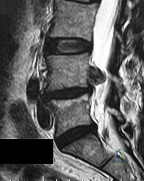
A 40-year-old male presents to the emergency room with acute, severe lower back pain, bilateral sciatica, and perianal numbness. He reports an inability to voluntarily void for the past 14 hours, and an ultrasound reveals a post-void residual volume of 650 mL.

MRI demonstrates a massive L4-L5 disc herniation filling the spinal canal. To maximize the probability of urologic and neurologic recovery, surgical decompression should ideally be performed within what maximum timeframe from the onset of symptoms?


Explanation

Cauda equina syndrome is a surgical emergency. The current literature strongly supports emergent decompression to maximize neurologic and urologic recovery. Historically and currently, intervention within 48 hours of symptom onset provides the greatest chance for return of bladder and bowel function, though many surgeons advocate for even earlier intervention (e.g., within 24 hours) if feasible.

Question 17

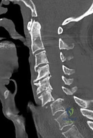
A 30-year-old male is involved in a high-speed motor vehicle collision.

Radiographs show a traumatic spondylolisthesis of the axis (Hangman's fracture). According to the Levine-Edwards classification, the fracture exhibits severe angulation and minimal translation, and the C2-C3 disc space is widened posteriorly (Type IIA). What is the primary mechanism of injury for this specific fracture pattern?


Explanation

A Levine-Edwards Type IIA Hangman's fracture is characterized by severe angulation with minimal translation. Unlike Type I and II fractures (which involve hyperextension/axial loading), the Type IIA mechanism is flexion-distraction. Crucially, applying cervical traction to a Type IIA fracture is contraindicated as it will worsen the deformity; it requires reduction with mild extension and axial compression.

Question 18

A 22-year-old collegiate wrestler presents to the emergency room with severe neck pain and a radicular pain shooting down his right arm after being taken down on his head.

Lateral cervical radiographs reveal approximately 25% anterior subluxation of the C5 vertebral body over C6. What is the classic mechanism of injury for a unilateral facet dislocation?


Explanation

A unilateral facet dislocation occurs due to a flexion-rotation mechanism. This typically results in less than 50% anterior translation (subluxation) of the superior vertebral body on the inferior one. In contrast, bilateral facet dislocations result from pure hyperflexion forces and typically cause greater than 50% anterior translation.

Question 19

During a posterior instrumented fusion of the lumbar spine, the surgeon is preparing to place a pedicle screw at the L4 level.

Which of the following describes the most accurate anatomic landmarks for establishing the starting point for a standard lumbar pedicle screw?


Explanation

The classic anatomic starting point for a lumbar pedicle screw is located at the intersection of two lines: a horizontal line that bisects the transverse process, and a vertical line that corresponds to the lateral border of the superior articular process (or the junction of the pars interarticularis). This intersection reliably leads to the center of the pedicle.

Question 20

A 72-year-old male presents with bilateral leg pain, heaviness, and cramping that predictably worsens after walking two blocks.

He states that leaning forward on a shopping cart completely relieves his pain, but standing completely upright without moving fails to relieve the symptoms. His pedal pulses are 2+ bilaterally. What is the most likely diagnosis?


Explanation

The clinical presentation is classic for neurogenic claudication secondary to lumbar spinal stenosis. A key differentiating factor between neurogenic and vascular claudication is the response to posture. Neurogenic claudication is relieved by lumbar flexion (e.g., leaning on a shopping cart, sitting) because flexion increases the cross-sectional area of the spinal canal. Standing upright still maintains lumbar extension, which continues to compress the neural elements, whereas vascular claudication is typically relieved simply by resting/standing still.

Question 21

A 7-year-old boy presents with painful torticollis 10 days after undergoing a routine tonsillectomy. On examination, his head is tilted to the right and his chin is rotated to the left. Neurologic examination is intact. What is the most appropriate initial management?


Explanation

This patient is presenting with Grisel's syndrome, which is a non-traumatic atlantoaxial subluxation most commonly seen in children following an upper respiratory infection or ENT surgery (like tonsillectomy). The pathophysiology involves inflammatory hyperemia spreading to the periodontoid vascular plexus, causing laxity of the transverse ligament. Initial management for early Fielding types (I and II) is medical, consisting of intravenous antibiotics, muscle relaxants, and a soft cervical collar.

Question 22

A 75-year-old male sustains a Type II odontoid fracture after a ground-level fall. Which of the following radiographic factors is MOST strongly associated with non-union if treated non-operatively in a halo vest?


Explanation

Type II odontoid fractures occur at the base of the dens. Risk factors for non-union with non-operative management include age > 50 years, initial displacement > 5 mm, angulation > 10 degrees, and a fracture gap > 1 mm. Given the patient's age and displacement, surgical stabilization (e.g., posterior C1-C2 fusion) is often indicated to avoid the high morbidity of halo vests in the elderly and the high rate of non-union.

Question 23

In a patient with advanced rheumatoid arthritis presenting for cervical spine evaluation, which of the following radiographic parameters is the most reliable predictor of impending neurologic deficit and indicates an urgent need for surgical stabilization?


Explanation

While an Anterior Atlanto-Dental Interval (ADI) > 3 mm is abnormal, it is the Posterior Atlanto-Dental Interval (PADI) that directly correlates with the space available for the spinal cord. A PADI of < 14 mm is highly predictive of neurologic deficit in rheumatoid patients and is an indication for surgical stabilization to prevent irreversible spinal cord injury.

Question 24

A 65-year-old male with a known history of ankylosing spondylitis presents to the emergency department after a low-speed motor vehicle collision. A CT scan demonstrates a fracture through the C6-C7 disc space extending into the posterior elements.

What is the most appropriate surgical treatment?


Explanation

Fractures in the ankylosed spine act as long lever arms, making them highly unstable shear injuries that inherently involve all three spinal columns. Short-segment fixation or isolated anterior plating frequently fails. The standard of care is long-segment posterior fixation, often supplemented with an anterior approach if additional stability is needed. Halo vests are poorly tolerated and have high complication rates in AS patients.

Question 25

A 35-year-old male is involved in a motor vehicle collision. Examination reveals normal motor and sensory function throughout his upper and lower extremities. CT imaging shows an L1 burst fracture with 15 degrees of local kyphosis and 30% canal compromise. MRI confirms an intact posterior ligamentous complex. According to the Thoracolumbar Injury Classification and Severity (TLICS) score, what is the indicated treatment?


Explanation

According to the TLICS system, a burst fracture scores 2 points for morphology. An intact posterior ligamentous complex (PLC) scores 0 points, and a neurologically intact exam scores 0 points. The total TLICS score is 2. A score of 3 or less is an indication for non-operative management, typically with a TLSO brace and early mobilization.

Question 26

A 55-year-old male presents with slowly progressive bowel and bladder dysfunction. MRI reveals a large, lobulated presacral mass. CT-guided core needle biopsy shows large cells with abundant, vacuolated cytoplasm (physaliferous cells) in a myxoid background. What is the most appropriate surgical management?


Explanation

The clinical and histologic description (physaliferous cells) is pathognomonic for a sacral chordoma. Chordomas are slow-growing, locally aggressive malignant tumors that are largely resistant to conventional chemotherapy and radiation. The standard of care is wide en bloc excision with negative margins to minimize the risk of local recurrence.

Question 27

In adult spinal deformity surgery, the concept of spinopelvic harmony is critical to achieving successful outcomes and minimizing adjacent segment disease.

According to standard matching parameters, a patient's lumbar lordosis (LL) should ideally be restored to within how many degrees of their pelvic incidence (PI)?


Explanation

Pelvic incidence (PI) is a fixed morphologic parameter (PI = Pelvic Tilt + Sacral Slope). To achieve spinopelvic harmony and optimal sagittal balance, the postoperative Lumbar Lordosis (LL) should be matched to within 10 degrees of the patient's PI (PI - LL < 10 degrees).

Question 28

A 12-year-old girl with cerebral palsy (GMFCS level V) has a rapidly progressing neuromuscular scoliosis measuring 85 degrees, with severe associated pelvic obliquity. She is non-ambulatory and has difficulty sitting in her wheelchair. Which surgical strategy is most critical for addressing her pelvic obliquity and restoring sitting balance?


Explanation

In non-ambulatory patients with severe neuromuscular scoliosis and pelvic obliquity (often causing "windswept" hips and seating difficulties), extending the posterior spinal fusion to the pelvis is crucial. Stopping at L5 fails to correct the pelvic obliquity and leaves a lever arm that often leads to hardware failure and recurrent deformity.

Question 29

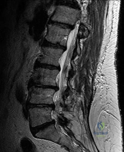
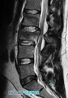
A 60-year-old male with poorly controlled diabetes mellitus presents with severe back pain, fever, and progressive bilateral lower extremity weakness over the past 24 hours. An urgent MRI confirms an anterior epidural abscess at L2-L3.

Blood cultures are drawn. What is the next best step in management?


Explanation

A spinal epidural abscess presenting with progressive neurologic deficit is a surgical emergency. Immediate surgical decompression (e.g., laminectomy) and debridement are required to preserve neurologic function. IV antibiotics are essential but insufficient alone when active neurologic deterioration is occurring.

Question 30

In a patient with cervical Ossification of the Posterior Longitudinal Ligament (OPLL), what does a "negative K-line" on a lateral radiograph imply regarding surgical planning?


Explanation

The K-line is drawn from the mid-point of the spinal canal at C2 to the mid-point at C7. If the OPLL mass exceeds this line (a negative K-line), the cervical alignment is often kyphotic or the mass is so large that posterior decompression (laminectomy/laminoplasty) will not allow the spinal cord to drift backward sufficiently. These patients typically require an anterior or combined approach for direct decompression.

Question 31

A 14-year-old female with Adolescent Idiopathic Scoliosis is being evaluated for surgery.

Radiographs demonstrate a main thoracic curve of 55 degrees and a lumbar curve of 35 degrees. On lateral bending films, the main thoracic curve corrects to 30 degrees, while the lumbar curve corrects to 15 degrees. According to the Lenke classification, what type of curve is this?


Explanation

In the Lenke classification, structural criteria define the curve type. A curve is non-structural if it bends to less than 25 degrees. Here, the lumbar curve bends to 15 degrees (non-structural), while the main thoracic curve remains > 25 degrees (structural). Thus, this is a Lenke 1 (Main Thoracic) curve.

Question 32

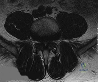
A 65-year-old male with neurogenic claudication is undergoing a decompressive laminectomy for central and lateral recess stenosis. During the approach, which specific anatomic structure must be undercut or partially resected to effectively decompress the traversing nerve root in the lateral recess?


Explanation

The lateral recess is bordered anteriorly by the vertebral body/disc, laterally by the pedicle, and posteriorly by the superior articular facet. Hypertrophy of the superior articular process is the primary osseous cause of lateral recess stenosis. Undercutting the medial aspect of the superior articular facet decompresses the traversing nerve root.

Question 33

A 30-year-old male sustains a C2 fracture in a motor vehicle collision. Radiographs demonstrate a fracture through the pars interarticularis of C2 with severe angular deformity but minimal translation. This is consistent with a Levine-Edwards Type IIA Hangman's fracture. What is the mechanism of injury, and what is the appropriate initial management?


Explanation

A Type IIA Hangman's fracture features severe angulation with minimal translation and is caused by a flexion-distraction mechanism. Because the posterior longitudinal ligament and disc are torn, longitudinal traction is strictly contraindicated as it will distract the fracture site and worsen the deformity. It requires gentle extension and compression for reduction, usually followed by halo immobilization or surgical fixation.

Question 34

A 25-year-old male involved in a high-speed MVC wearing a lap belt presents with severe abdominal bruising. A spine CT reveals a transverse fracture line propagating through the spinous process, pedicles, and vertebral body of L2.

Which of the following injuries has the strongest association with this specific fracture pattern?


Explanation

The fracture described is a Chance fracture (a bony flexion-distraction injury), classic for lap-belt injuries. There is a high association (up to 40-50%) between Chance fractures and intra-abdominal injuries, particularly hollow viscus injuries (e.g., bowel perforations), which must be carefully evaluated by general surgery.

Question 35

A 40-year-old female sustains a U-shaped sacral fracture with spinopelvic dissociation after a fall from a height. According to the Denis classification of sacral fractures, which zone is primarily involved in determining the highest risk of neurologic injury, and what is the approximate historical rate of neurologic injury associated with this zone?


Explanation

The Denis classification divides sacral fractures into three zones. Zone I (alar) has a 5% rate of nerve injury. Zone II (foraminal) has a 28% rate. Zone III (central canal) involves the spinal canal, including transverse and U-type fractures with spinopelvic dissociation, and carries the highest rate of neurologic injury, historically cited as 57%.

Question 36

A 28-year-old male presents with a spinal cord injury following a knife wound to the right side of his mid-thoracic back. Neurological examination reveals complete loss of motor function and proprioception in his right lower extremity, and a loss of pain and temperature sensation in his left lower extremity. Which spinal cord syndrome describes this pattern?


Explanation

Brown-Séquard syndrome results from a functional hemisection of the spinal cord. It presents with ipsilateral loss of motor function (corticospinal tract) and proprioception/vibratory sense (dorsal columns), and contralateral loss of pain and temperature sensation (spinothalamic tract), typically starting a few levels below the lesion.

Question 37

On a sagittal MRI of the lumbar spine, a vertebral body endplate adjacent to a degenerated disc shows hypointense signal on T1-weighted images and hyperintense signal on T2-weighted images. This finding corresponds to which Modic type, and what does it pathologically represent?


Explanation

Modic changes describe MRI signal intensity variations in vertebral body endplates. Modic Type 1 shows T1 hypointensity and T2 hyperintensity, representing bone marrow edema and acute fibrovascular inflammation. Type 2 is T1 hyperintense and T2 hyperintense/isointense (fatty replacement). Type 3 is T1 and T2 hypointense (sclerosis).

Question 38

A 16-year-old male presents with a stiff, painful thoracic hyperkyphosis that does not correct upon hyperextension. Standing lateral radiographs reveal anterior wedging of 4 consecutive thoracic vertebrae by 6 degrees each, along with irregular endplates and prominent Schmorl's nodes. Which condition is the most likely diagnosis?


Explanation

Scheuermann's disease is an structural kyphosis of the thoracic or thoracolumbar spine. The diagnostic Sorensen criteria require at least 3 consecutive vertebrae, each with at least 5 degrees of anterior wedging. Associated findings include irregular endplates, Schmorl's nodes, and narrowing of the intervertebral disc spaces.

Question 39

A 75-year-old female with severe osteoporosis complains of unremitting back pain 6 months after a minor fall. Her pain worsens significantly when standing and improves when supine. A lateral radiograph demonstrates an intravertebral vacuum cleft sign within a severely compressed T12 vertebral body. What is the eponymous name of this condition?


Explanation

Kümmell disease is delayed post-traumatic avascular necrosis of a vertebral body. It presents as a progressive vertebral collapse following a minor injury, often after an asymptomatic period. The hallmark radiographic finding is the intravertebral vacuum cleft sign on extension films, representing nitrogen gas filling the necrotic cavity.

Question 40

A 62-year-old male undergoes a C3-C6 posterior laminectomy and fusion for multilevel cervical spondylotic myelopathy. On post-operative day 2, he develops profound weakness of the right deltoid and biceps (MRC grade 2/5) without new sensory deficits or lower extremity weakness.

What is the most widely accepted primary pathomechanical explanation for this complication?


Explanation

This patient has developed a C5 palsy, a well-known complication after cervical decompression (especially posterior laminectomy/laminoplasty). The most widely accepted mechanism is the 'tethering effect': as the decompressed spinal cord drifts posteriorly, it places traction on the short, relatively horizontal C5 nerve roots, causing a neurapraxia.

Question 41

In the surgical planning and evaluation of a patient undergoing correction for adult spinal deformity, what is the widely accepted target goal for the relationship between lumbar lordosis (LL) and pelvic incidence (PI) to achieve optimal sagittal balance?


Explanation

The Pelvic Incidence (PI) is a fixed anatomical parameter (PI = Pelvic Tilt + Sacral Slope). To achieve a harmonious spino-pelvic alignment and reduce the risk of adjacent segment disease and mechanical failure, the Lumbar Lordosis (LL) should be matched to the Pelvic Incidence. The widely accepted goal, described by Schwab et al., is that PI minus LL should be less than or equal to 10 degrees.

Question 42

A 68-year-old male presents to the emergency department after falling forward and striking his chin, sustaining a hyperextension injury to his cervical spine. On examination, he exhibits bilateral upper extremity weakness (motor grade 2/5) but is able to move his lower extremities against resistance (motor grade 4/5). He also has patchy sensory deficits. Which spinal cord syndrome is most likely, and what is its typical prognosis for future ambulation?


Explanation

This classic presentation represents Central Cord Syndrome, which typically occurs in elderly patients with pre-existing cervical spondylosis who sustain a hyperextension injury. It affects the centrally located cervical tracts supplying the upper extremities more than the peripherally located tracts supplying the lower extremities. The prognosis for ambulation is generally good, with most patients regaining the ability to walk, although fine motor function in the hands often remains impaired.

Question 43

The modified Tokuhashi scoring system is a widely utilized tool to predict life expectancy in patients with metastatic spine disease and to guide surgical decision-making. Which of the following parameters is specifically evaluated in this scoring system?


Explanation

The modified Tokuhashi score evaluates six parameters: 1) General condition (Karnofsky performance status), 2) Number of extraspinal bone metastases, 3) Number of vertebral metastases, 4) Metastases to major internal organs, 5) Primary tumor site (e.g., thyroid/breast/prostate score higher than lung/stomach), and 6) Severity of spinal cord palsy. Age, BMD, and chemotherapy history are not specific components of the score.

Question 44

A 15-year-old male presents with a progressive rounding of his back. Lateral radiographs are taken to evaluate for Scheuermann's kyphosis. According to the classic Sorensen criteria, which of the following radiographic findings confirms the diagnosis?


Explanation

The classic Sorensen criteria for the diagnosis of Scheuermann's disease include a thoracic kyphosis greater than 45 degrees and the presence of anterior wedging of at least 5 degrees in three or more consecutive vertebrae. Other findings like Schmorl's nodes and endplate irregularities are common but are not the primary defining diagnostic criteria.

Question 45

A 55-year-old female undergoes a complex 10-hour posterior spinal fusion for degenerative scoliosis. During the surgery, there is significant estimated blood loss requiring massive transfusion, and prolonged hypotensive anesthesia is maintained. Upon waking in the recovery room, she complains of bilateral, painless vision loss. What is the most common cause of postoperative vision loss (POVL) in this specific clinical scenario?


Explanation

Ischemic Optic Neuropathy (ION) is the most common cause of postoperative vision loss (POVL) following major prone spine surgery. Risk factors include prolonged surgical time, large blood loss, intraoperative hypotension, and prone positioning which increases venous pressure in the head. Central retinal artery occlusion is less common and is typically unilateral due to direct globe compression.

Question 46

Ossification of the posterior longitudinal ligament (OPLL) is a frequent cause of myelopathy. Which of the following describes the most common anatomical location and the most characteristic patient demographic for this condition?


Explanation

OPLL is most prevalent in East Asian populations (particularly Japanese, where the prevalence can be 2-3%). It predominantly affects the cervical spine, leading to cervical myelopathy due to progressive anterior compression of the spinal cord by the ossified ligament.

Question 47

A 30-year-old male is evaluated in the trauma bay following a high-speed rollover motor vehicle collision. AP and lateral cervical radiographs reveal a unilateral facet dislocation at C5-C6 on the right side. What is the primary mechanism of injury, and what is the typical radiographic appearance on the AP view?


Explanation

Unilateral facet dislocations are primarily caused by a flexion-rotation mechanism. On an AP radiograph, the spinous process of the dislocated vertebra appears deviated toward the side of the dislocated facet. This is because the anterior vertebral body rotates away from the dislocated side, causing the posterior elements (spinous process) to swing toward the dislocated side.

Question 48

Recombinant human bone morphogenetic protein-2 (rhBMP-2) possesses potent osteoinductive properties and is used off-label in various spinal fusion procedures. If utilized in anterior cervical spine surgery (e.g., ACDF), what is a well-documented, potentially life-threatening complication that prompted an FDA warning?


Explanation

The off-label use of rhBMP-2 in anterior cervical spine surgery has been strongly linked to exaggerated prevertebral soft-tissue swelling and severe dysphagia, which can precipitate airway compromise. Consequently, the FDA issued a public health advisory warning against its routine use in the anterior cervical spine.

Question 49

According to the Fielding and Hawkins classification for atlantoaxial rotatory subluxation (AARS) in pediatric patients, what radiographic parameter defines a Type II injury?


Explanation

The Fielding and Hawkins classification for AARS is: Type I: Rotatory fixation with no anterior displacement (ADI < 3 mm); transverse ligament intact. Type II: Rotatory fixation with anterior displacement of 3-5 mm; transverse ligament ruptured but alar ligaments intact. Type III: Anterior displacement > 5 mm; rupture of transverse and alar ligaments. Type IV: Posterior displacement of the atlas.

Question 50

A 14-year-old female gymnast presents with insidious onset lower back pain. Imaging confirms an L5-S1 spondylolisthesis secondary to bilateral stress fractures of the pars interarticularis. According to the Wiltse classification of spondylolisthesis, which type does this represent?


Explanation

The Wiltse classification categorizes spondylolisthesis by etiology. Type I is Dysplastic (congenital anomaly). Type II is Isthmic (lesion in the pars interarticularis, common in gymnasts). Type III is Degenerative. Type IV is Traumatic (fracture in areas other than the pars). Type V is Pathologic (generalized or localized bone disease). Type VI is Iatrogenic (post-surgical).

Question 51

Pyogenic spondylodiscitis is a serious spinal infection that most frequently affects the lumbar spine in adult patients. What is the most common route of pathogen dissemination leading to this condition?


Explanation

While retrograde venous spread via Batson's plexus was historically emphasized in older literature, contemporary understanding establishes that hematogenous spread via the arterial system is the most common route for pyogenic spondylodiscitis in adults. The infection typically lodges in the highly vascularized subchondral bone adjacent to the endplate before spreading to the disc.

Question 52

A 40-year-old male presents with severe mechanical back pain. Standing lateral radiographs reveal an isthmic spondylolisthesis at L5-S1. The L5 vertebral body has slipped anteriorly by 60% of the width of the S1 endplate. According to the Meyerding classification, what grade is this slip?


Explanation

The Meyerding classification grades spondylolisthesis based on the percentage of anterior slip: Grade 1 (0-25%), Grade 2 (26-50%), Grade 3 (51-75%), Grade 4 (76-100%), and Grade 5 (>100%, also known as spondyloptosis). A 60% slip falls into the Grade 3 category.

Question 53

A 45-year-old male presents with severe radiating leg pain. MRI of the lumbar spine reveals a far lateral (extraforaminal) disc herniation at the L4-L5 level. Which nerve root is predominantly compressed by a herniation in this specific location?


Explanation

In the lumbar spine, a far lateral (extraforaminal) disc herniation compresses the exiting nerve root at that level. Therefore, at L4-L5, a far lateral disc herniation will compress the L4 nerve root. In contrast, a central or paracentral disc herniation at L4-L5 would compress the traversing L5 nerve root.

Question 54

A 65-year-old male with long-standing, advanced ankylosing spondylitis presents to the emergency department after a low-energy mechanical fall. He complains of severe neck pain but has a normal neurological examination. Radiographs and CT reveal a highly unstable, extension-type fracture through the C5-C6 disc space extending into the posterior elements. What is the most appropriate definitive management?


Explanation

Spinal fractures in patients with ankylosing spondylitis are highly unstable, often traversing all three columns, and the fused spine acts as a long lever arm. Conservative management (collar or halo) has a high failure rate and significant complication profile in these patients. The gold standard treatment is rigid internal fixation, typically involving a long-segment posterior fusion (often 3 levels above and below the fracture) to resist the extreme lever-arm forces.

Question 55

Degenerative spondylolisthesis most commonly occurs at the L4-L5 level in older adults, particularly females. Which anatomical feature is most strongly associated with the predisposition for developing degenerative spondylolisthesis at this specific level?


Explanation

The development of degenerative spondylolisthesis is strongly associated with a more sagittal orientation of the facet joints at the L4-L5 level. Normally, L4-L5 facet joints have a more coronal orientation, which biomechanically resists anterior translation. A more sagittally oriented facet joint provides less resistance to shear forces, predisposing the segment to anterior slippage as the disc degenerates.

Question 56

A 40-year-old trauma patient requires the application of a halo-vest for a cervical spine injury. To minimize the risk of iatrogenic injury to the supraorbital and supratrochlear nerves, what is the optimal anatomical placement for the anterior halo pins?


Explanation

Safe placement of anterior halo pins requires positioning them in the lateral one-third of the eyebrow (or lateral to the supraorbital notch), approximately 1 cm superior to the orbital rim. This location avoids the supratrochlear and supraorbital nerves, which run medially. Placing pins medially risks nerve injury, and placing them over the frontal sinus or glabella risks penetration of the thin bone and sinus.

Question 57

A 3-year-old child is diagnosed with congenital scoliosis. Radiographs demonstrate a unilateral unsegmented bar with a contralateral hemivertebra at the same level. What is the expected natural history of this specific congenital spinal anomaly?


Explanation

The combination of a unilateral unsegmented bar and a contralateral hemivertebra at the same level has the highest risk of rapid and severe curve progression among congenital scoliosis anomalies. Because growth is tethered on one side and accelerated on the other, early surgical intervention (often early fusion or excision) is almost universally required, as bracing is ineffective for congenital rigid curves.

Question 58

Fractures of the odontoid process (dens) are classified into three types by Anderson and D'Alonzo. Type II fractures are notorious for having a high rate of nonunion. What is the primary anatomical reason for this high nonunion rate?


Explanation

Type II odontoid fractures occur at the base (junction) of the dens and the body of C2. This region represents a vascular watershed zone between the blood supply provided by the apical arcade (via the alar ligaments) and the vessels supplying the body of C2. This tenuous blood supply, combined with the small surface area and high mobility of the segment, leads to a high rate of nonunion if not adequately immobilized or surgically fixed.

Question 59

A 25-year-old male is involved in a high-speed motor vehicle collision while wearing only a lap belt. He sustains a classic Chance fracture of the L2 vertebra. Based on the Denis three-column classification of the spine, which columns are involved in the pathomechanics of a classic Chance fracture?


Explanation

A classic Chance fracture (seatbelt fracture) is a flexion-distraction injury. The axis of rotation is anterior to the vertebral body (at the abdominal wall where the seatbelt sits). This causes tension failure extending through the posterior, middle, and anterior columns of the spine. Therefore, all three Denis columns are disrupted (typically bony failure through the spinous process, pedicles, and vertebral body).

Question 60

In the evaluation of adult spinal deformity, achieving neutral global sagittal balance is a key surgical objective. What is the generally accepted threshold for a normal Sagittal Vertical Axis (SVA), measured as the distance from the C7 plumb line to the posterior superior corner of S1?


Explanation

The Sagittal Vertical Axis (SVA) is a measure of global sagittal alignment. A normal or well-compensated SVA is generally considered to be less than 5 cm (i.e., the C7 plumb line falls within 5 cm anterior or posterior to the posterior-superior corner of the S1 endplate). An SVA greater than 5 cm indicates positive sagittal malalignment, which correlates strongly with poor health-related quality of life (HRQOL) scores.

Question 61

A 24-year-old male arrives at the trauma bay following a high-speed motor vehicle collision. Neurologic examination reveals no voluntary motor function or sensation below the T4 dermatome. His rectal tone is flaccid, and the bulbocavernosus reflex is absent. Which of the following statements regarding his neurologic prognosis is most accurate?


Explanation

The absence of the bulbocavernosus reflex indicates the patient is in a state of spinal shock. During spinal shock, the true extent of the spinal cord injury cannot be accurately determined. Prognostication and ASIA classification must be deferred until the reflex returns, marking the resolution of spinal shock.

Question 62

A 45-year-old female presents with sharp, radiating right arm pain. Examination demonstrates significant weakness in right wrist extension, numbness over the dorsal aspect of the thumb and index finger, and a diminished brachioradialis reflex. An MRI of the cervical spine is most likely to show a posterolateral disc herniation at which of the following levels?


Explanation

The patient's findings (weak wrist extension, numbness in the thumb/index finger, diminished brachioradialis reflex) correspond to a C6 radiculopathy. In the cervical spine, exiting nerve roots exit above their corresponding pedicle. Therefore, a C5-C6 posterolateral disc herniation impinges the exiting C6 nerve root.

Question 63

A 35-year-old male presents after a rollover motor vehicle crash. Imaging demonstrates a bilateral pars interarticularis fracture of C2 with severe angulation and 1 mm of anterior translation (Levine-Edwards Type IIA Hangman's fracture). What maneuver is strictly contraindicated in the management of this specific fracture pattern?


Explanation

A Type IIA Hangman's fracture involves severe angulation with minimal translation and is caused by a flexion-distraction injury with an intact anterior longitudinal ligament but torn posterior disc space. Longitudinal traction is strictly contraindicated as it can exacerbate the distraction and lead to catastrophic neurologic injury. Treatment requires gentle extension and compression, often utilizing a halo vest.

Question 64

A 62-year-old male with a 20-year history of ankylosing spondylitis presents to the emergency department complaining of new-onset, severe neck pain after a minor fall from a chair. He has no neurologic deficits. Plain radiographs of the cervical spine demonstrate extensive syndesmophytes but no obvious fracture. What is the most appropriate next step in management?


Explanation

Patients with ankylosing spondylitis have rigid, osteopenic spines that are highly susceptible to unstable fractures from low-energy trauma, which may be occult on plain radiographs. A CT scan of the entire spine is critical to rule out a fracture. Flexion-extension views are dangerous and contraindicated in this setting due to the risk of iatrogenic spinal cord injury.

Question 65

A 40-year-old construction worker falls from a ladder and sustains an L1 burst fracture. He is neurologically intact. An MRI of the lumbar spine confirms complete disruption of the posterior ligamentous complex (PLC). According to the Thoracolumbar Injury Classification and Severity (TLICS) score, what is the most appropriate treatment recommendation?


Explanation

The TLICS score for this patient is 5 (Burst morphology = 2, PLC disruption = 3, Neurologically intact = 0). A TLICS score of 4 is indeterminate, while a score of 5 or greater is an indication for surgical stabilization. Non-operative management is generally reserved for a score of 3 or less.

Question 66

In the preoperative planning for a 65-year-old female undergoing surgical correction for adult degenerative scoliosis, the surgeon calculates a pelvic incidence (PI) of 55 degrees. According to the SRS-Schwab classification, what is the ideal radiographic target for her postoperative lumbar lordosis (LL)?


Explanation

A key principle in adult spinal deformity surgery is restoring sagittal balance by minimizing the PI-LL mismatch. The ideal postoperative lumbar lordosis should be within 10 degrees of the patient's pelvic incidence (PI-LL < 10 degrees). Therefore, a target LL of approximately 45 to 65 degrees is optimal for a PI of 55 degrees.

Question 67

A 68-year-old male undergoes a C3-C7 posterior cervical laminectomy and instrumented fusion for severe cervical spondylotic myelopathy. On postoperative day two, he develops isolated 2/5 weakness in right shoulder abduction and elbow flexion, with no sensory deficits or long tract signs. An MRI shows adequate decompression with no hematoma. What is the most appropriate initial management?


Explanation

This patient has developed a C5 palsy, a known complication following cervical decompression (especially posterior laminectomy), thought to be caused by posterior spinal cord shift and subsequent traction on the short C5 nerve roots. In the absence of an epidural hematoma or inadequate decompression on MRI, the best management is observation and supportive physical therapy, as most cases spontaneously improve over several months.

Question 68

A 55-year-old male with a history of renal cell carcinoma presents with progressive lower extremity weakness and a solitary, highly destructive lytic lesion at T8 causing spinal cord compression. He is planned for an urgent anterior corpectomy and stabilization. Which of the following preoperative interventions is highly recommended to reduce perioperative morbidity?


Explanation

Renal cell carcinoma and thyroid carcinoma metastases to the spine are classically hypervascular. Preoperative transarterial embolization of the feeding vessels is highly recommended to significantly reduce intraoperative blood loss and perioperative morbidity. Radiation is typically reserved for radiosensitive tumors or postoperative adjuvant therapy.

Question 69

A 22-year-old male is evaluated after a diving accident. An open-mouth odontoid radiograph demonstrates a C1 ring fracture (Jefferson fracture). Which of the following radiographic measurements on the open-mouth view strongly suggests an incompetent transverse atlantal ligament (TAL)?


Explanation

The Rule of Spence states that a combined lateral mass overhang of C1 on C2 of greater than 6.9 mm on an open-mouth radiograph suggests rupture of the transverse atlantal ligament (TAL). A ruptured TAL renders the fracture highly unstable, often necessitating surgical stabilization or a rigid halo, rather than simple collar immobilization.

Question 70

A 15-year-old female gymnast complains of chronic lower back pain and tightness in her hamstrings. Imaging reveals a Meyerding Grade II isthmic spondylolisthesis at L5-S1. Despite 6 months of dedicated physical therapy, bracing, and activity modification, her symptoms severely limit her daily activities. What is the most appropriate surgical intervention?


Explanation

For a symptomatic Grade II isthmic spondylolisthesis at L5-S1 that fails conservative management, L5-S1 posterior instrumented fusion is the gold standard treatment. Pars repair (Buck's repair) is typically reserved for young patients with a pars defect but no significant slip (Grade 0 or early Grade I) at levels above L5-S1. Laminectomy alone in a pediatric patient with an unstable slip is contraindicated.

Question 71

A 4-year-old boy is brought to the emergency department after falling from a trampoline. He is moving all extremities but guards his neck. A lateral cervical spine radiograph shows 3 mm of anterior displacement of C2 on C3. The Swischuk line (drawn from the anterior aspect of the C1 posterior arch to the C3 posterior arch) passes 1 mm anterior to the anterior aspect of the C2 spinous process. What is the most appropriate next step?


Explanation

This presentation is classic for pseudosubluxation of C2 on C3, a normal physiologic variant common in children under 8 years of age due to ligamentous laxity and horizontal facet joints. A Swischuk line passing within 2 mm of the anterior aspect of the C2 spinous process confirms this is a benign, physiologic variant rather than a true traumatic subluxation. Reassurance and discharge are appropriate.

Question 72

A 16-year-old male presents with cosmetic concerns regarding a "hunchback" posture. Radiographs reveal hyperkyphosis of the thoracic spine. To establish a formal radiographic diagnosis of Scheuermann's kyphosis (Sorensen criteria), what specific parameters must be met?


Explanation

The classic Sorensen criteria for diagnosing Scheuermann's disease requires anterior wedging of greater than 5 degrees in at least 3 consecutive thoracic vertebrae. Additional supportive findings often include Schmorl's nodes, endplate irregularities, and narrowing of the intervertebral disc spaces.

Question 73

A spine surgeon is performing an anterior cervical discectomy and fusion (ACDF) at C6-C7. Depending on the side of the approach, there are differing risks regarding the recurrent laryngeal nerve (RLN). Which of the following statements regarding the RLN anatomy is true?


Explanation

The right recurrent laryngeal nerve has a more variable course and loops under the right subclavian artery, making it susceptible to being a 'non-recurrent' laryngeal nerve (arising directly from the vagus in the neck) in about 1% of the population. The left RLN has a more consistent, protected course looping under the aortic arch, leading some surgeons to prefer a left-sided approach for lower cervical levels.

Question 74

A 70-year-old male complains of bilateral leg and buttock pain that progressively worsens after walking two blocks. The pain is rapidly relieved when he sits or pushes a shopping cart. Pedal pulses are bounding. This classic presentation of neurogenic claudication is primarily caused by hypertrophy of which of the following structures?


Explanation

Neurogenic claudication is the hallmark symptom of lumbar spinal stenosis. It is typically caused by degenerative hypertrophy of the ligamentum flavum and facet joint osteoarthropathy, leading to central canal narrowing. Symptoms are relieved by lumbar flexion (e.g., sitting or leaning on a shopping cart), which increases the cross-sectional area of the spinal canal.

Question 75

An 80-year-old female sustains a Type II odontoid fracture after a ground-level fall. Her family prefers conservative management over surgery due to her severe cardiac comorbidities. Which of the following is the strongest risk factor for non-union of a Type II odontoid fracture treated with a rigid cervical collar?


Explanation

The strongest risk factors for non-union in Type II odontoid fractures include initial displacement > 5 mm, posterior displacement, age > 50 years, and a delay in diagnosis or treatment. Among the choices provided, initial displacement > 5 mm is a classic, highly predictive factor for non-union.

Question 76

A 72-year-old male with type 2 diabetes presents with progressive dysphagia. Lateral cervical spine radiographs demonstrate flowing, continuous ossification along the anterior aspect of five contiguous vertebral bodies, with preservation of the intervertebral disc spaces. His sacroiliac joints are radiographically normal. What is the most likely diagnosis?


Explanation

Diffuse idiopathic skeletal hyperostosis (DISH) is characterized by flowing anterior ossification over at least four contiguous vertebral levels with preserved disc heights. Unlike ankylosing spondylitis, DISH does not involve the sacroiliac joints and typically presents in older patients, often with metabolic syndrome or diabetes. Dysphagia is a known complication due to massive anterior cervical osteophytes.

Question 77

A 35-year-old male falls from a ladder and sustains localized thoracolumbar pain. He is neurologically intact. CT and MRI confirm an L1 burst fracture with 15 degrees of kyphosis, 30% canal compromise, and an intact posterior ligamentous complex.

Based on the Thoracolumbar Injury Classification and Severity (TLICS) score, what is the most appropriate management?


Explanation

The patient's TLICS score is 2 (Burst fracture = 2, Neurologically intact = 0, PLC intact = 0). A score of 3 or less indicates non-operative management, typically with a TLSO.

Question 78

A 68-year-old female with adult spinal deformity presents with progressive back pain and forward truncal lean. Her spinopelvic parameters reveal a Pelvic Incidence (PI) of 60 degrees. To achieve a harmonious sagittal profile postoperatively, what is the most appropriate target for her Lumbar Lordosis (LL)?


Explanation

The formula PI - LL mismatch < 10 degrees is standard for restoring sagittal balance. With a PI of 60, an ideal postoperative LL target is approximately 60 degrees.

Question 79

A 50-year-old male with severe, long-standing ankylosing spondylitis presents to the emergency department with new-onset neck pain after a low-speed motor vehicle collision. Neurological examination is normal. An initial cross-table lateral radiograph of the cervical spine is read as negative for acute fracture. What is the most appropriate next step in management?


Explanation

Patients with ankylosing spondylitis are at high risk for highly unstable occult fractures even from minor trauma. Standard plain films are often inadequate; therefore, a CT of the cervical spine (or whole spine) is mandatory if there is clinical suspicion.

Question 80

A 55-year-old Asian male presents with progressive clumsiness in his hands and a wide-based gait. Imaging reveals ossification of the posterior longitudinal ligament (OPLL) from C3 to C6. The sagittal MRI shows that the OPLL mass crosses the K-line (K-line negative), and the cervical spine has 15 degrees of local kyphosis. Which surgical approach is most appropriate?


Explanation

A negative K-line and local kyphosis are contraindications to pure posterior indirect decompression techniques like laminoplasty. An anterior approach (or combined approach) is necessary to directly decompress the cord and correct the kyphosis.

Question 81

A 45-year-old male presents with acute, severe right leg radicular pain. MRI of the lumbar spine reveals a far lateral (extra-foraminal) disc herniation at the L4-L5 level on the right side. Which nerve root is most likely compressed, and what clinical finding is expected?


Explanation

A far lateral disc herniation at L4-L5 compresses the exiting L4 nerve root. Compression of the L4 root presents with quadriceps weakness (knee extension) and a diminished patellar reflex.

Question 82

A 65-year-old male undergoes a C3-C6 posterior laminectomy and fusion for cervical spondylotic myelopathy. On postoperative day 2, he develops profound weakness of bilateral deltoid and biceps muscles without new sensory deficits. Lower extremity function remains intact. What is the most likely etiology of this complication?


Explanation

C5 palsy is a well-known complication after posterior cervical decompression. It is most commonly attributed to a posterior shift of the spinal cord, leading to traction and tethering of the relatively short C5 nerve roots.

Question 83

A 24-year-old male is involved in a high-speed collision and sustains a Levine-Edwards Type IIa traumatic spondylolisthesis of the axis (Hangman's fracture). Radiographs show significant angular deformity with minimal translation. What is the most appropriate management?


Explanation

Type IIa Hangman's fractures feature severe angulation with flexion-distraction injury of the C2-3 disc. Traction is strictly contraindicated as it causes over-distraction; treatment consists of a Halo vest with slight compression and extension.

None

Dr. Mohammed Hutaif
Medically Verified Content by
Prof. Dr. Mohammed Hutaif
Consultant Orthopedic & Spine Surgeon
Chapter Index