Orthopedic Pathology Review | Dr Hutaif Basic Science R -...
Key Takeaway
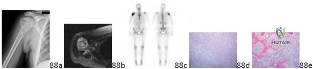
Discover the latest medical recommendations for ORTHOPEDIC MCQS ONLINE 011 PATHOLOGY. Orthopedic imaging, as seen in figures, aids in diagnosing musculoskeletal conditions. Synovial chondromatosis presents with calcified loose bodies and a painful ankle mass. Benign distal femoral enchondromas, often incidental findings, typically require only radiographic follow-up if asymptomatic and non-aggressive. Differentiating these from malignant tumors like chondrosarcoma relies on specific clinical and imaging features.
Orthopedic Pathology Review | Dr Hutaif Basic Science R -...
Comprehensive 100-Question Exam
00:00
Start Quiz
Question 1
An 11-year-old boy presents with a painful mass in the diaphysis of his femur. Radiographs show an aggressive, permeative lytic lesion with an 'onion-skin' periosteal reaction. Biopsy reveals small round blue cells. Which of the following immunohistochemical markers and translocations are most characteristic of this lesion?
Explanation
Question 2
A 28-year-old woman presents with knee pain. Radiographs demonstrate an eccentrically located lytic lesion in the distal femoral epiphysis that extends to the subchondral bone. Histology reveals mononuclear cells and multinucleated giant cells. Which of the following best describes the true neoplastic cells in this lesion?
Explanation
Question 3
A 25-year-old man presents with a slow-growing, deep-seated mass in his popliteal fossa. Imaging reveals a soft tissue mass with punctate calcifications. Biopsy demonstrates a biphasic spindle cell neoplasm with epithelial components. What is the pathognomonic chromosomal translocation associated with this tumor?
Explanation
Question 4
A 14-year-old girl is evaluated for a 'shepherd's crook' deformity of her proximal femur. Radiographs show a ground-glass intramedullary lesion. She also has multiple café-au-lait spots with irregular borders and a history of precocious puberty. What is the underlying genetic mechanism of her disease?
Explanation
Question 5
A 22-year-old man presents with an anterior tibial bowing deformity and a multi-loculated, lytic 'soap-bubble' lesion in the anterior cortex of the tibial diaphysis. Biopsy shows islands of epithelial cells surrounded by fibrous stroma. Immunohistochemistry is positive for cytokeratin. What is the most appropriate definitive management?
Explanation
Question 6
An 18-year-old male complains of severe, progressively worsening nocturnal thigh pain that is completely relieved by ibuprofen. Radiographs reveal a cortical thickening with a 7mm radiolucent nidus. Which of the following is true regarding the pathophysiology of his pain?
Explanation
Question 7
A 65-year-old man presents with back pain, fatigue, and hypercalcemia. Radiographs demonstrate multiple 'punched-out' lytic lesions in his skull and pelvis. A technetium-99m bone scan is reportedly 'cold' in the areas of these lesions. What is the primary reason for the lack of uptake on the bone scan?
Explanation
Question 8
A 32-year-old woman presents with a firm mass in her foot, firmly attached to the plantar fascia. Biopsy shows nests of uniform cells with clear cytoplasm separated by fibrous septa. The cells are strongly positive for S-100 and HMB-45. Which of the following translocations is diagnostic for this tumor?
Explanation
Question 9
A 25-year-old male presents with multiple asymmetric cartilaginous tumors in his hands and long bones. He also has multiple soft tissue hemangiomas on his trunk and extremities. What is his most likely underlying diagnosis, and what gene mutation is most commonly associated with it?
Explanation
Question 10
A 72-year-old man with increasing hat size, hearing loss, and bowing of his tibias presents for evaluation. Radiographs show thickened cortices and a 'cotton wool' appearance of the skull. A bone biopsy in the sclerotic phase would most likely reveal which of the following histological features?
Explanation
Question 11
A 6-year-old boy presents with back pain and is found to have a severe compression fracture (vertebra plana) of T8 on radiographs. Biopsy of the lesion shows histiocytes with grooved, 'coffee-bean' nuclei, mixed with eosinophils. Electron microscopy reveals Birbeck granules. Which marker is most reliably positive on immunohistochemistry?
Explanation
Question 12
An 18-year-old female sustains a pathologic fracture of her proximal humerus. Radiographs show an expansile, eccentrically placed, radiolucent lesion with 'fluid-fluid' levels on MRI. Biopsy shows blood-filled spaces lacking an endothelial lining and scattered giant cells. Which genetic alteration is primarily responsible for the primary form of this lesion?
Explanation
Question 13
A 15-year-old boy presents with knee pain. Radiographs demonstrate a 2 cm lytic lesion strictly confined to the proximal tibial epiphysis with a thin sclerotic rim. Histological examination shows mononuclear cells with grooved nuclei and areas of 'chicken-wire' calcification. Which of the following is the most appropriate treatment?
Explanation
Question 14
A 35-year-old man presents with recurrent bloody effusions and chronic swelling in his knee. MRI reveals a large, lobulated intra-articular mass with low signal intensity on both T1 and T2 weighted images, with prominent blooming artifact on gradient-echo sequences. What is the characteristic genetic translocation and resulting molecular driver of this condition?
Explanation
Question 15
A 45-year-old male undergoes wide resection of a massive, painless, deep-seated soft tissue mass in his thigh. Pathology reveals a uniform proliferation of round cells, signet-ring lipoblasts, and a prominent plexiform ('chicken-wire') capillary network in a myxoid stroma. Which genetic abnormality is highly specific for this sarcoma?
Explanation
Question 16
A 9-year-old boy falls and sustains a pathologic fracture of his proximal humerus. Radiographs reveal a centrally located, completely lytic lesion in the metaphysis that abuts the physis. A bone fragment is seen resting at the dependent portion of the cyst. Which of the following statements about the cyst fluid is most accurate?
Explanation
Question 17
A 55-year-old man presents with a pathologic fracture of his right femur. Radiographs reveal an aggressive, blastic (sclerotic) lesion in the proximal femur. Which of the following primary malignancies is the most likely source of this metastasis?
Explanation
Question 18
A 25-year-old woman presents with a slow-growing mass on the posterior aspect of her distal femur. Radiographs show a heavily ossified, broad-based mass originating from the cortical surface without medullary involvement. Histology reveals low-grade spindle cells with abundant osteoid formation. What is her expected 5-year survival with wide surgical resection alone?
Explanation
Question 19
A 16-year-old boy presents with a rapidly enlarging mass in his forearm. Biopsy shows small round blue cells arranged in nests separated by fibrous septa, with a loss of central cellular cohesion mimicking pulmonary alveoli. Immunohistochemistry is strongly positive for MyoD1 and myogenin. Which translocation implies the worst prognosis for this specific tumor type?
Explanation
Question 20
A 30-year-old Ashkenazi Jewish man presents with severe, acute left thigh pain and fever, initially mimicking osteomyelitis. Radiographs of the femur show an 'Erlenmeyer flask' deformity of the distal femur with scattered lytic and sclerotic areas. He also has hepatosplenomegaly. Deficiency of which of the following enzymes is responsible for his skeletal manifestations?
Explanation
Question 21
A 45-year-old male presents with chronic knee pain. Radiographs reveal a lytic lesion in the proximal tibial epiphysis with faint mineralization. Biopsy shows lobules of cells with abundant clear cytoplasm and distinct boundaries, interspersed with reactive woven bone formation. Which of the following is the most likely diagnosis?
Explanation
Question 22
A 28-year-old female undergoes resection of a deep-seated soft tissue mass near her knee joint. Histology demonstrates a biphasic pattern of spindle cells and epithelial-like glandular structures. Which of the following chromosomal translocations is characteristic of this tumor?
Explanation
Question 23
A 14-year-old girl is evaluated for a 'shepherd's crook' deformity of her proximal femur. Biopsy reveals irregular woven bone trabeculae lacking osteoblastic rimming set in a bland fibrous stroma. Which of the following gene mutations is most strongly associated with this condition?
Explanation
Question 24
A 24-year-old male presents with anterior lower leg pain. Radiographs show a multicentric, eccentric, lytic lesion in the anterior cortex of the tibial diaphysis. Histopathology reveals clusters of basaloid epithelial cells arranged in nests within a fibrous stroma. What is the most appropriate definitive management?
Explanation
Question 25
A 30-year-old female presents with a destructive, expansile lytic lesion in the distal femoral epiphysis. Biopsy confirms a giant cell tumor of bone. Molecular testing of the neoplastic cells is most likely to reveal a mutation in which of the following genes?
Explanation
Question 26
A 25-year-old female presents with a painless posterior knee mass. Radiographs show a densely ossified mass attached to the posterior cortex of the distal femur by a broad base, with a distinct radiolucent cleft separating it from the underlying bone. Genetic analysis of this lesion will most likely demonstrate amplification of which of the following genes?
Explanation
Question 27
A 20-year-old male presents with a painful lytic lesion in the proximal tibial metaphysis. Radiographs show an eccentric, radiolucent defect with a sclerotic margin. Histology demonstrates lobules of stellate and spindle cells in a myxoid stroma, with increased cellularity at the periphery of the lobules. Multinucleated giant cells are present. What is the most likely diagnosis?
Explanation
Question 28
A 45-year-old man undergoes resection of a large intramuscular mass in his thigh. Pathology reveals a multinodular tumor with a myxoid background, an arborizing 'chicken-wire' capillary network, and small uniform lipoblasts. What chromosomal translocation is diagnostic for this tumor?
Explanation
Question 29
A 7-year-old boy presents with back pain. Radiographs demonstrate a flattened vertebral body (vertebra plana) at T10. Biopsy reveals a proliferation of mononuclear cells with grooved nuclei mixed with eosinophils. Immunohistochemistry will most likely be positive for which of the following markers?
Explanation
Question 30
A 68-year-old male with a long history of increasing hat size and bowing of his femurs presents with new, severe mid-thigh pain and a rapidly enlarging mass. Radiographs show a destructive lytic lesion in the bowed femur. What is the most likely histologic finding in the pre-existing bone?
Explanation
Question 31
An 11-year-old boy presents with fever, weight loss, and thigh pain. Radiographs show a permeative, diaphyseal femoral lesion with an 'onion-skin' periosteal reaction. Histology reveals sheets of small, round, blue cells. Which immunohistochemical marker is characteristically strongly positive in this condition?
Explanation
Question 32
A 25-year-old woman is diagnosed with a high-grade bone tumor of the pelvis. Biopsy demonstrates a highly cellular tumor exhibiting a biphasic pattern: sheets of undifferentiated small round blue cells interspersed with distinct, abrupt islands of well-differentiated hyaline cartilage. Hemangiopericytoma-like vascular patterns are also noted. What is the most likely diagnosis?
Explanation
Question 33
A 16-year-old boy presents with worsening night pain in his tibia that is dramatically relieved by NSAIDs. Radiographs show a 1 cm radiolucent nidus surrounded by dense reactive sclerosis. If excised, histologic examination of the nidus is most likely to demonstrate which of the following?
Explanation
Question 34
A 72-year-old man undergoes a biopsy of a densely sclerotic lesion in his L4 vertebral body. Pathology reveals irregular bone trabeculae lined by uniform atypical cells with prominent nucleoli forming back-to-back glands. Immunohistochemistry is strongly positive for PSA. Which of the following factors is primarily responsible for the osteosclerotic (osteoblastic) appearance of these metastases?
Explanation
Question 35
A 14-year-old boy presents with a rapidly growing soft tissue mass in his forearm. Biopsy reveals small round blue cells arranged in clusters divided by fibrous septa, forming pseudoalveolar spaces. Cytogenetics identifies a t(2;13) chromosomal translocation. This translocation results in the fusion of which two genes?
Explanation
Question 36
A 12-year-old girl presents with a painful, rapidly expanding mass in the posterior elements of her cervical spine. Radiographs reveal an expansile, multicystic lesion. Biopsy shows blood-filled spaces lacking endothelial lining, separated by fibrous septa containing osteoclast-like giant cells. Which of the following genetic alterations is diagnostic of the primary form of this lesion?
Explanation
Question 37
A 60-year-old male presents with bowel and bladder incontinence and chronic lower back pain. Imaging shows a large, destructive midline mass involving the sacrum. Biopsy reveals lobules of large cells with prominent intracytoplasmic vacuoles in a myxoid background. Which immunohistochemical marker is highly sensitive and specific for confirming this diagnosis?
Explanation
Question 38
A 28-year-old female presents with a lytic lesion in her distal radius. Biopsy demonstrates uniform, spindle-shaped fibroblasts separated by abundant collagenous matrix, without cytologic atypia, pleomorphism, or mitosis. Immunohistochemistry reveals nuclear positivity for beta-catenin. What is the most likely diagnosis?
Explanation
Question 39
A 24-year-old male presents with a painless, slow-growing nodule on the volar aspect of his index finger. Initial excision was read as a necrotizing granuloma, but the mass recurred locally. Re-evaluation of the histology shows nodules of plump epithelioid cells surrounding central areas of necrosis. Immunohistochemistry shows loss of SMARCB1 (INI1) expression. What is the diagnosis?
Explanation
Question 40
A 15-year-old male with a history of multiple soft-tissue hemangiomas presents with a newly symptomatic, expansile cartilaginous lesion in his right humerus. He is diagnosed with Maffucci syndrome. The pathogenesis of his skeletal and vascular lesions is most strongly associated with somatic mosaic mutations in which of the following genes?
Explanation
Question 41
A 25-year-old male presents with chronic leg pain. Radiographs reveal a 'soap-bubble' multiloculated osteolytic lesion in the anterior tibial diaphysis. Biopsy reveals islands of epithelial cells surrounded by a bland fibrous stroma. Immunohistochemistry is strongly positive for cytokeratin. Which of the following is the most appropriate management for this condition?
Explanation
Question 42
A 55-year-old male presents with constipation and lower back pain. Imaging shows a large, destructive midline sacral mass. Biopsy reveals lobules of cells with prominent vacuolated cytoplasm in a myxoid background. To differentiate this lesion from a chondrosarcoma, which of the following immunohistochemical markers is the most specific?
Explanation
Question 43
A 15-year-old boy presents with knee pain. Radiographs reveal a well-circumscribed, eccentrically located radiolucent lesion in the proximal tibial epiphysis. Biopsy demonstrates mononuclear cells with longitudinal nuclear grooves and areas of fine, intercellular 'chicken-wire' calcifications. Which of the following gene mutations is most characteristically associated with this tumor?
Explanation
Question 44
A 45-year-old male presents with progressive hip pain. Radiographs show a distinct, heavily calcified radiolucent lesion in the proximal femoral epiphysis. Biopsy reveals large cells with distinct borders, central round nuclei, and abundant optically clear cytoplasm, mixed with areas of conventional chondrosarcoma and reactive woven bone. What is the most likely diagnosis?
Explanation
Question 45
A 35-year-old woman with recurrent, extensive pigmented villonodular synovitis (PVNS) of the knee is deemed unsuitable for further surgical resection. Systemic medical therapy is planned. This therapy most likely targets which of the following pathways?
Explanation
Question 46
A 12-year-old boy presents with severe, aching pain in his mid-thigh that is significantly worse at night and dramatically relieved by ibuprofen. Radiographs show cortical thickening with a small radiolucent nidus. The symptomatic relief provided by NSAIDs is primarily due to the inhibition of which biochemical mediator produced by the tumor?
Explanation
Question 47
Fibrous dysplasia is associated with a post-zygotic, somatic activating mutation in the GNAS gene. This genetic alteration directly results in the constitutive activation of which of the following intracellular signaling mechanisms?
Explanation
Question 48
Denosumab is an effective systemic treatment for surgically unsalvageable or metastatic Giant Cell Tumor of Bone (GCTB). It works by binding to RANKL. In the pathophysiology of GCTB, which cellular population is the primary source of RANKL expression?
Explanation
Question 49
A 10-year-old girl presents with a large, destructive, permeative diaphyseal lesion of the femur with an 'onion-skin' periosteal reaction. A biopsy is performed. The diagnosis of Ewing sarcoma is supported by a strong, diffuse membranous staining for CD99. To further distinguish this from other small blue cell tumors, which of the following immunohistochemical markers is currently considered the most highly sensitive and specific adjunct?
Explanation
Question 50
A 30-year-old male presents with a slowly enlarging, deep-seated soft tissue mass in the thigh near the knee joint. Biopsy reveals a biphasic tumor comprised of both epithelial components (glandular structures) and a spindle cell mesenchymal stroma. Which of the following cytogenetic abnormalities is diagnostic for this tumor?
Explanation
Question 51
A 28-year-old female presents with a painless, hard mass on the posterior aspect of her distal thigh. Radiographs demonstrate a dense, lobulated, heavily ossified mass arising from the posterior cortex of the distal femur, with a thin radiolucent 'cleft' separating the bulk of the tumor from the underlying cortex. Biopsy reveals a low-grade fibroblastic stroma with parallel trabeculae of woven bone. Amplification of which gene is the molecular hallmark of this entity?
Explanation
Question 52
A 70-year-old man with advanced prostate cancer develops multiple dense, osteoblastic metastatic lesions in his lumbar spine and pelvis. The characteristic osteosclerotic nature of these metastatic bone lesions is primarily driven by tumor cell secretion of which of the following factors?
Explanation
Question 53
Multiple myeloma bone disease is characterized by purely osteolytic lesions with virtually no reactive new bone formation, leading to a negative ('cold') bone scan in many cases. The profound suppression of osteoblast function in these lesions is primarily mediated by myeloma cell secretion of which molecule?
Explanation
Question 54
A 60-year-old man presents with an increasing hat size and bowing of his tibiae. Radiographs show thickened, disorganized trabeculae and cortical thickening ('cotton wool' appearance). A genetic evaluation is performed due to a strong family history. An activating mutation in which of the following genes is most commonly associated with the familial form of this disease?
Explanation
Question 55
A 22-year-old male is evaluated for multiple bony deformities and is diagnosed with Ollier disease, characterized by multiple enchondromas. He is counseled regarding the risk of malignant transformation. Somatic mosaic mutations in which of the following genes are the primary driver for both Ollier disease and Maffucci syndrome?
Explanation
Question 56
A 16-year-old boy presents with a rapidly expanding, painful distal femoral lytic lesion. MRI reveals multiple fluid-fluid levels, mimicking an Aneurysmal Bone Cyst (ABC). A biopsy is performed to rule out Telangiectatic Osteosarcoma. Which of the following histologic features definitively establishes the diagnosis of Telangiectatic Osteosarcoma over an ABC?
Explanation
Question 57
A 7-year-old boy presents with back pain. Radiographs reveal a completely flattened T8 vertebral body (vertebra plana). Needle biopsy demonstrates an infiltrate of eosinophils mixed with prominent histiocytes that have folded, 'coffee-bean' shaped nuclei. Which of the following immunohistochemical markers is the most highly specific for the primary pathologic cell in this condition?
Explanation
Question 58
A 5-year-old boy presents with a painless anterior bowing of his right tibia. Radiographs reveal a multilocular, intracortical radiolucent lesion of the anterior diaphyseal tibia. A biopsy is obtained to differentiate this from fibrous dysplasia. Which of the following histologic findings is characteristic of Osteofibrous Dysplasia and absent in classical Fibrous Dysplasia?
Explanation
Question 59
A 22-year-old male presents with chronic knee pain. Imaging reveals an eccentric, well-demarcated lytic lesion with a sclerotic rim in the proximal tibial metaphysis. Biopsy exhibits a distinct lobular architecture with a myxoid and chondroid background. The periphery of the lobules is highly cellular with spindle-shaped cells, whereas the center is hypocellular with stellate cells. What is the most likely diagnosis?
Explanation
Question 60
A 35-year-old woman presents with a locally aggressive, destructive radiolucent lesion in the mandibular ramus, extending into the soft tissues. Biopsy reveals a dense proliferation of uniform, elongated spindle cells producing abundant collagen without nuclear atypia, mitosis, or any osteoid or chondroid matrix formation. Molecular analysis demonstrates a mutation in the CTNNB1 gene. This lesion is the intraosseous counterpart to which of the following soft tissue tumors?
Explanation
Question 61
A 14-year-old boy presents with aching pain in his proximal tibia that is significantly worse at night and rapidly relieved by ibuprofen. Radiographs demonstrate a radiolucent nidus less than 1.5 cm surrounded by reactive sclerosis. Which biochemical mediator is produced in exceptionally high quantities by the cells within this nidus?
Explanation
Question 62
A 25-year-old female presents with a slow-growing, painless mass on the posterior aspect of her distal femur. Radiographs reveal a dense, heavily ossified mass attached to the cortex via a broad base with a 'string sign' indicating a radiolucent cleft. Genetic analysis of the tumor cells is most likely to reveal which of the following abnormalities?
Explanation
Question 63
A 32-year-old female presents with multiple asymmetrical, expanding cartilaginous tumors in the phalanges of her hands, accompanied by soft tissue hemangiomas exhibiting phleboliths on radiographs. Which gene mutation is most likely responsible for her underlying syndrome?
Explanation
Question 64
A 30-year-old man presents with a slow-growing, deep-seated soft tissue mass in his foot. Biopsy reveals nests of pale-staining spindle cells separated by fibrous septa. Immunohistochemistry is strongly positive for HMB-45, Melan-A, and S-100. Which chromosomal translocation defines this neoplasm?
Explanation
Question 65
A 40-year-old male undergoes excision of a deep thigh mass. Histopathology shows a proliferation of uniform round cells, signet-ring lipoblasts, and a prominent branching capillary network resembling 'chicken wire' in a myxoid stroma. What is the characteristic genetic translocation associated with this sarcoma?
Explanation
Question 66
A 12-year-old boy presents with a 'shepherd's crook' deformity of his proximal femur. Radiographs demonstrate an expansive, intramedullary ground-glass lesion. What is the underlying cellular mechanism driving the pathogenesis of this osseous lesion?
Explanation
Question 67
A 6-year-old boy presents with localized back pain. Radiographs demonstrate a 'vertebra plana' in the thoracic spine. Biopsy of the lesion shows a proliferation of cells with distinct nuclear grooves, admixed with eosinophils. Electron microscopy identifies tennis-racket shaped organelles. Which immunohistochemical marker will definitively identify the lesional cells?
Explanation
Question 68
A 28-year-old male presents with dull, aching anterior lower leg pain. Radiographs show a multicystic, eccentric, expansile lytic lesion in the anterior tibial diaphysis. Histopathology reveals islands and nests of epithelial cells surrounded by a bland fibrous stroma. Which immunohistochemical stain is most likely positive in the lesional cells?
Explanation
Question 69
A 55-year-old male presents with chronic constipation and saddle anesthesia. Imaging reveals a large, destructive midline sacral mass. Biopsy shows large, vacuolated cells arranged in lobules and cords within a prominent myxoid stroma. Which immunohistochemical marker is highly specific for confirming this diagnosis?
Explanation
Question 70
A 35-year-old female undergoes MRI for recurrent, unexplained bloody knee effusions. The imaging reveals a nodular synovial mass demonstrating low signal intensity on both T1 and T2 sequences, with a 'blooming' artifact on gradient echo. What is the primary pathogenic mechanism driving the growth of this mass?
Explanation
Question 71
A 9-year-old boy presents with a proximal humerus fracture after a minor fall. Radiographs show a centrally located, completely lytic metaphyseal lesion with a 'fallen leaf' sign. Aspiration of the fluid from this lesion would most likely reveal pathologically high levels of which of the following?
Explanation
Question 72
A 68-year-old man presents with severe generalized back pain, normocytic anemia, and hypercalcemia. Radiographs show multiple punched-out lytic skull lesions. Which of the following imaging modalities is generally considered the LEAST sensitive for detecting the extent of skeletal involvement in this specific disease process?
Explanation
Question 73
A 14-year-old female presents with acute knee pain. MRI shows an eccentric, expansile metaphyseal lesion in the distal femur containing multiple fluid-fluid levels. Biopsy confirms an aneurysmal bone cyst (ABC). Which specific gene rearrangement is considered the primary neoplastic driver of this primary lesion?
Explanation
Question 74
A 45-year-old man presents with chronic knee catching and swelling. Radiographs demonstrate dozens of small, uniform, radiopaque loose bodies confined to the joint space. This condition is primarily characterized by which of the following pathophysiologic processes?
Explanation
Question 75
A 14-year-old girl presents with a rapidly enlarging, painful mass in her proximal tibia. Radiographs demonstrate an eccentric, expansile, purely lytic metaphyseal lesion with thinning of the surrounding cortex. MRI shows multiple fluid-fluid levels. Biopsy reveals blood-filled cystic spaces separated by fibrous septa containing giant cells, without significant atypia. Which of the following genetic alterations is the primary driver of this neoplasm?
Explanation
Question 76
A 65-year-old man presents with severe back pain and fatigue. Laboratory studies reveal hypercalcemia, anemia, and an elevated serum creatinine. Radiographs show multiple punched-out lytic lesions in his skull and spine. A technetium-99m bone scan is notable for a lack of uptake in the corresponding lytic areas. What is the primary mechanism of bone destruction in this condition?
Explanation
Question 77
A 55-year-old man presents with chronic low back pain and recent-onset bowel and bladder incontinence. MRI demonstrates a destructive, lobulated midline mass arising from the sacrum. Biopsy reveals nests of large, vacuolated cells in a myxoid stroma. Which of the following immunohistochemical markers is most specific for diagnosing this lesion?
Explanation
Question 78
A 16-year-old boy presents with right shoulder pain. Radiographs show a well-circumscribed lytic lesion in the proximal humeral epiphysis with central calcifications. Histologic examination demonstrates mononuclear cells, scattered osteoclast-like giant cells, and a 'chicken-wire' pattern of pericellular calcification. Which genetic mutation is classically associated with this tumor?
Explanation
Question 79
A 30-year-old man presents with anterior bowing of his lower leg. Radiographs reveal an eccentric, multiloculated 'soap-bubble' lytic lesion in the anterior diaphysis of the tibia. Biopsy shows a biphasic tumor with both epithelial and osteofibrous components. Which of the following is true regarding this pathology?
Explanation
Question 80
A 45-year-old woman presents with a deep-seated thigh mass. MRI shows a well-defined, multi-lobulated soft tissue mass. Histology demonstrates an abundant myxoid stroma, a rich branching capillary network resembling 'chicken-wire', and small lipoblasts. What is the characteristic chromosomal translocation associated with this sarcoma?
Explanation
Question 81
A 7-year-old boy presents with severe localized thoracic back pain. Radiographs reveal uniform flattening of the T7 vertebral body (vertebra plana) with preserved adjacent disc spaces. Biopsy shows a cellular infiltrate including eosinophils and mononuclear cells with grooved, 'coffee-bean' nuclei. Electron microscopy is most likely to reveal which of the following?
Explanation
Question 82
A 24-year-old woman notes a painless, slow-growing mass on the posterior aspect of her distal thigh. Radiographs demonstrate a heavily ossified, broad-based mass attached to the posterior cortex of the distal femur, with a radiolucent cleft separating a portion of the tumor from the underlying bone (string sign). What molecular abnormality is the hallmark of this lesion?
Explanation
Question 83
A 15-year-old boy presents with thigh pain and a destructive metaphyseal lesion in his distal femur. MRI reveals fluid-fluid levels, mimicking an aneurysmal bone cyst. However, biopsy of the septal walls demonstrates highly pleomorphic spindle cells producing fine, lace-like osteoid. Which of the following best describes the clinical behavior of this lesion?
Explanation
Question 84
A 12-year-old girl is evaluated for multiple bony deformities and numerous soft tissue masses. Radiographs reveal multiple enchondromas in her hands and long bones. Physical exam confirms the presence of several bluish, compressible soft tissue nodules representing hemangiomas. Patients with this specific syndrome are at the highest risk for developing which of the following malignancies?
Explanation
Question 85
A 26-year-old man presents with a firm, painless nodule on the volar aspect of his wrist. Initial biopsy was read as a necrotizing granuloma, but the mass recurred and enlarged, and he now has palpable axillary lymphadenopathy. Repeat deep biopsy shows sheets of eosinophilic epithelial-appearing cells transitioning into spindle cells. Which immunohistochemical finding is diagnostic of this aggressive sarcoma?
Explanation
Question 86
A 48-year-old man presents with hip pain. Radiographs reveal a lytic lesion in the proximal femoral epiphysis with distinct sclerotic margins. Biopsy shows sheets of large cells with abundant clear cytoplasm and distinct cell membranes, interspersed with areas of hyaline cartilage. What is the most appropriate management for this lesion?
Explanation
Question 87
A 16-year-old girl is diagnosed with a high-grade soft tissue mass in her distal leg. Molecular testing demonstrates a t(2;13)(q35;q14) translocation resulting in a PAX3-FOXO1 fusion transcript. Based on these findings, which histological pattern is most likely to be seen on biopsy?
Explanation
Question 88
A 32-year-old woman presents with recurrent hemorrhagic effusions of her left knee. MRI reveals a nodular synovial mass with significant blooming artifact on gradient-echo sequences. The pathogenesis of this disease is primarily driven by a t(1;2) translocation causing the overexpression of which of the following?
Explanation
Question 89
A 19-year-old man presents with localized nighttime pain in his proximal femur that is dramatically relieved by ibuprofen. CT imaging reveals a 1-cm radiolucent nidus surrounded by dense reactive sclerosis. The nidus of this lesion is characterized by high levels of which of the following enzymes?
Explanation
Question 90
A 35-year-old man with a known history of Neurofibromatosis type 1 (NF-1) presents with rapid enlargement and new-onset severe resting pain in a long-standing, palpable thigh mass. A biopsy confirms a high-grade spindle cell sarcoma. Which genetic event is most strongly associated with the malignant transformation of his pre-existing lesion?
Explanation
Question 91
A 14-year-old boy completes neoadjuvant chemotherapy and undergoes a wide resection of a conventional osteosarcoma in his distal femur. Pathological analysis of the resected specimen is performed. According to the Rosen grading system, what histological finding in the resected specimen is the most powerful predictor of long-term survival?
Explanation
Question 92
A 22-year-old man presents with an eccentric, radiolucent lesion in the proximal tibial metaphysis with a well-defined sclerotic rim. Biopsy reveals a lobular architecture with stellate and spindle cells embedded in an abundant myxoid and chondroid background. Multinucleated giant cells are present at the lobular peripheries. Which diagnosis is most consistent with these findings?
Explanation
Question 93
A 9-year-old boy with multiple café-au-lait macules with irregular borders ('coast of Maine') presents with a limp. Radiographs demonstrate a classic 'shepherd’s crook' deformity of the proximal femur with a ground-glass appearance of the medullary canal. The fundamental cellular defect in this condition leads to which intracellular abnormality?
Explanation
Question 94
An 8-year-old child presents with multiple painless bony masses near the joints of the knees, ankles, and shoulders. Radiographs reveal multiple broad-based, cartilage-capped bony outgrowths continuous with the medullary cavity of the parent bone. The pathogenesis of this condition most directly involves abnormal function in which signaling pathway component?
Explanation
None
