Orthopedic Anatomy Imag Review | Dr Hutaif Basic Scienc -...
Key Takeaway
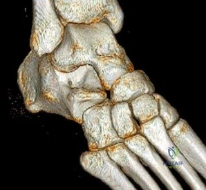
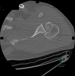
This article provides essential research regarding ORTHOPEDIC MCQS ONLINE 014 ANATOMY IMAGING. In orthopedic self-assessment modules, the preferred response figure identifies the correct diagnosis for imaging studies. For instance, '1' may indicate a normal foot, '2' a calcaneonavicular coalition, and '3' a talocalcaneal middle facet coalition. This figure guides learners through accurate identification of conditions presented in the medical imagery questions.
Orthopedic Anatomy Imag Review | Dr Hutaif Basic Scienc -...
Comprehensive 100-Question Exam
00:00
Start Quiz
Question 1
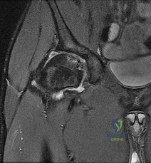
You are reviewing an axial cross-section of the shoulder during a pre-operative imaging review for a posterior approach.
What structure forms the superior boundary of the quadrangular space, and what is its primary innervation?
Explanation
Question 2
A trauma patient undergoes a pelvic series after a high-speed motor vehicle collision.
On an obturator oblique radiograph (Judet view) of the pelvis, which two osseous acetabular structures are best profiled?
Explanation
Question 3
A surgeon plans a volar (Henry) approach to the proximal radius for open reduction internal fixation of a diaphyseal fracture.
What are the innervations of the muscles defining the proximal internervous plane in this approach?
Explanation
Question 4
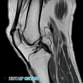
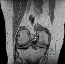
A sagittal MRI of the knee is obtained to evaluate a suspected multi-ligamentous injury.
Which of the following best describes the femoral origin and tibial insertion of the anteromedial (AM) bundle of the anterior cruciate ligament (ACL)?
Explanation
Question 5
An axial MRI of the ankle reveals pathology in the posteromedial compartment.
Immediately posterior/lateral to the flexor digitorum longus (FDL) tendon at the level of the medial malleolus, which anatomic structure is located?
Explanation
Question 6
During a posterior (Kocher-Langenbeck) approach to the hip, protecting the primary blood supply to the adult femoral head is paramount. In this region, where is the deep branch of the medial femoral circumflex artery (MFCA) consistently found?
Explanation
Question 7
A 45-year-old competitive cyclist presents with isolated weakness of the dorsal interossei, but normal sensation in the little finger and normal hypothenar muscle strength. Compression of the ulnar nerve is suspected in Guyon's canal (Zone III). Which of the following structures forms the floor of Guyon's canal?
Explanation
Question 8
Which of the following is the most common site of ulnar nerve compression leading to cubital tunnel syndrome?
Explanation
Question 9
A patient presents with weakness in wrist extension and altered sensation over the dorsal web space between the thumb and index finger, consistent with a C6 radiculopathy. In the cervical spine, the C6 nerve root exits through which neural foramen?
Explanation
Question 10
A 24-year-old marathon runner develops severe lateral and posterior leg pain, concerning for exertional compartment syndrome. Which of the following structures is exclusively located within the deep posterior compartment of the leg?
Explanation
Question 11
During a complicated resection of a palmar tumor, the vascular supply of the hand is meticulously dissected. The deep palmar arch is primarily formed by the terminal continuation of which artery?
Explanation
Question 12
During a midfoot surgical approach, the surgeon identifies the 'Master Knot of Henry' on the plantar aspect of the foot. At this location, what is the specific anatomical relationship between the traversing long flexor tendons?
Explanation
Question 13
A saphenous nerve block is planned within the adductor (Hunter's) canal for post-operative analgesia following knee surgery. Besides the superficial femoral artery and vein, what other structures travel through the adductor canal?
Explanation
Question 14
To anatomically reconstruct the posterolateral corner of the knee, understanding the spatial relationship on the lateral femoral epicondyle is critical. What is the relative position of the origin of the fibular collateral ligament (FCL) compared to the popliteus tendon insertion?
Explanation
Question 15
A 65-year-old woman sustains a displaced, non-operatively managed distal radius fracture. Eight weeks later, she presents with inability to actively extend her thumb interphalangeal joint, secondary to tendon rupture around Lister's tubercle. In which extensor compartment does this ruptured tendon normally travel?
Explanation
Question 16
The stability of the distal tibiofibular syndesmosis is dependent on several ligamentous structures. Which of the following ligaments provides the greatest contribution to the strength of the syndesmotic complex?
Explanation
Question 17
Avascular necrosis (AVN) is a well-known complication of proximal pole scaphoid fractures. The primary intraosseous blood supply to the proximal pole of the scaphoid enters the bone at which anatomical location?
Explanation
Question 18
A 35-year-old carpenter suffers a deep laceration at the level of the proximal carpal tunnel, completely transecting the median nerve. Which of the following intrinsic muscles of the hand would lose its innervation as a direct result?
Explanation
Question 19
Meniscal tears are a common knee pathology. In basic science review of meniscal anatomy, which of the following statements is true regarding the medial meniscus compared to the lateral meniscus?
Explanation
Question 20
The coracoacromial arch is an important anatomical restraint that prevents superior translation of the humeral head and is often implicated in subacromial impingement syndrome. The coracoacromial ligament attaches to which two osseous structures?
Explanation
Question 21
An orthopedic surgeon is performing a posterolateral approach to the tibial plateau. To adequately expose the joint, the fibular collateral ligament may need to be visualized. What nerve is most at risk during the distal extent of this exposure, and where does it typically cross the fibula?
Explanation
Question 22
During a standard ilioinguinal approach for an anterior column acetabular fracture, the surgeon is working in the middle window. Which of the following neurovascular structures is primarily found in this window?
Explanation
Question 23
A 35-year-old male sustains a midshaft humerus fracture and develops a secondary radial nerve palsy after closed reduction. If the surgeon decides to explore the nerve via a posterior approach, between which two muscle bellies does the radial nerve most reliably emerge from the spiral groove?
Explanation
Question 24
When reviewing an MRI for a patient with suprascapular neuropathy, a cyst is identified at the spinoglenoid notch. Which of the following clinical findings is most likely associated with compression at this specific anatomical location?
Explanation
Question 25
While performing an extended volar approach to the radiocarpal joint (Henry approach), the surgeon retracts the flexor carpi radialis (FCR) tendon. To minimize the risk of injury to the palmar cutaneous branch of the median nerve (PCBMN), in which direction should the FCR tendon be retracted and where does the PCBMN typically lie?
Explanation
Question 26
During a lateral approach to the fibula for a distal third fracture, a surgeon must be careful to avoid the superficial peroneal nerve. Where does this nerve typically pierce the deep fascia to become subcutaneous?
Explanation
Question 27
You are reviewing axial CT imaging of the upper extremity. In the proximal forearm, the median nerve passes between the two heads of the pronator teres. Which of the following structures passes between the ulnar and humeral heads of the flexor carpi ulnaris (FCU)?
Explanation
Question 28
When examining an axial T1-weighted MRI of the lumbar spine at the L4-L5 disc level, which nerve root is typically located within the lateral recess and is most susceptible to compression from a paracentral disc herniation at this level?
Explanation
Question 29
A surgeon is utilizing the Smith-Petersen approach to the hip. This approach exploits an internervous plane between which of the following muscles superficially?
Explanation
Question 30
You are evaluating an axial cervical spine MRI of a 45-year-old male with neck pain.
Which structure is located immediately anterior to the normal exiting C6 nerve root as it passes through the intervertebral foramen?
Explanation
Question 31
A 24-year-old overhead athlete presents with painless weakness of external shoulder rotation. MRI demonstrates a paralabral cyst causing compression at the spinoglenoid notch. Which of the following represents the expected clinical and anatomical findings?
Explanation
Question 32
A surgeon is planning a lateral collateral ligament (LCL) reconstruction for a patient with severe posterolateral rotatory instability (PLRI) of the elbow.
The lateral ulnar collateral ligament (LUCL), the primary restraint to PLRI, originates on the lateral epicondyle and inserts on which of the following structures?
Explanation
Question 33
During open reduction and internal fixation of a scaphoid nonunion via a dorsal approach, the surgeon must carefully preserve the blood supply to the proximal pole. What is the primary arterial supply to the proximal pole of the scaphoid?
Explanation
Question 34
A 32-year-old carpenter suffers a deep penetrating injury to the mid-palm, resulting in pulsatile bleeding. Surgical exploration reveals a lacerated deep palmar arch. This vascular structure is primarily formed by the terminal continuation of which of the following?
Explanation
Question 35
An axial MRI of the proximal forearm is reviewed to plan for a volar approach. The anterior interosseous nerve (AIN) and artery are identified. Which two muscle bellies directly border the AIN as it courses distally through the mid-forearm?
Explanation
Question 36
Reviewing an axial T1 MRI of the mid-thigh, you identify the neurovascular structures within the adductor (Hunter's) canal. What muscle forms the anterolateral boundary of this space?
Explanation
Question 37
A cervical spine CT angiogram is ordered for a patient with a facet dislocation. The radiologist traces the vertebral artery. In a normal anatomic variant, the vertebral artery typically first enters the transverse foramen at which cervical level?
Explanation
Question 38
An MRI of the shoulder demonstrates a paralabral cyst at the suprascapular notch, causing nerve compression. What is the normal anatomic relationship of the suprascapular nerve and artery to the superior transverse scapular ligament?
Explanation
Question 39
A trauma patient is evaluated with a pelvic radiograph series after a fall from a height. On an iliac oblique radiograph (Judet view), which two osseous structures of the acetabulum are best profiled?
Explanation
Question 40
A coronal MRI of the knee is reviewed prior to posterolateral corner reconstruction.
What is the precise femoral attachment site of the fibular collateral ligament (FCL) relative to the popliteus tendon insertion?
Explanation
Question 41
An axial MRI of the ankle at the level of the medial malleolus demonstrates the contents of the tarsal tunnel. From anterior to posterior, the posterior tibial artery and tibial nerve are located between which two tendons?
Explanation
Question 42
During a pre-operative imaging review for a severe cubital tunnel syndrome extending into the wrist, an axial MRI highlights Guyon's canal. Which structure forms the floor of this anatomic tunnel?
Explanation
Question 43
While executing a deltopectoral approach for a total shoulder arthroplasty, the coracobrachialis is retracted.
To avoid iatrogenic injury, the surgeon must remember that the musculocutaneous nerve typically enters the coracobrachialis at what average distance distal to the coracoid process?
Explanation
Question 44
An MRI of the hip reveals an intra-articular mass requiring a surgical dislocation via a posterior approach. To protect the deep branch of the medial circumflex femoral artery (MCFA), the tendon of which muscle must be preserved or carefully handled?
Explanation
Question 45
When planning an anterolateral approach to the distal humerus, an MRI shows the radial nerve piercing the lateral intermuscular septum. At what average distance proximal to the lateral epicondyle does this anatomically occur?
Explanation
Question 46
An axial T2 MRI of the L4-L5 level shows a far lateral (extraforaminal) disc herniation.
Which nerve root is most likely compressed, and what is the primary expected clinical motor deficit?
Explanation
Question 47
An axial CT of a distal tibia pilon fracture demonstrates a classic 3-part articular fragment pattern. The anterolateral (Tillaux-Chaput) fragment is avulsed by its attachment to which critical ligament?
Explanation
Question 48
In a coronal MRI of the brachial plexus evaluating a traction injury, the posterior cord is visualized. Which of the following is a direct terminal branch of the posterior cord?
Explanation
Question 49
A sagittal MRI of the knee highlights the meniscofemoral ligaments originating from the posterior horn of the lateral meniscus.
The ligament of Wrisberg passes in what anatomical relationship to the posterior cruciate ligament (PCL)?
Explanation
Question 50
During a modified Stoppa approach for an acetabular fracture, an aberrant vascular anastomosis termed the 'corona mortis' is encountered over the superior pubic ramus. This structure connects which two vascular systems?
Explanation
Question 51
A sagittal oblique MRI of the shoulder demonstrates the boundaries of the rotator interval.
Which of the following structures is NOT considered a standard component within the rotator interval?
Explanation
Question 52
The Kocher-Langenbeck approach to the acetabulum does not utilize a true internervous plane. What is the primary innervation of the gluteus maximus muscle, which is split during the superficial dissection of this approach?
Explanation
Question 53
A surgeon is performing an anterolateral approach to the distal tibia. Which nerve is most at risk during the superficial dissection, and what compartment of the leg does it primarily motor innervate?
Explanation
Question 54
When performing a posterior approach to the humerus, the radial nerve must be identified and protected. Approximately how far proximal to the lateral epicondyle does the radial nerve pierce the lateral intermuscular septum?
Explanation
Question 55
In the anterior approach to the hip (Smith-Petersen), the superficial internervous plane lies between two muscles. What are the respective innervations of these two muscles?
Explanation
Question 56
In a patient undergoing an MRI for quadrilateral space syndrome, which vascular structure is typically compressed alongside the axillary nerve?
Explanation
Question 57
On an axial MRI of the mid-calf, a soft tissue sarcoma is identified strictly confined within the deep posterior compartment. Which nerve runs in this compartment, and what is its primary sensory distribution?
Explanation
Question 58
Reviewing an axial MRI of the shoulder, you identify a retracted full-thickness tear of the subscapularis tendon. What is the dual motor innervation of this muscle?
Explanation
Question 59
When planning pedicle screw placement in the lumbar spine, standard anatomic landmarks are utilized. The optimal starting point for an L4 pedicle screw is located at the intersection of the pars interarticularis, the midpoint of the transverse process, and what other bony landmark?
Explanation
Question 60
A patient presents with a suspected anterior column acetabular fracture. Which standard radiographic view best profiles the anterior column of the acetabulum and the posterior edge of the iliac wing?
Explanation
Question 61
In evaluating an axial MRI of the wrist for ulnar tunnel syndrome, which anatomical structure serves as the floor of Guyon's canal?
Explanation
Question 62
During harvest of a hamstring autograft, the pes anserinus is exposed via an anteromedial tibial incision. From anterior to posterior, what is the correct order of the tendinous insertions?
Explanation
Question 63
In the anterior approach to the lower cervical spine (Smith-Robinson), why is a left-sided approach theoretically preferred by many surgeons regarding cranial nerve safety?
Explanation
Question 64
During an in situ decompression of the ulnar nerve at the elbow, the surgeon must release a distinct fascial band that bridges the two heads of the flexor carpi ulnaris (FCU) from the medial epicondyle to the olecranon. What is the name of this structure?
Explanation
Question 65
On an axial MRI of the distal third of the thigh, multiple hamstring muscles are visualized. Which of these muscles is uniquely innervated by the common peroneal division of the sciatic nerve?
Explanation
Question 66
A deep laceration to the palm severs the deep motor branch of the ulnar nerve. This injury will result in direct denervation of which of the following lumbrical muscles?
Explanation
Question 67
Reviewing a sagittal MRI of the knee, the normal anatomy of the posterior cruciate ligament (PCL) is best visualized. Which bundle of the PCL becomes tightest as the knee goes into deep flexion?
Explanation
Question 68
The deltopectoral approach utilizes a true internervous plane to access the anterior shoulder. What are the respective innervations of the two muscles that define this plane?
Explanation
Question 69
In an axial MRI evaluating tarsal tunnel syndrome, the posterior tibial neurovascular bundle is situated between the tendons of which two muscles at the level of the medial malleolus?
Explanation
None
