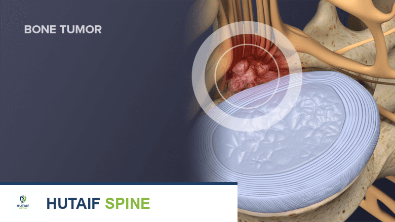
Elastofibroma Dorsi: Clinical Presentation, Imaging, and Diagnostic Pitfalls in a Geriatric Patient
Key Takeaway
Elastofibroma Dorsi is a benign soft tissue tumor often presenting as a deep infrascapular mass, commonly in elderly patients. Diagnosis relies on clinical findings like a slowly enlarging, firm mass with mechanical symptoms, confirmed by characteristic MRI and ultrasound imaging, which helps differentiate it from malignant lesions.
Patient Presentation and History
A 72-year-old male, retired construction worker, presented with a 2-year history of a slowly enlarging, deep-seated mass in the left posterior thoracic region, inferior to the scapula. The patient reported a dull ache and a persistent sensation of pressure, particularly when lying supine or during overhead activities. He also described an intermittent "snapping" or "grinding" sensation with scapular movement, which had become more pronounced over the last 6 months. There was no history of acute trauma to the area; however, the patient recalled a minor, non-impact fall approximately 3 years prior where he landed on his back, experiencing transient muscle soreness that resolved spontaneously. He had initially attributed his symptoms to age-related musculoskeletal changes or a persistent muscle strain.
His past medical history was significant for well-controlled hypertension and hyperlipidemia. He was a former smoker (quit 20 years prior) and consumed alcohol occasionally. There was no personal or family history of malignancy. He had no constitutional symptoms such as fever, night sweats, or unintentional weight loss. He denied any neurological deficits or paresthesias in the ipsilateral upper extremity. The progressive nature of the mass and the onset of mechanical symptoms prompted him to seek medical evaluation. His primary care physician, concerned about the differential diagnosis of a deep soft tissue mass in an elderly patient, referred him to Orthopedic Oncology.
Clinical Examination
Inspection
On visual inspection, a subtle fullness was noted in the left infrascapular region. There was no overlying skin erythema, discoloration, ulceration, or obvious skin changes. No prominent venous patterns were observed. The scapula appeared to be in a normal resting position, and there was no obvious winging or asymmetry at rest. Symmetry of the shoulder girdles and overall posture were unremarkable.
Palpation
Palpation revealed a firm, ill-defined, non-tender, deep-seated mass measuring approximately 8 x 6 cm in the left infrascapular region, deep to the latissimus dorsi and rhomboid musculature. The mass felt somewhat rubbery with a distinct fibrous consistency. It was relatively mobile relative to the overlying skin and superficial fascia but exhibited restricted mobility relative to the chest wall and underlying deeper structures. There was no warmth or crepitus on palpation. Deep palpation elicited the patient's described pressure sensation but no sharp pain. A "clunking" sensation was appreciated with passive and active scapular protraction and retraction, suggesting impingement or friction between the mass and the scapula and rib cage.
Range of Motion
Cervical Spine demonstrated full active and passive range of motion and was non-painful.
Left Shoulder active range of motion showed forward flexion 160 degrees, abduction 150 degrees, external rotation 60 degrees with the arm at the side, and internal rotation to T8. End-range movements, particularly abduction and forward flexion, were limited by a deep pressure sensation, not acute pain. The snapping sensation was reproducible during active scapulothoracic motion. Passive range of motion was symmetric with active range, indicating no significant capsular restriction or adhesive capsulitis. Right Shoulder demonstrated full and painless active and passive range of motion.
Neurological and Vascular Assessment
Left Upper Extremity Neurological assessment showed intact motor function (MRC grade 5/5) in all myotomes (C5-T1). Intact sensation to light touch and pinprick in all dermatomes. Deep tendon reflexes (biceps, triceps, brachioradialis) were 2+ and symmetric bilaterally. No pathological reflexes were elicited. Left Upper Extremity Vascular assessment revealed strong and symmetric radial and ulnar pulses. Capillary refill was brisk (under 2 seconds). No evidence of edema or trophic changes. Axillary lymph nodes were non-palpable.
Imaging and Diagnostics
Initial Radiographic Evaluation
Standard orthogonal radiographs of the left shoulder and scapula, including anteroposterior, lateral, and axillary views, were obtained. The osseous structures appeared unremarkable, with no evidence of periosteal reaction, cortical destruction, or focal osseous lesions in the scapula or underlying ribs. A subtle, non-specific soft tissue density was appreciated in the infrascapular region, but no intralesional calcifications or phleboliths were identified. Radiographs primarily served to rule out primary bone pathology, such as osteochondroma of the ventral scapula, which can also present with mechanical snapping.
Advanced Cross Sectional Imaging
Magnetic Resonance Imaging with and without intravenous gadolinium contrast was performed to characterize the soft tissue mass. The MRI revealed a poorly circumscribed, unencapsulated, lenticular-shaped mass located deep to the lower pole of the serratus anterior and latissimus dorsi, adjacent to the posterior thoracic cage.
The lesion demonstrated a highly characteristic fascicular or "layered" architecture. On T1-weighted and T2-weighted sequences, the mass exhibited alternating bands of intermediate-to-low signal intensity (isointense to skeletal muscle, representing dense fibrous tissue) and high signal intensity (isointense to subcutaneous fat, representing entrapped adipose tissue). Following contrast administration, the fibrous components demonstrated mild, heterogeneous enhancement. There was no evidence of peritumoral edema, cystic degeneration, or invasion into the adjacent ribs or intercostal musculature. The contralateral subscapular region was evaluated, revealing a smaller, asymptomatic 2 cm lesion with identical imaging characteristics, confirming bilateral disease.
Histopathological Diagnostic Criteria
Given the pathognomonic MRI findings, a core needle biopsy was deemed unnecessary, and the decision was made to proceed with marginal excision for symptomatic relief and definitive histopathological confirmation.
Grossly, the excised specimen was an ill-defined, rubbery, fibrofatty mass. Microscopic examination revealed a mixture of mature adipose tissue and dense, hypocellular bands of collagenous connective tissue. The hallmark histological feature was the presence of numerous thick, fragmented, and heavily eosinophilic elastic fibers embedded within the collagen stroma. These fibers were beautifully highlighted using a Verhoeff-Van Gieson stain, displaying a classic "beaded," "globular," or "pipe-cleaner" appearance. The spindle cells present lacked nuclear atypia, pleomorphism, or mitotic activity. Immunohistochemistry was unremarkable, with spindle cells staining positive for vimentin and CD34, but negative for S100, desmin, and smooth muscle actin, definitively ruling out liposarcoma, desmoid fibromatosis, and smooth muscle neoplasms.
Introduction and Epidemiology
Elastofibroma dorsi is an uncommon, benign, slow-growing, unencapsulated soft tissue pseudotumor. First described by Järvi and Saxén in 1961, it is currently classified by the World Health Organization within the category of benign fibroblastic and myofibroblastic tumors. Despite its nomenclature, it is widely considered a reactive, non-neoplastic process rather than a true neoplasm.
The condition is predominantly observed in the geriatric population, with the vast majority of cases diagnosed in individuals over the age of 55. There is a documented female predilection, with female-to-male ratios reported ranging from 5 to 1 up to 13 to 1 in various institutional series. While the patient in this vignette is male, the presentation remains classic in terms of age and anatomical location.
Epidemiological studies utilizing autopsy data and asymptomatic CT screening suggest that the true prevalence of elastofibroma dorsi is significantly higher than clinically reported, potentially affecting up to 2 percent of the population over 60 years of age. The lesion is bilateral in 10 to 66 percent of cases. When unilateral, it exhibits a slight predilection for the right side, a phenomenon often attributed to right-handed dominance and the associated increased mechanical stress on the dominant shoulder girdle.
The pathogenesis is fundamentally linked to mechanical friction. Repetitive microtrauma between the inferior angle of the scapula and the posterior chest wall leads to reactive hyperproliferation of fibroblastic tissue. This mechanical stress induces abnormal elastogenesis, resulting in the deposition of structurally altered, fragmented elastic fibers. The patient's occupational history as a construction worker provides a classic biomechanical substrate for this reactive process.
Surgical Anatomy and Biomechanics
A profound understanding of the periscapular anatomy and scapulothoracic articulation is requisite for the safe evaluation and surgical management of elastofibroma dorsi.
Muscular and Fascial Boundaries
Elastofibroma dorsi classically arises in the connective tissue spaces of the subscapular and infrascapular regions. The mass is typically situated deep to the rhomboid major and latissimus dorsi muscles, and superficial to the serratus anterior and the periosteum of the 6th to 8th ribs. The auscultatory triangle—bounded medially by the lateral border of the trapezius, laterally by the medial border of the scapula, and inferiorly by the superior border of the latissimus dorsi—often serves as a superficial anatomical landmark, as the mass frequently bulges through or deep to this fascial weakness during scapular protraction.
Neurovascular Considerations
Surgical excision requires navigation around several critical neurovascular structures. The dorsal scapular nerve and artery descend along the medial border of the scapula, deep to the rhomboid muscles. While the mass is usually inferior to the main trunk of this nerve, aggressive medial dissection or injudicious retraction can result in neuropraxia or structural injury, leading to rhomboid paralysis and subtle lateral scapular winging. Deep to the mass, the intercostal neurovascular bundles run along the inferior margins of the respective ribs. Because elastofibroma dorsi is unencapsulated and can adhere to the external intercostal fascia, deep dissection must be meticulous to avoid pleural violation or intercostal neuralgia.
Biomechanics of the Scapulothoracic Articulation
The scapulothoracic joint is not a true synovial joint but a physiological articulation dependent entirely on the coordinated action of the periscapular musculature. Normal scapulothoracic rhythm requires smooth gliding of the concave anterior surface of the scapula over the convex posterior thoracic cage, facilitated by the bursa and loose areolar tissue in the fascial planes between the subscapularis, serratus anterior, and chest wall.
The presence of a space-occupying lesion like an elastofibroma disrupts this gliding mechanism. As the mass enlarges, it increases the resting tension on the latissimus dorsi and rhomboids and alters the kinematic tracking of the scapula. During active overhead elevation (which requires upward rotation, posterior tilt, and external rotation of the scapula), the inferior angle of the scapula impinges upon the mass. This mechanical impingement generates the palpable and audible "clunking" or "snapping" described by the patient, and the localized pressure leads to ischemic pain in the overlying musculature.
Indications and Contraindications
The management of elastofibroma dorsi is dictated by the presence of symptoms, the size of the lesion, and the diagnostic certainty obtained from imaging. Because the lesion has zero malignant potential and does not undergo sarcomatous dedifferentiation, prophylactic excision of asymptomatic lesions is strongly discouraged.
| Clinical Scenario | Management Strategy | Rationale and Specific Criteria |
|---|---|---|
| Symptomatic Mass | Operative (Marginal Excision) | Indicated for persistent pain, severe mechanical snapping, or functional limitation of shoulder ROM that impairs activities of daily living. |
| Large Lesion Over 5 cm | Operative (Marginal Excision) | Lesions exceeding 5 cm are highly likely to become symptomatic due to mass effect and altered scapular kinematics. |
| Diagnostic Uncertainty | Operative (Excisional Biopsy) | Indicated if MRI findings are atypical (e.g., lack of alternating fat/fibrous bands, presence of necrosis, rapid growth) to definitively rule out soft tissue sarcoma. |
| Asymptomatic Incidentaloma | Non-Operative (Observation) | No malignant potential. Serial clinical evaluation is sufficient. MRI follow-up is only necessary if rapid growth occurs. |
| High Surgical Risk Geriatric Patient | Non-Operative (Observation) | In patients with severe cardiopulmonary comorbidities where general anesthesia poses an unacceptable risk, conservative management is preferred unless symptoms are debilitating. |
Absolute and Relative Contraindications
Absolute contraindications to surgical excision include active overlying soft tissue infection, severe cardiopulmonary instability precluding general or regional anesthesia, and uncorrectable coagulopathy. Relative contraindications include asymptomatic presentation (regardless of size), small lesions (under 4 cm) with minimal symptoms, and patients who cannot comply with postoperative activity restrictions, which are critical for preventing seroma formation.
Pre Operative Planning and Patient Positioning
Thorough preoperative planning is essential to minimize morbidity and optimize functional outcomes.
Preoperative Imaging Review
The surgeon must meticulously review the MRI to define the exact anatomical boundaries of the mass. The relationship of the tumor to the inferior angle of the scapula, the depth of invasion into the serratus anterior fascia, and the proximity to the pleural space must be established. Furthermore, the surgeon must explicitly check the contralateral side on the imaging, as bilateral disease is common. If a contralateral asymptomatic lesion is present, the patient must be counseled preoperatively about its existence and the rationale for leaving it undisturbed.
Anesthesia and Patient Positioning
The procedure is typically performed under general anesthesia to ensure complete muscle relaxation, which is vital for scapular mobilization and deep soft tissue retraction.
The patient is placed in the prone position or the lateral decubitus position. The prone position is generally preferred for bilateral excisions or when the mass is located very medially. The ipsilateral arm is placed in a "chicken wing" position—abducted, internally rotated, and the dorsum of the hand rested on the lumbar spine. This specific maneuver forces the scapula into protraction and lateral translation, lifting the inferior angle away from the midline and maximizing the exposure of the subscapular space and the underlying mass.
All bony prominences must be meticulously padded to prevent pressure necrosis or peripheral neuropathies, a particularly critical step in the geriatric population. The surgical field is prepped and draped widely to allow for intraoperative manipulation of the upper extremity if further scapular mobilization is required.
Detailed Surgical Approach and Technique
The surgical objective is a marginal excision. Because elastofibroma dorsi lacks a true capsule and blends imperceptibly with the surrounding musculature, attempting a wide oncologic resection will result in unnecessary morbidity and functional deficit. The goal is macroscopic removal of the pseudotumor while preserving the integrity of the latissimus dorsi, rhomboids, and serratus anterior.
Incision and Superficial Dissection
A linear or slightly oblique incision is planned over the most prominent portion of the mass, typically following Langer's lines or paralleling the medial border of the scapula to optimize cosmesis. The incision is carried down through the skin and subcutaneous adipose tissue using electrocautery. Hemostasis is maintained meticulously to preserve visualization.
Deep Dissection and Muscle Retraction
Upon reaching the deep fascia, the superior border of the latissimus dorsi and the inferior border of the trapezius are identified. Depending on the exact location of the mass, the latissimus dorsi may need to be split in line with its fibers or mobilized and retracted inferiorly. If the mass extends superiorly, the rhomboid major may also require mobilization.
The mass is typically encountered immediately deep to these muscle bellies. It presents as a firm, poorly circumscribed, rubbery mass with a characteristic grey-white fibrous appearance interspersed with yellow adipose tissue.
Marginal Excision of the Pseudotumor
Dissection commences at the most accessible margin of the mass, utilizing a combination of blunt dissection and electrocautery. Traction sutures (e.g., 0-Vicryl or 2-0 Ethibond) can be placed directly into the dense fibrous tissue of the mass to provide continuous upward traction. This traction facilitates the identification of the plane between the reactive fibrous tissue and the normal skeletal muscle.
The dissection proceeds circumferentially. Careful attention is paid to the deep margin, where the mass adheres to the fascia of the serratus anterior and the periosteum of the ribs. The surgeon must remain superficial to the intercostal muscles to avoid entering the pleural cavity or injuring the intercostal neurovascular bundles.
At the superior-medial pole of the dissection, the surgeon must be cognizant of the dorsal scapular nerve and vessels. Retraction in this area should be gentle, and the use of monopolar electrocautery should be minimized near the nerve's anticipated trajectory.
Hemostasis and Dead Space Management
Once the mass is removed en bloc, the wound bed is copiously irrigated with sterile saline. Achieving absolute hemostasis is a critical step, as the highly vascularized muscle beds are prone to postoperative bleeding.
Because the excision of a large elastofibroma leaves a significant potential dead space, meticulous closure techniques are mandatory to prevent seroma formation. The use of closed-suction drains (e.g., Jackson-Pratt) is highly recommended and should be placed deep into the excision cavity and brought out through a separate stab incision laterally.
Furthermore, the surgeon should employ quilting sutures. This technique involves placing multiple interrupted absorbable sutures (e.g., 2-0 Vicryl) to tack the overlying latissimus dorsi and subcutaneous tissue down to the underlying serratus anterior and chest wall fascia, effectively obliterating the dead space.
Layered Closure
The fascial layers and muscle splits are approximated using interrupted absorbable sutures. The deep dermal layer is closed with buried interrupted sutures, and the skin is closed with a running subcuticular suture or surgical staples, depending on surgeon preference and skin tension. A sterile compressive dressing is applied.
Complications and Management
While marginal excision of elastofibroma dorsi is generally safe and curative, complications can occur, primarily related to soft tissue healing and the large dead space created by the resection.
| Complication | Estimated Incidence | Etiology and Risk Factors | Management and Salvage Strategies |
|---|---|---|---|
| Postoperative Seroma | 15% to 30% | Large dead space, inadequate tissue quilting, early excessive shoulder motion, premature drain removal. | Prolonged use of closed-suction drains. Serial ultrasound-guided aspirations. For refractory cases, sclerotherapy or surgical re-exploration with aggressive quilting. |
| Postoperative Hematoma | 2% to 5% | Inadequate intraoperative hemostasis, rebound vasodilation, coagulopathy. | Small hematomas can be observed. Expanding or painful hematomas require urgent surgical evacuation, irrigation, and cauterization of bleeding vessels. |
| Surgical Site Infection | 1% to 3% | Geriatric age, diabetes, prolonged operative time, hematoma/seroma acting as a nidus. | Oral or intravenous antibiotics based on culture. Superficial infections treated with local wound care; deep infections require surgical debridement and washout. |
| Nerve Injury (Dorsal Scapular) | Under 1% | Overzealous medial retraction, thermal injury from electrocautery. | Usually a transient neuropraxia that resolves with observation and physical therapy. Permanent injury may result in mild rhomboid weakness and subtle winging. |
| Tumor Recurrence | Under 2% | Incomplete marginal excision of the reactive tissue. | Re-evaluation with MRI. Observation if asymptomatic. Revision marginal excision is rarely necessary unless severe mechanical symptoms return. |
Post Operative Rehabilitation Protocols
Postoperative rehabilitation is carefully phased to balance the need for tissue healing—specifically preventing seroma formation by limiting shearing forces across the surgical dead space—with the prevention of adhesive capsulitis and periscapular muscle atrophy.
Phase One Acute Tissue Healing
This phase encompasses postoperative weeks 0 through 2. The primary goals are pain control, wound healing, and seroma prevention.
* Immobilization: The patient is placed in a standard arm sling for comfort and to restrict excessive shoulder motion.
* Range of Motion: Active and active-assisted range of motion of the shoulder is strictly limited. Specifically, forward flexion and abduction are restricted to less than 90 degrees. Cross-body adduction and extreme internal rotation (which stretch the posterior incision) are prohibited.
* Distal Joints: Active range of motion of the elbow, wrist, and hand is encouraged immediately to prevent distal edema and stiffness.
* Drain Management: Drains remain in place until output is consistently less than 20 to 30 cc over a 24-hour period, which typically occurs between postoperative days 3 and 7.
Phase Two Early Mobilization
This phase encompasses postoperative weeks 2 through 6. The primary goals are restoring baseline range of motion and initiating gentle muscle activation.
* Sling Discontinuation: The sling is gradually weaned and discontinued.
* Range of Motion: Progression to full active and passive range of motion in all planes. Gentle stretching is initiated to prevent contracture of the latissimus dorsi and rhomboids.
* Strengthening: Submaximal, pain-free isometric exercises for the rotator cuff and periscapular stabilizers are introduced. Scapular retraction and depression exercises (e.g., scapular squeezes) are initiated to restore scapulothoracic rhythm.
Phase Three Progressive Strengthening
This phase encompasses postoperative weeks 6 through 12. The primary goal is a return to baseline functional activities.
* Strengthening: Isotonic strengthening of the periscapular musculature (rhomboids, latissimus dorsi, serratus anterior, trapezius) is advanced. Resistance bands and light weights are incorporated.
* Functional Training: Integration of proprioceptive and kinetic chain exercises. For patients returning to manual labor, work-specific simulated tasks are introduced.
* Return to Activity: Patients are typically cleared for full, unrestricted activity, including heavy lifting and overhead labor, by 10 to 12 weeks postoperatively, provided they have achieved full, painless range of motion and symmetric strength.
Summary of Key Literature and Guidelines
The academic understanding of elastofibroma dorsi has evolved significantly since its initial description. Current orthopedic oncology guidelines emphasize the importance of non-invasive diagnosis and conservative management where appropriate.
The seminal work by Järvi and Saxén in 1961 established the histopathological criteria that remain the gold standard today. They correctly hypothesized the reactive, friction-based etiology of the lesion, which has been supported by decades of subsequent biomechanical and epidemiological research.
Modern radiological guidelines, supported by extensive literature from musculoskeletal radiologists, dictate that MRI is the definitive imaging modality. The pathognomonic "layered" or "fascicular" appearance of alternating fibrous and adipose tissue on T1 and T2 sequences is considered highly specific. Consequently, current consensus guidelines from major orthopedic oncology societies suggest that if the clinical presentation and MRI findings are classic, a preoperative biopsy is unnecessary and potentially introduces unnecessary morbidity and risk of tumor bed contamination.
Surgical literature consistently highlights the high incidence of postoperative seroma. Studies evaluating closure techniques have demonstrated a statistically significant reduction in seroma formation when closed-suction drainage is combined with aggressive fascial quilting sutures, compared to drainage alone. Therefore, dead space obliteration remains the most critical technical pearl in the surgical management of this condition.
In summary, elastofibroma dorsi is a benign, reactive pseudotumor with a classic clinical and radiological profile. In the geriatric patient presenting with mechanical scapulothoracic symptoms, accurate diagnosis via MRI prevents unnecessary biopsies. When surgical intervention is indicated for symptomatic relief, meticulous marginal excision combined with rigorous dead space management yields excellent functional outcomes and a negligible recurrence rate.
