Intracapsular Hip Fractures in Young Patients: Epidemiology, Anatomy, Biomechanics & Management
Key Takeaway
Intracapsular hip fractures in young, neurologically intact patients are high-energy injuries primarily challenging due to the risk of avascular necrosis (AVN) and nonunion. Management emphasizes anatomic reduction and stable fixation to preserve the native hip. Understanding femoral head blood supply and fracture classifications (Garden, Pauwels) is crucial for optimal outcomes and preventing devastating complications.
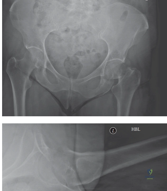
You are in the emergency department. A 35-year-old patient presents following a high-energy motorcycle accident. Clinical and radiographic evaluation confirms a displaced intracapsular femoral neck fracture. How do you approach the initial assessment and planning for this patient?
Candidate: I would follow ATLS protocols to rule out other injuries, then obtain AP and lateral radiographs of the hip. I would classify the fracture using the Garden and Pauwels systems. I would plan for urgent surgery, likely internal fixation with cannulated screws, aiming for anatomic reduction.
Failing to emphasize the "time is bone" concept. A poor answer focuses only on the hip and neglects to mention that the patient needs a full trauma screen or that the timing of surgery is a prognostic factor for AVN and nonunion.
The candidate structures the response: 1) Systemic: ATLS clearance for associated high-energy injuries. 2) Imaging: AP/Lateral pelvis and hip, CT for fracture morphology/comminution. 3) Classification: Garden for displacement and Pauwels for shear/stability. 4) Management: Urgent surgery (ideally <6-12 hours) to decompress the hematoma and stabilize the joint. 5) Goal: Anatomic reduction to minimize AVN and nonunion risk.
You have taken the patient to the operating room. You have performed a closed reduction maneuver, but the post-reduction imaging shows residual displacement of 3mm on the AP view. How do you proceed, and what radiographic parameters are you looking for?
Candidate: If the reduction is >2mm, I would perform an open reduction using an anterior approach, such as the Smith-Petersen approach. On imaging, I am looking for the restoration of Shenton’s line and proper neck-shaft alignment.
Suggesting multiple, forceful closed reduction attempts. These repeated maneuvers are known to damage the retinacular blood supply and worsen the prognosis for the femoral head.
State clearly: "I would not persist with closed maneuvers, as these risk further vascular insult." Move to an open reduction using an anterior approach for direct visualization and hematoma decompression. Radiographic benchmarks for anatomical reduction include restoration of Shenton's line, femoral head height symmetry, and alignment of the posterior cortex on the lateral view.
Looking at Figure 1, describe the ideal configuration for cannulated screws. Why is this specific arrangement used, and what biomechanical purpose does it serve?
Candidate: The screws should be in an inverted triangle configuration. One screw is inferior, near the calcar, and two are superior. This arrangement provides stability against shear and rotational forces.
Failing to mention the "calcar" screw. The inferior screw is the primary load-bearing construct; a candidate who misses the importance of calcar support shows a lack of understanding of hip biomechanics.
Identify the 'inverted triangle' configuration. The inferior screw must be placed along the dense calcar femorale to act as the primary weight-bearing support. The superior screws provide rotational stability. Emphasize that they must be placed parallel to each other and parallel to the femoral neck axis to allow for fracture settling/compression without 'backing out' or cutting through the head.
