Femoral Neck Fracture Case Study: Comprehensive Clinical and Diagnostic Approach
Key Takeaway
Femoral neck fractures are diagnosed through detailed clinical examination revealing pain, inability to bear weight, limb shortening, and external rotation, confirmed by plain radiographs. They are classified using systems like Garden (e.g., Type III for displaced fractures with valgus impaction) and Pauwels (e.g., Type II for significant shear forces) to guide treatment and prognosis.
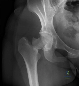
A 78-year-old female presents following a fall from standing height. She is non-ambulatory and holds her leg in the position shown below. Describe your initial assessment and the radiographic findings.
Candidate: The patient has a displaced right femoral neck fracture. Clinically, she presents with shortening and external rotation due to muscle pull. Radiographically, it is a Garden III fracture. I would perform a neurovascular exam, ensure the patient is medically optimized, and plan for arthroplasty given her age and displacement.
Candidates often fail to describe the mechanism of the deformity (the specific muscles responsible, like the iliopsoas for external rotation), ignore the distinction between intra- and extracapsular fractures, or fail to mention the importance of checking the contralateral hip for templating. They often neglect to mention the urgency of surgical intervention in the geriatric population.
The candidate should immediately recognize the "shortened, externally rotated" limb as a classic proximal femur fracture. They should categorize the fracture as intracapsular (Garden III/Pauwels II), explain the biomechanics (proximal migration due to rectus femoris/hamstrings; external rotation due to gravity and iliopsoas pull), and state that surgical treatment is mandatory. Crucially, they should mention: 1) Systemic optimization (DVT/delirium risk), 2) The necessity of early mobilization, and 3) That for an independent elderly patient, THA is preferred over hemiarthroplasty to restore function.
You have decided to proceed with surgery. The patient has a history of long-term bisphosphonate use and osteoporosis. How does this influence your implant choice and technical planning?
Candidate: I would consider a cemented femoral stem. Since she has osteoporotic bone (Dorr Type B or C), cement provides better initial fixation and reduces the risk of periprosthetic fractures during implantation compared to press-fit stems.
Ignoring the "cementing technique" is a major error. A candidate who suggests cement but forgets to mention the distal plug, pulsatile lavage, or the risk of "Bone Cement Implantation Syndrome" (BCIS) demonstrates a lack of surgical maturity.
The ideal response: "In the setting of osteoporotic (Dorr B/C) bone, a cemented femoral stem is the gold standard to ensure immediate macro-interlock and reduce the risk of iatrogenic periprosthetic fracture. I must emphasize the meticulous technique: 1) Pulsatile lavage and drying to ensure the cement-bone interface is dry, 2) Use of a distal plug to allow pressurization, and 3) Awareness of BCIS (hypotension/hypoxia) during insertion. I would also note that while bisphosphonates are used, this is a standard fragility fracture, not an atypical subtrochanteric fracture."
The patient's initial radiographs were equivocal for a fracture, but your clinical index of suspicion remains high. What is your next step in management?
Candidate: I would order an MRI of the hip. It is the gold standard for occult fractures, specifically looking for marrow edema on STIR sequences.
Delaying for a repeat X-ray in 24 hours is a significant error. Borderline candidates often suggest "wait and see," which risks the patient further falling and displacing a potentially stable, non-displaced fracture.
The candidate must display decisiveness: "If the initial radiographs are negative but the patient cannot weight bear, I must rule out an occult fracture. MRI (T1/STIR) is the gold standard. If MRI is unavailable or contraindicated, a CT scan is a reasonable second choice, though less sensitive for trabecular stress injuries. The goal is to prevent displacement, which would change the management from in-situ fixation to arthroplasty."
