Fixation of Femoral Neck Fractures with Cannulated Screws: A Comprehensive Surgical Guide
Key Takeaway
Cannulated screw fixation remains a cornerstone technique for managing undisplaced or reducible displaced femoral neck fractures in physiologically appropriate patients. This comprehensive surgical guide details the biomechanical principles, precise patient positioning, closed reduction maneuvers, and the step-by-step execution of the inverted triangle screw configuration. Mastery of this technique, including accurate guide pin placement and avoidance of subtrochanteric stress risers, is essential for optimizing fracture stability and minimizing postoperative complications such as nonunion or avascular necrosis.
Comprehensive Introduction and Patho-Epidemiology
Pathophysiology of Femoral Neck Fractures
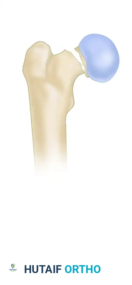
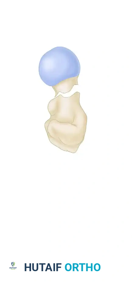
Femoral neck fractures represent a profound challenge in the realm of orthopedic trauma, characterized by their unique anatomical environment and precarious biological healing capacity. Unlike extracapsular proximal femur fractures, femoral neck fractures are strictly intracapsular. This anatomical distinction is critical because the femoral neck lacks a cambium layer within its periosteum, meaning that fracture healing is entirely dependent on endosteal callus formation. Furthermore, the fracture hematoma is contained within the joint capsule, exposing the fracture site to synovial fluid. The enzymatic properties of synovial fluid, particularly its high concentration of fibrinolysins, actively inhibit the initial stages of hematoma consolidation and osteogenesis, thereby predisposing the fracture to nonunion.
The mechanism of injury typically follows a bimodal distribution. In the physiologically young demographic, these fractures are almost exclusively the result of high-energy trauma, such as motor vehicle collisions or falls from significant heights. These high-energy injuries are frequently associated with vertically oriented fracture lines (high Pauwels angle), significant comminution, and concomitant systemic injuries. Conversely, in the elderly population, femoral neck fractures are predominantly fragility fractures resulting from low-energy mechanisms, such as a fall from standing height. The underlying pathophysiology in this cohort is driven by osteopenia or osteoporosis, which severely compromises the trabecular architecture of the proximal femur, particularly within Ward's triangle.
Epidemiological Considerations
The epidemiological burden of femoral neck fractures is staggering and continues to escalate in tandem with the globally aging population. Current projections suggest that the worldwide incidence of hip fractures will exceed 6 million annually by the year 2050. The mortality associated with these fractures in the geriatric population is well-documented, with one-year mortality rates ranging from 20% to 30%, largely secondary to cardiopulmonary complications, thromboembolic events, and prolonged immobility. In younger patients, while mortality is lower, the morbidity associated with treatment failure—specifically avascular necrosis (AVN) and nonunion—can lead to lifelong disability and the need for multiple complex reconstructive surgeries.
Evolution of Cannulated Screw Fixation
The surgical management of femoral neck fractures has evolved significantly over the past century. Early attempts at internal fixation utilized the tri-flanged Smith-Petersen nail, which, while revolutionary at the time, frequently failed due to inadequate rotational control and massive bone loss upon removal. The advent of multiple partially threaded cancellous screws marked a paradigm shift. Today, the utilization of 6.5 mm or 7.0 mm cannulated screws represents the gold standard for the fixation of undisplaced fractures across all age groups and displaced fractures in young patients. The cannulated design allows for precise, minimally invasive placement over guide wires, minimizing soft tissue stripping and preserving the tenuous residual blood supply to the femoral head. The partially threaded nature of these screws is paramount, as it allows the threads to purchase entirely within the dense subchondral bone of the femoral head, facilitating dynamic interfragmentary compression across the fracture site as the screw head engages the lateral femoral cortex.
Detailed Surgical Anatomy and Biomechanics
Osseous Architecture and Trabecular Patterns
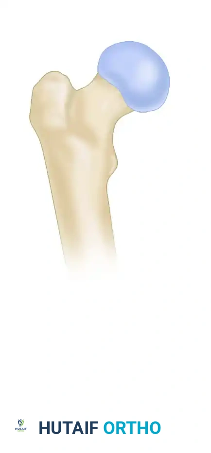
A profound understanding of the proximal femoral osseous architecture is mandatory before undertaking internal fixation. The internal structure of the femoral neck is defined by distinct trabecular patterns that align with the primary mechanical stresses acting upon the hip joint. The primary compressive trabeculae extend from the medial cortex of the femoral shaft into the superior aspect of the femoral head, while the primary tensile trabeculae arc from the lateral cortex to the inferior femoral head. The intersection of these trabecular systems creates a central area of relative structural weakness known as Ward's triangle. The calcar femorale, a dense vertical plate of bone originating from the posteromedial aspect of the femoral shaft and extending into the posterior femoral neck, serves as the primary structural buttress. Fixation constructs must leverage the dense bone of the calcar to achieve mechanical stability, particularly in the inferior and posterior quadrants.
Vascular Anatomy and the Retinacular Vessels
The vascular supply to the proximal femur is famously precarious and is the primary determinant of biological success or failure following a femoral neck fracture. The primary blood supply to the femoral head is derived from the medial circumflex femoral artery (MCFA). The MCFA gives rise to the lateral epiphyseal artery, which traverses the posterosuperior aspect of the femoral neck within the retinacular folds before penetrating the femoral head. A secondary, highly variable supply is provided by the artery of the ligamentum teres, though this is rarely sufficient to sustain the femoral head in the event of MCFA disruption. Surgical approaches and implant placement must meticulously respect this anatomy. Errant superior or posterior guide pin placement can cause iatrogenic vascular insult, drastically increasing the risk of postoperative AVN. Furthermore, the intracapsular hematoma generates a tamponade effect, elevating intracapsular pressures and potentially occluding the retinacular vessels; hence, capsulotomy is advocated by many trauma surgeons in young patients to decompress the joint and restore perfusion.
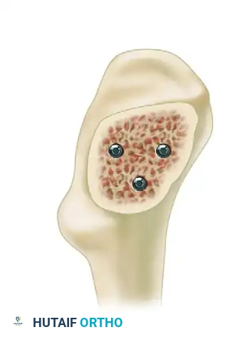
Biomechanical Principles of Screw Configuration
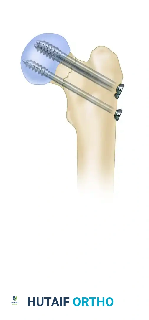
The goal of multiple cannulated screw fixation is to provide stable, compression-yielding osteosynthesis that resists the shear, varus, and rotational forces acting across the femoral neck. The standard construct utilizes three partially threaded screws placed in an inverted triangle configuration.
The inferocentral screw acts as the primary load-bearing strut. It must be placed immediately adjacent to the calcar femorale to provide inferior cortical support and resist varus displacement. The posterosuperior and anterosuperior screws provide rotational stability and anterior/posterior cortical support. In a biomechanical model, screw configuration has been shown to heavily influence the occurrence of iatrogenic subtrochanteric femoral fractures. Fractures fixed with an apex-distal configuration (where the inferior screw is the most distal and central) exhibit a significantly greater load to failure compared to an apex-proximal configuration. Always strive for an apex-distal inverted triangle to minimize stress risers in the lateral cortex.
Lowell's "S" Curve and Radiographic Alignment
Radiographic assessment of anatomical alignment relies heavily on the restoration of normal osseous contours. As described by Lowell, the concave outline of the femoral neck meets the convex outline of the femoral head in an "S" or reversed "S" curve superiorly, inferiorly, anteriorly, and posteriorly. Restoration of these "S" signs on both the anteroposterior (AP) and lateral fluoroscopic views is the definitive radiographic indicator of anatomical alignment. Acceptance of a reduction that fails to restore these curves, particularly one leaving the fracture in varus or retroversion, significantly increases the risk of mechanical failure, as it places the fixation construct under excessive bending moments that it is not designed to withstand.
Exhaustive Indications and Contraindications
Patient Selection Criteria
The decision to proceed with cannulated screw fixation hinges on a complex interplay of the patient's physiological age, baseline functional status, bone quality, and fracture morphology. In the physiologically young patient (generally defined as under 60-65 years of age, though physiological age supersedes chronological age), joint preservation is the absolute priority. In this demographic, native hip preservation is mandated regardless of fracture displacement, necessitating urgent anatomical reduction and stable internal fixation. In the elderly, low-demand patient with a displaced fracture (Garden III or IV), the unacceptably high rates of AVN and nonunion associated with internal fixation make arthroplasty (hemiarthroplasty or total hip arthroplasty) the definitive treatment of choice.
Fracture Classification and Treatment Algorithms
The Garden and Pauwels classifications are the primary frameworks guiding surgical decision-making. The Garden classification assesses the degree of displacement on the AP radiograph. Garden I (incomplete/valgus impacted) and Garden II (complete, undisplaced) fractures are universally treated with in situ cannulated screw fixation across all age groups, given their inherently high rates of union and low rates of AVN. The Pauwels classification, however, evaluates the fracture line's orientation relative to the horizontal plane. A high Pauwels angle (Type III, >50 degrees) indicates a highly vertical fracture line subjected to massive shear forces. In these vertically oriented fractures, simple cannulated screws may fail to resist shear, and surgeons must strongly consider more rigid constructs, such as a dynamic hip screw (DHS) with an anti-rotation screw, or a specifically designed length-stable implant.
Indications and Contraindications Table
| Category | Parameters | Clinical Rationale / Notes |
|---|---|---|
| Absolute Indications | Undisplaced fractures (Garden I, II) in all ages. | High union rates, minimal AVN risk, preserves native joint. |
| Absolute Indications | Displaced fractures (Garden III, IV) in physiologically young patients. | Arthroplasty is contraindicated due to lifespan and activity level; joint preservation is mandatory. |
| Relative Indications | Basicervical fractures in young patients with excellent bone stock. | DHS is often preferred due to biomechanical instability at the base of the neck, but multiple screws can be used if perfectly reduced. |
| Absolute Contraindications | Displaced fractures (Garden III, IV) in elderly, osteoporotic patients. | High risk of fixation failure, nonunion, and AVN. Arthroplasty is the standard of care. |
| Absolute Contraindications | Active joint infection or severe overlying soft tissue compromise. | High risk of deep space infection; requires source control prior to definitive skeletal management. |
| Relative Contraindications | Pre-existing severe osteoarthritis of the affected hip. | Fixation may heal the fracture but leaves the patient with a painful, arthritic joint. Total hip arthroplasty is preferred. |
| Relative Contraindications | Pauwels Type III (highly vertical) shear fractures. | High shear forces frequently lead to screw cutout or varus collapse. A fixed-angle device (DHS) is biomechanically superior. |
Pre-Operative Planning, Templating, and Patient Positioning
Imaging Modalities and Preoperative Templating
Meticulous preoperative planning is the cornerstone of a successful osteosynthesis. Standard imaging must include an AP view of the pelvis and a cross-table lateral view of the affected hip. The AP pelvis allows for comparison with the contralateral, uninjured hip to assess native neck-shaft angle and offset. Traction-internal rotation views can be highly beneficial in the emergency department to better define the fracture pattern and assess the potential for closed reduction. In cases of high-energy trauma or suspected basicervical extension, a fine-cut computed tomography (CT) scan with 3D reconstructions is invaluable. CT imaging accurately delineates the degree of posterior comminution—a critical factor that may necessitate the use of a four-screw construct to prevent postoperative retroversion. Digital templating should be performed to estimate screw lengths and anticipate the optimal starting point on the lateral cortex.
Operating Room Setup and Table Selection
The choice of operating table and room configuration significantly impacts the flow of the procedure. The patient is typically placed supine on a radiolucent orthopedic fracture table. The fracture table allows for the application of controlled, sustained longitudinal traction and precise rotational adjustments, which are essential for achieving and maintaining closed reduction. A radiolucent flat table may be used by surgeons who prefer manual traction or who plan to proceed directly to an open reduction via a Smith-Petersen or Watson-Jones approach, but this requires dedicated, experienced assistants to maintain the reduction during fixation.
Patient Positioning and Fluoroscopic Optimization
Positioning must facilitate unimpeded fluoroscopic visualization in both the AP and true lateral planes. We advocate for the "scissoring" technique on the fracture table. The injured extremity is secured in the traction boot and positioned in neutral extension. The contralateral, uninjured limb is extended, abducted, and dropped posteriorly. This creates a clear window for the C-arm to swing into a true lateral position without interference from the contralateral limb or the central post. Alternatively, the uninjured leg can be placed in a well-leg holder in a lithotomy position (flexed, abducted, and externally rotated). The C-arm is positioned between the patient's legs or entering from the contralateral side. The surgeon must ensure that perfect AP and true lateral views of the femoral neck can be obtained with minimal adjustment of the C-arm base before the patient is prepped and draped.
Step-by-Step Surgical Approach and Fixation Technique
Closed Reduction Techniques and Assessment
Anatomical reduction is absolutely non-negotiable. Fixation of a femoral neck fracture in varus or retroversion leads to unacceptably high rates of nonunion and implant failure due to the exponential increase in shear forces across the fracture site.
Closed reduction is typically achieved using the Whitman or Leadbetter maneuvers. The standard sequence involves:
1. Application of longitudinal traction to disimpact the fracture fragments and restore functional leg length.
2. Slight flexion of the hip to relax the anterior capsule, followed by slow, controlled internal rotation (typically 15 to 20 degrees) to correct the external rotation deformity and align the distal fragment with the proximal head.
3. Abduction of the limb to lock the fracture fragments under tension against the intact superior capsule.
The reduction is then rigorously evaluated using fluoroscopy. On the AP view, the surgeon must confirm a normal neck-shaft angle (130-135 degrees) or slight valgus. Varus is strictly unacceptable. On the lateral view, there must be no anterior or posterior angulation (sagittal tilt), and the anterior and posterior cortices must be collinear. If closed reduction is unsuccessful after two or three gentle attempts, the surgeon must abandon closed maneuvers to avoid further iatrogenic damage to the retinacular vessels and proceed immediately to an open reduction.
Surgical Approach and Soft Tissue Dissection
Following successful reduction, the lateral proximal femur is prepped and draped. Fluoroscopy is utilized to localize the starting point for the inferocentral wire. A 3 to 5 cm longitudinal skin incision is made over the lateral aspect of the greater trochanter, extending distally. The fascia lata is incised in line with the skin incision. Using a Cobb elevator or blunt retractors, the fibers of the vastus lateralis are split longitudinally down to the lateral cortex of the femur. The muscle belly is swept anteriorly and posteriorly, and a self-retaining retractor is placed to maintain exposure of the lateral cortex. Meticulous hemostasis is achieved using electrocautery.
Guide Wire Trajectory and Placement
The placement of the first guide wire—the inferocentral wire—is the most critical step of the procedure, as it dictates the spatial orientation of the entire construct.
The starting point must be carefully selected on the lateral cortex. It is imperative to start above the level of the lesser trochanter. Starting at or below the lesser trochanter creates a massive stress riser in the subtrochanteric region, significantly increasing the risk of a catastrophic postoperative subtrochanteric fracture.
The inferocentral wire is advanced under fluoroscopic guidance. On the AP view, it should run parallel to and immediately superior to the calcar femorale. On the lateral view, it must be perfectly central within the femoral neck. The wire is advanced into the subchondral bone of the femoral head, stopping 2 to 3 mm short of the articular surface.
Once the inferocentral wire is perfectly positioned, a parallel drill guide is utilized to place the posterosuperior and anterosuperior pins, creating the apex-distal inverted triangle. These pins should be spread as widely as possible within the neck to maximize cortical support and rotational stability, while remaining entirely intraosseous.
Drilling, Measurement, and Screw Insertion
With all three guide wires in place, the length of each wire is measured using a cannulated depth gauge. A crucial technical pearl is to subtract 5 mm from the measured length to determine the final screw length. This allows for dynamic fracture compression during screw insertion and postoperative weight-bearing without the risk of the screw tip penetrating the articular cartilage.
In young patients with dense cortical bone, the lateral cortex must be predrilled over the guide wires using a cannulated drill bit to prevent the screw threads from binding in the lateral cortex, which could inadvertently distract the fracture.
Self-drilling, self-tapping partially threaded screws (typically 6.5 mm or 7.0 mm) are then inserted. The use of washers is highly recommended, particularly in osteopenic bone, to prevent the screw heads from countersinking through the lateral cortex and losing compression.
The screws are advanced sequentially. Traction is slightly released just prior to final tightening to allow the fracture fragments to compress dynamically. Final fluoroscopic images are obtained through a full arc of rotation to ensure no screws have violated the joint space and that all threads have completely bypassed the fracture line, residing entirely within the femoral head epiphysis.
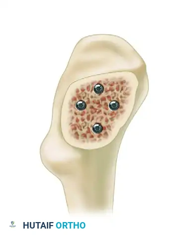
Advanced Configurations: The Diamond Construct
While the three-screw inverted triangle is the standard of care, specific fracture morphologies require augmented fixation strategies.
In patients with significant posterior comminution, the intrinsic stability of the posterior neck is lost. This makes the standard three-screw construct highly susceptible to retroversion and failure under physiological loads. In such scenarios, a fourth screw, creating a diamond configuration, is indicated. The fourth screw is placed centrally and posteriorly to provide additional structural rigidity, effectively acting as a substitute for the deficient posterior cortical buttress. When utilizing a four-screw construct, the surgeon must be exceedingly careful not to crowd the lateral cortex, ensuring adequate bone bridges remain between the screw entry points to prevent lateral wall blowout.
Complications, Incidence Rates, and Salvage Management
Avascular Necrosis and Nonunion
Avascular necrosis (AVN) and nonunion are the most devastating biological complications following femoral neck fracture fixation. AVN occurs due to the disruption of the medial circumflex femoral artery at the time of injury or iatrogenically during forceful reduction maneuvers. The incidence of AVN ranges from 5% to 10% in undisplaced fractures and skyrockets to 20% to 30% in displaced fractures. It may present clinically anywhere from 6 months to 2 years postoperatively. Nonunion, occurring in up to 15% to 20% of displaced fractures, is primarily a biomechanical failure driven by poor initial reduction (specifically varus alignment), failure to achieve interfragmentary compression (e.g., screw threads crossing the fracture site), or lack of biological healing capacity.
Iatrogenic Subtrochanteric Fractures and Hardware Failure
Iatrogenic subtrochanteric fractures are a catastrophic mechanical complication directly linked to surgical technique. They occur when multiple drill holes are made in the lateral cortex during failed attempts at guide wire placement, or when the starting point for the inferior screw is placed at or below the level of the lesser trochanter. This creates a massive stress riser in an area subjected to high tensile forces. Hardware failure, such as screw cutout into the joint or screw back-out, is typically secondary to fracture nonunion, profound osteopenia, or failure to utilize washers in soft bone.
Complications, Incidence, and Salvage Strategies Table
| Complication | Estimated Incidence | Pathophysiology / Risk Factors | Salvage Management / Treatment |
|---|---|---|---|
| Avascular Necrosis (AVN) | 10-30% (Displaced) 5-10% (Undisplaced) |
Disruption of MCFA retinacular vessels. Risk increases with displacement, delay to surgery, and non-anatomic reduction. | Young Patients: Core decompression (early), proximal femoral osteotomy, or THA. Elderly: Total Hip Arthroplasty (THA). |
| Nonunion | 10-20% (Displaced) | Varus malreduction, inadequate compression, threads crossing fracture line, poor bone biology. | Young Patients: Valgus-producing subtrochanteric osteotomy to convert shear forces to compressive forces. Elderly: THA. |
| Screw Cutout / Migration | 5-10% | Secondary to nonunion, varus collapse, or profound osteoporosis. | Hardware removal and conversion to THA or hemiarthroplasty depending on patient age and acetabular cartilage status. |
| Iatrogenic Subtrochanteric Fracture | 1-3% | Stress riser from drill holes below the lesser trochanter or apex-proximal screw configuration. | Long cephalomedullary nailing or fixed-angle plate fixation spanning the subtrochanteric defect. |
| Deep Surgical Site Infection | < 1-2% | Hematoma formation, prolonged operative time, medical comorbidities (diabetes, obesity). | Aggressive surgical debridement, hardware retention (if stable and fracture healing), culture-directed IV antibiotics. |
Phased Post-Operative Rehabilitation Protocols
Immediate Postoperative Phase and Weight-Bearing
The immediate postoperative phase focuses on medical optimization, pain control, and the prevention of systemic complications. Chemical and mechanical deep vein thrombosis (DVT) prophylaxis is mandatory and should be tailored to the patient's individual risk profile, typically continuing for 28 to 35 days postoperatively.
Weight-bearing protocols following cannulated screw fixation remain a subject of intense debate among orthopedic surgeons. For physiologically young patients with excellent bone quality and a biomechanically stable, anatomically reduced construct, many surgeons now advocate for immediate weight-bearing as tolerated (WBAT). The rationale is that physiological loading promotes dynamic compression across the fracture site, enhancing endosteal healing. Conversely, in elderly patients with osteopenic bone, or in cases where the fixation is deemed tenuous, restricted weight-bearing (toe-touch or partial weight-bearing) for 6 to 8 weeks is traditionally advised, although literature suggests that elderly patients are notoriously non-compliant with restricted weight-bearing instructions.
Intermediate Rehabilitation and Functional Restoration
Physical therapy is initiated on postoperative day one. The intermediate phase of rehabilitation (weeks 2 through 6) emphasizes the restoration of active and passive range of motion of the hip, knee, and ankle. Specific attention is directed toward strengthening the hip abductor musculature (gluteus medius and minimus), which is often inhibited postoperatively due to the lateral surgical approach and underlying pain. Gait training with appropriate assistive devices (walker or crutches) is essential to normalize the gait pattern and prevent compensatory musculoskeletal issues.
Long-Term Monitoring and Return to Activity
Long-term radiographic surveillance is critical to identify late-onset complications, specifically AVN and delayed union. Serial radiographs (AP and cross-table lateral) should be obtained at 2 weeks, 6 weeks, 3 months, 6 months, 1 year, and 2 years postoperatively. The patient must be educated on the symptoms of AVN, primarily the insidious onset of groin pain with weight-bearing. Return to high-impact activities or heavy manual labor in young patients is generally restricted until definitive radiographic union is confirmed, typically between 3 to 6 months postoperatively.
Summary of Landmark Literature and Clinical Guidelines
The FAITH Trial and Construct Superiority
The Fixation using Alternative Implants for the Treatment of Hip fractures (FAITH) trial is a landmark multicenter randomized controlled trial that significantly influenced modern practice. The trial compared the outcomes of sliding hip screws (DHS) versus multiple cancellous screws in the treatment of femoral neck fractures. The FAITH trial demonstrated that while both constructs yielded similar overall rates of reoperation, the use of a sliding hip screw was associated with a lower risk of AVN and reoperation in the specific subset of patients with displaced femoral neck fractures and those who were active smokers. However, for undisplaced fractures, multiple cannulated screws remain the preferred, less invasive option with excellent outcomes.
Current AAOS and OTA Guidelines
The American Academy of Orthopaedic Surgeons (AAOS) and the Orthopaedic Trauma Association (OTA) have established rigorous clinical practice guidelines for the management of hip fractures. A critical consensus point is the timing of surgery. Guidelines strongly recommend that surgical fixation of femoral neck fractures be performed within 24 to 48 hours of admission. Expedited surgery has been definitively linked to decreased mortality, lower rates of systemic complications (such as pneumonia and pressure ulcers), and a shorter length of hospital stay. In the young patient cohort, while the literature is mixed regarding whether urgent surgery (< 6-8 hours) definitively decreases the rate of AVN, it is universally accepted that these injuries should be treated as urgencies to optimize the biological environment for joint preservation.
