High-Energy Schatzker Type VI Tibial Plateau Fracture: A Case Study with Neurovascular Compromise
Key Takeaway
A Schatzker Type VI tibial plateau fracture from high-energy trauma presents with gross deformity, severe pain, and critical neurovascular deficits like peroneal nerve palsy and diminished ABI. X-rays and CT scans with 3D reconstructions are crucial to confirm complex bicondylar comminution, articular depression, and metaphyseal-diaphyseal dissociation, guiding urgent orthopedic management.
A 35-year-old construction worker presents following a high-energy dashboard injury. On inspection, you note significant swelling, ecchymosis, and a 3x2 cm non-blanchable blister over the lateral aspect of the knee. Describe your immediate clinical assessment priorities and the rationale behind your soft tissue grading.
Candidate: I would perform a secondary survey, assess neurovascular status—specifically checking pedal pulses and looking for peroneal nerve palsy—and monitor for compartment syndrome. The blister indicates a Tscherne Grade III injury, which means the skin is severely compromised and I must delay definitive surgery until the tissues are ready.
Candidates often jump straight to "I'll order a CT scan" or "I'll book them for surgery in a few days." They frequently forget to mention the specific vascular examination (ABI), the serial assessment for compartment syndrome, or fail to define the injury grade, which is critical for surgical planning.
A high-scoring answer addresses the "ABCs" of the limb: 1. Neurovascular: ABI measurement and formal neurologic exam (peroneal nerve function). 2. Compartment Syndrome: Establishing a baseline for potential acute compartment syndrome (ACS), noting firm compartments, and committing to serial exams. 3. Soft Tissue Classification: Correctly identifying this as a Tscherne Grade III injury (non-blanchable fracture blisters, subcutaneous degloving). 4. Decision Framework: Stating that this requires a staged approach: temporary external fixation to restore the mechanical axis, followed by definitive ORIF only when the "wrinkle sign" appears (typically 10-14 days).
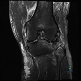
The radiographs and CT confirm a Schatzker VI fracture with significant posteromedial displacement. Looking at this imaging, why is the posteromedial fragment considered the "keystone" of the knee, and how does this influence your surgical approach?
Candidate: The posteromedial fragment is vital because it supports the medial plateau. If you don't fix it properly, the knee will develop a varus deformity and the tibia will subluxate posteriorly. I would use a posteromedial approach to buttress this fragment.
Candidates often treat the fracture as a single lateral column injury. They fail to mention the sequence of reduction. In Schatzker VI fractures, if the lateral side is fixed first, the medial side is often impossible to reduce secondarily due to the rigidity of the lateral construct.
The candidate must articulate the following: 1. Pathomechanics: The posteromedial fragment is the primary restraint to posterior subluxation. Failure to fix it results in mechanical failure of the construct. 2. Sequence: The posteromedial column MUST be addressed first. Once this is anatomically reduced and buttressed with an anti-glide plate, it provides a stable medial platform. 3. Approach: Use a dual-incision technique (posteromedial and anterolateral) rather than an extensile anterior approach to reduce the risk of soft tissue sloughing and wound breakdown.
During your preoperative planning, you note the ABI is 0.82 and the CTA shows a non-flow-limiting intimal flap in the popliteal artery. How does this finding change your management of the tibial plateau fracture?
Candidate: I would immediately consult vascular surgery. I would proceed with emergent external fixation to restore the mechanical axis, which might relieve the traction on the artery. I would monitor the limb closely for signs of ischemia or evolving thrombosis.
Suggesting systemic anticoagulation is a dangerous error in a patient with a high-energy fracture and impending compartment syndrome risk. Another common mistake is ignoring the external fixator's role in "realigning" the vessel to stop the intimal injury from progressing.
The candidate demonstrates high-level judgment by: 1. Prioritizing Restoration: Using the external fixator for emergent ligamentotaxis to restore limb length and alignment, which often corrects popliteal kinking. 2. Multidisciplinary Care: Formal vascular surgery consultation for the intimal flap. 3. Conservative Management of Vessel: Avoiding anticoagulation due to the elevated risk of compartment syndrome and surgical hemorrhage. 4. Serial Monitoring: High-frequency checks for distal perfusion and compartment pressures, acknowledging that the arterial injury is a "watch and wait" if distal flow is preserved.
