High-Energy Schatzker Type VI Tibial Plateau Fracture: Clinical Case Study & Diagnostics
Key Takeaway
Diagnosing a high-energy Schatzker Type VI tibial plateau fracture involves detailed clinical assessment, observing presentation, deformity, and neurovascular status. Comprehensive imaging, including X-rays and CT with 3D reconstructions, is vital. These diagnostics accurately characterize articular depression, comminution, and condylar widening, essential for precise classification and surgical planning.
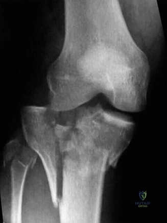
A 42-year-old male presents following a high-speed MVC with a deformed, swollen left knee. The patient is neurovascularly intact. This is the initial AP radiograph. How would you classify this fracture, and what are the primary clinical concerns at this stage?
Candidate: This is a Schatzker Type VI tibial plateau fracture. It is a bicondylar injury with metaphyseal-diaphyseal dissociation. My primary concerns are the soft tissue envelope, the risk of compartment syndrome, and ruling out vascular injury given the high-energy mechanism.
Candidates often jump straight to "I will perform dual plating." This ignores the "Damage Control" phase. Failing to mention a thorough neurovascular assessment, the high risk of compartment syndrome, or the timing of surgery based on soft tissue status will lead to a failure in the viva.
A structured response is essential. 1) Classification: Schatzker VI / AO 41-C3. 2) Immediate Priorities: ATLS survey, distal neurovascular status, and assessment for Acute Compartment Syndrome (ACS). 3) Soft Tissue Management: Acknowledge the Tscherne grade and state that definitive fixation is contraindicated until soft tissues are quiescent (typically 10-21 days). 4) Damage Control: Propose a spanning external fixator to restore length, alignment, and rotation while avoiding future incision zones.
You mention the "three-column concept" for this fracture. Why is this superior to the Schatzker system for pre-operative planning, and what specific fragment are you looking for on the CT scan?
Candidate: The three-column classification, described by Luo, is better because it accounts for the posterior column, which Schatzker does not. I am specifically looking for a posteromedial shear fragment. If this is present, it changes my surgical approach because it requires a posteromedial buttress plate to prevent varus collapse.
Candidates who fail to acknowledge the biomechanical importance of the posterior column often struggle to justify the need for a dual-incision approach. Saying "I would just plate the lateral side" is a classic error that leads to varus malalignment and implant failure.
The candidate should state: "Schatzker is purely 2D/radiographic and fails to characterize the posterior articular involvement. Luo's system utilizes axial CT to identify the medial, lateral, and posterior columns. Recognition of a posteromedial coronal shear fragment is critical; it is the 'cornerstone' of the medial column. If ignored, the construct will fail in varus as the medial plateau collapses, regardless of how strong the lateral lateral locking plate is."
The soft tissues have now settled at 14 days. You are in the OR for definitive fixation. You have reduced the articular surface through an anterolateral approach. What is the role of bone graft/substitutes in this scenario, and how do you achieve medial stability?
Candidate: I use a structural allograft or calcium phosphate cement to fill the metaphyseal void created by elevating the depressed articular fragments. This provides early compressive strength. For the medial side, I use a posteromedial approach to apply an anti-glide plate to buttress that posterior fragment.
Failing to mention the why of the calcium phosphate (early compressive strength allowing earlier ROM) or suggesting the medial plate can be applied through an anterior approach. An anterior approach to the posteromedial fragment is anatomically dangerous due to the neurovascular structures and does not provide an effective buttress.
The candidate should emphasize: 1) The Void: "Elevating depressed fragments creates a structural defect; bone substitute is required to prevent secondary subsidence." 2) The Medial Buttress: "The posteromedial fragment acts as a wedge. I use a posteromedial approach with an anti-glide plate. The plate must be placed on the posterior aspect of the medial tibia to act as a buttress, preventing the vertical shear force from displacing the fragment posteriorly and medially."
