Tibialis Posterior Tendon Debridement: A Comprehensive Review of Indications, Anatomy, and Outcomes in PTTD Management
Key Takeaway
Tibial tendon debridement is a surgical procedure for chronic posterior tibial tendon dysfunction (PTTD), often called adult-acquired flatfoot. It's indicated for recalcitrant tendinosis, partial tears, or inflammatory tenosynovitis when non-operative treatments fail, particularly in Stage I PTTD. It may also be an adjunctive procedure in more advanced reconstructive strategies, restoring foot mechanics.
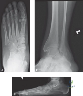
A 55-year-old female presents with a 9-month history of medial ankle pain and progressive flattening of her left foot. Examination reveals a positive "too many toes" sign and inability to perform a single-limb heel rise. She has failed a comprehensive 6-month trial of orthotics, physical therapy, and NSAIDs. Radiographs confirm a flexible flatfoot. What is your clinical staging, and how do you approach the decision-making process for this patient?
Candidate: This patient has Stage II PTTD. Since she failed non-operative treatment, I would discuss surgical reconstruction. This would involve an FDL tendon transfer to the navicular and likely a medializing calcaneal osteotomy to correct the hindfoot valgus.
Jumping directly to the surgical plan without confirming the "flexibility" of the deformity or neglecting the clinical examination findings (like equinus). Failing to mention that the FDL transfer is done in conjunction with bony work, not as a standalone procedure.
This is Stage II PTTD (Johnson & Strom). I would confirm the diagnosis with an MRI to assess tendon integrity and spring ligament status. The decision-making is structured: 1) Confirm the deformity is flexible on physical exam. 2) Address the 'deforming forces' (equinus contracture via gastroc-soleus recession). 3) Address the mechanical collapse (medializing calcaneal osteotomy). 4) Augment the dynamic stabilizer (FDL tendon transfer). I would explicitly state that isolated debridement is insufficient at this stage, as the biomechanical instability must be addressed.
Look at the MRI image provided. You are in the operating theatre. You have performed the medial exposure and opened the tendon sheath. Describe your systematic approach to the debridement and what specific intra-operative findings would change your surgical plan?
Candidate: I would identify the neurovascular bundle, protect the saphenous nerve and vein, and then perform a synovectomy. I would inspect the tendon for longitudinal tears and remove any mucoid, degenerate tissue.
Ignoring the "Spring Ligament." A common mistake is focusing only on the tendon while failing to palpate the spring ligament complex, which is often attenuated in PTTD and requires repair/reefing to prevent recurrence.
I would perform a systematic synovectomy to improve visualization. I'd inspect the full length of the tendon from the retromalleolar groove to the navicular insertion. If I find a longitudinal tear, I would debride to healthy tissue and repair with non-absorbable sutures. Crucially, I would probe the spring ligament for insufficiency. If the tendon is shredded beyond functional repair, I would abandon hope of a direct PTT repair and convert to an FDL transfer to the navicular as the primary reconstructive strategy, rather than attempting a primary repair of the degenerate PTT.
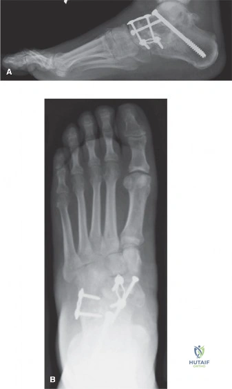
The surgery went well, and you have performed an FDL transfer and a medializing calcaneal osteotomy as seen in the intra-operative view. How do you structure the post-operative rehabilitation for this specific patient to ensure success?
Candidate: I'd put them in a plaster cast for 6 weeks non-weight bearing, followed by a walking boot and physiotherapy.
Lack of nuance. Simply saying "6 weeks NWB" is standard but misses the importance of protecting the transfer site (FDL to navicular) and the gradual transition to strengthening, which is the most common reason for secondary failure.
The rehab must be staged: 1) 0-6 weeks: Strict immobilization in a short leg cast/splint, non-weight bearing, with the foot in slight plantarflexion/inversion to offload the transfer. 2) 6-12 weeks: Transition to a CAM boot, weight-bearing as tolerated. Begin passive range of motion, but delay heavy resisted inversion/plantarflexion until 8-12 weeks to allow biological incorporation of the FDL graft. 3) 3-6 months: Focus on proprioceptive training and eccentric strengthening. Long-term, I emphasize the need for orthotics to protect the reconstruction from recurrent mechanical overload.
