Flexor Hallucis Longus (FHL) Tendon Transfer for Chronic Achilles Rupture: Comprehensive Review
Key Takeaway
Flexor Hallucis Longus (FHL) transfer is a gold standard surgical technique for chronic Achilles tendon ruptures, particularly for large defects. The FHL tendon is harvested, often proximal to the Knot of Henry, and used to reconstruct the Achilles, restoring plantarflexion strength. This method leverages the FHL's expendability and anatomical benefits to bridge tendon gaps.
A 52-year-old active male presents with a 3-month history of persistent calf weakness and difficulty with push-off after a fall at a recreational tennis match. He was treated initially with a compression bandage in the ED for an "ankle sprain." Examination reveals a palpable gap in the Achilles tendon and an inability to perform a single-limb heel rise. What is your clinical diagnosis, and how do you classify the severity of this injury?
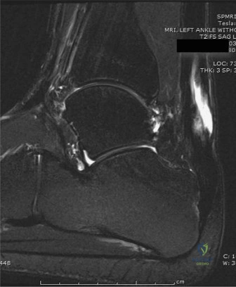
Candidate: The patient has a chronic Achilles tendon rupture. The diagnosis is confirmed by the clinical finding of a gap and the inability to perform a single-limb heel raise. I would use the Kuwada classification to determine the size of the defect, which is critical for planning the reconstructive approach.
Failing to emphasize the "chronic" nature of the presentation or missing the diagnostic nuance—candidates often focus only on the physical exam findings without addressing the diagnostic delay. Furthermore, failing to explicitly mention the differential (e.g., misdiagnosed acute vs. true chronic) or the specific clinical tests (Thompson test may be falsely negative here) shows a lack of seniority.
The candidate should state: "This is a chronic Achilles tendon rupture, defined by a presentation >4-6 weeks post-injury. The diagnosis is supported by the palpable gap and the pathognomonic loss of single-limb heel rise, noting that the Thompson test may be unreliable due to accessory plantarflexors. I utilize the Kuwada Classification for planning: Type I (partial), Type II (<3cm), Type III (3-6cm), and Type IV (>6cm). Given the chronic presentation and potential for significant retraction, I would anticipate a Type III or IV defect requiring reconstructive intervention."
You have decided to proceed with an FHL tendon transfer for this patient. Explain your rationale for choosing this graft over other options, and describe the relevant anatomical relationships you must respect during the harvest.
Candidate: I choose the FHL because it is the second strongest plantarflexor and it is "in-phase," meaning it fires with the triceps surae during gait, so no retraining is needed. Anatomically, I must be careful of the neurovascular bundle, which lies medial to the FHL, and ensure I harvest enough length from the midfoot.
Failing to mention the vascular contribution. A high-scoring candidate recognizes that the FHL muscle belly provides a vascularized bed to the "watershed" zone of the Achilles, which is a major advantage for graft incorporation in chronic, scarred tissue environments.
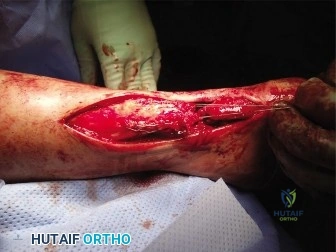
Structure the answer into Biomechanical and Anatomical buckets: 1. Biomechanical: The FHL is the second-strongest plantarflexor (30% of power), "in-phase" firing, and allows for biological restoration rather than just a static bridge. 2. Vascularity: The low-lying FHL muscle belly acts as a "living patch" providing critical blood supply to the avascular posterior ankle. 3. Anatomy: Crucial to identify the neurovascular bundle (tibial nerve/posterior tibial artery) lying medially to the muscle belly. I would perform a medial approach, keeping the dissection deep to the flexor retinaculum, and utilize a single incision, harvesting distally to the Master Knot of Henry if extra length is required.
During the procedure, you have prepped your FHL graft. How do you decide on the appropriate tension for the transfer to ensure the patient regains functional push-off without causing equinus contracture?
Candidate: I would tension the graft while the ankle is in neutral or slight plantarflexion. I would compare it to the contralateral side to ensure there is no excessive tightness that would lead to a fixed equinus deformity.
Vague references to "clinical feel." Examiners want to hear the specific degree of position (15-20 degrees of plantarflexion) and the use of the contralateral side as a gold-standard reference for resting tension.
Tensioning is the "make or break" step. I would fix the ankle in 15–20 degrees of plantarflexion. This ensures the graft is not under-tensioned, which leads to "push-off weakness," nor over-tensioned, which results in a post-operative equinus contracture. I use the contralateral limb's resting tension as the benchmark. Fixation should be rigid, using a cannulated interference screw in the calcaneal tunnel, followed by a side-to-side anastomosis to the proximal Achilles stump to provide supplemental stability and vascularity.
