Beyond PTTD: Comprehensive Differential Diagnosis of Hindfoot Pain in Orthopedics
Key Takeaway
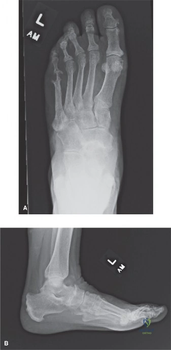
Beyond PTTD, common hindfoot pain causes include subtalar arthritis, peroneal tendon pathologies, sinus tarsi syndrome, and posterior ankle impingement. Other etiologies are calcaneal stress fractures and flexor hallucis longus tenosynovitis. Accurate diagnosis requires a deep understanding of hindfoot anatomy and biomechanics to guide effective treatment strategies.
A 45-year-old female presents with chronic medial hindfoot pain. She has been managed for suspected PTTD for 6 months without improvement. She describes a "popping" sensation and localized tenderness just distal to the medial malleolus. You are considering a differential diagnosis. What are your key clinical and imaging priorities to differentiate this from PTTD?
Candidate: I would start by performing a thorough physical exam, specifically checking the "too many toes" sign and the single-heel rise test. If these are negative, I'd consider tarsal tunnel syndrome or FHL tenosynovitis. I would order an MRI to look at the tendons, and perhaps weight-bearing X-rays to assess alignment.
The candidate focuses too heavily on PTTD-specific tests. Failing to mention specific provocative tests for non-PTTD pathologies (like Tinel's over the tarsal tunnel, FHL excursion pain, or sural nerve sensitivity) indicates a narrow focus. They also often fail to mention the Saltzman view for hindfoot alignment.
I would structure my assessment into clinical examination and targeted imaging. Clinically, I would evaluate for tarsal tunnel syndrome via a Tinel's sign over the medial malleolus and check for FHL stenosing tenosynovitis by assessing pain during hallux IP joint ROM. I would also palpate the spring ligament and the sinus tarsi. For imaging, I would request weight-bearing radiographs including a Saltzman view to rule out structural varus/valgus malalignment. An MRI is the modality of choice to differentiate between PTTD, occult bony pathology, and neurovascular entrapment.
You have diagnosed a patient with chronic, symptomatic subtalar arthritis refractory to conservative management. They are scheduled for a subtalar arthrodesis. Discuss your surgical approach and the key anatomical structures you must protect.
Candidate: I would perform a lateral sinus tarsi approach. I'd make an incision over the sinus tarsi, carefully retracting the extensor digitorum brevis. I would excise the articular cartilage and fix it with two screws. I'd make sure to watch out for the nerves in that area.
Vague references like "watch out for nerves" are unacceptable at this level. A failing candidate ignores the specific plane of dissection and fails to name the sural nerve as the primary structure at risk, or misidentifies the muscle belly being retracted.
I use the lateral sinus tarsi approach. The critical structure at risk is the sural nerve, which courses subcutaneously posterior and lateral to the incision. I dissect down to the extensor digitorum brevis (EDB) muscle belly, which I reflect plantarly to access the sinus tarsi. I excise the cervical and interosseous talocalcaneal ligaments to visualize the subtalar joint. I emphasize that correct joint preparation—denuding cartilage to bleeding subchondral bone—is vital to prevent non-union, and I aim for a neutral to 5-degree valgus alignment using compression screws.
A 30-year-old dancer presents with persistent posterior ankle pain. She is told she has "posterior impingement." When would you advise surgery, and what are the specific risks of the arthroscopic approach?
Candidate: If they fail physiotherapy and activity modification, I would offer surgery. Arthroscopy is better because it is less invasive, but you have to worry about damaging the nerves around the ankle.
Failing to distinguish between the posterolateral and posteromedial portals and the specific structures at risk (sural nerve vs. tibial neurovascular bundle) is a major deficiency. Candidates often forget to mention FHL tendon safety.
Surgery is indicated when there is mechanical blockage (e.g., symptomatic os trigonum) or FHL tenosynovitis that remains painful despite 3-6 months of conservative management. The primary risk of arthroscopic intervention is injury to the neurovascular structures: the sural nerve with the posterolateral portal and the tibial nerve and posterior tibial artery with the posteromedial portal. I also emphasize the risk of iatrogenic injury to the FHL tendon, which must be identified and protected during the debridement of the posterior talar process.
