Painful Bones & Joints? Find Causes & Effective Treatments
Key Takeaway
Here are the crucial details you must know about Painful Bones & Joints? Find Causes & Effective Treatments. Orthopedic conditions are disorders impacting the musculoskeletal system, including the bones and joints, muscles, tendons, and ligaments. They can manifest as pain, stiffness, inflammation, or deformity. Common examples include osteoarthritis, rheumatoid arthritis, fractures, and sprains, which often require expert diagnosis and treatment to restore function and alleviate discomfort.
Introduction and Epidemiology
Orthopedic conditions represent a vast spectrum of musculoskeletal disorders encompassing traumatic, degenerative, inflammatory, metabolic, and neoplastic etiologies affecting bones, joints, muscles, tendons, and ligaments. These pathologies are a leading cause of pain, functional impairment, and disability globally, imposing significant burdens on healthcare systems and patient quality of life. The prevalence of these conditions necessitates a deep understanding of their pathophysiology, accurate diagnostic algorithms, and evidence-based management strategies, including advanced surgical interventions.
Epidemiological Highlights
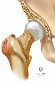
- Osteoarthritis (OA): The most prevalent joint disease, affecting hundreds of millions worldwide. Its incidence increases with age, with a substantial proportion of individuals over 65 exhibiting radiographic evidence of OA in at least one joint. Risk factors include advanced age, obesity, prior joint trauma, genetic predisposition, and occupational stressors. The knee and hip are the most commonly affected large joints requiring surgical intervention.
- Rheumatoid Arthritis (RA): An autoimmune inflammatory arthropathy affecting approximately 0.5-1% of the adult population. While systemic medical therapy is the cornerstone of management, surgical interventions such as synovectomy, arthrodesis, and arthroplasty remain crucial for pain relief, deformity correction, and improved function in advanced disease.
- Gout: A crystal-induced arthropathy affecting approximately 4% of adults in the U.S., with increasing prevalence. While primarily managed medically, surgical intervention may be indicated for chronic tophaceous gout causing joint destruction, nerve compression, or skin ulceration.
- Bursitis and Tendinitis: Common inflammatory conditions, often related to overuse, trauma, or systemic diseases. While typically self-limiting or responsive to conservative measures, chronic or recalcitrant cases may warrant surgical decompression, debridement, or bursectomy.
- Sprains and Strains: Acute soft tissue injuries of ligaments and muscles, respectively. Account for a significant percentage of emergency department visits. While most are managed non-operatively, severe ligamentous ruptures (e.g., ACL, syndesmosis, collateral ligaments) or muscle avulsions often necessitate surgical repair or reconstruction to restore stability and function.
- Fractures and Dislocations: Traumatic disruptions of bone integrity and joint congruity. Fractures alone represent a substantial portion of orthopedic workload, with incidence peaking in younger males (high-energy trauma) and older females (osteoporotic fragility fractures). Dislocations often accompany fractures or occur in isolation, demanding prompt reduction and often surgical stabilization depending on associated injuries and joint stability.
The increasing longevity of the population and improvements in diagnostic capabilities contribute to the growing incidence of degenerative conditions requiring surgical solutions, particularly arthroplasty. Concurrently, advancements in trauma care continue to refine surgical approaches for acute injuries, emphasizing anatomical reduction, stable fixation, and early mobilization.
Surgical Anatomy and Biomechanics
A profound understanding of regional surgical anatomy and joint biomechanics is paramount for any orthopedic intervention, guiding incision placement, internervous plane dissection, implant selection, and functional restoration. For the purpose of detailing surgical technique, we will focus on the knee joint, a complex diarthrodial hinge joint, as a representative example relevant to both degenerative (OA) and traumatic conditions.
Osteology of the Knee
- Bones: Distal femur, proximal tibia, and patella.
- Femur: Medial and lateral condyles (articulating surfaces), intercondylar notch (for cruciate ligaments), epicondyles (for collateral ligament attachment), and the trochlear groove (for patellofemoral articulation). The medial condyle extends further distally and is larger than the lateral condyle, a critical factor in the screw-home mechanism.
- Tibia: Medial and lateral plateaus separated by the tibial eminence. The medial plateau is concave and bears more physiologic load, while the lateral plateau is convex and more mobile. The tibial tubercle serves as the insertion site for the patellar tendon.
- Patella: The largest sesamoid bone in the human body, embedded within the extensor mechanism. It features a thick articular cartilage layer with medial and lateral facets divided by a vertical ridge.
Ligamentous and Meniscal Structures
The stability of the knee relies heavily on its static and dynamic soft tissue restraints.
* Cruciate Ligaments: The Anterior Cruciate Ligament (ACL) prevents anterior translation of the tibia on the femur and provides rotational stability. The Posterior Cruciate Ligament (PCL) prevents posterior translation and is the primary stabilizer of the knee in flexion.
* Collateral Ligaments: The Medial Collateral Ligament (MCL) consists of superficial and deep components, serving as the primary restraint to valgus stress. The Lateral Collateral Ligament (LCL) is a primary restraint to varus stress and forms part of the posterolateral corner (PLC).
* Menisci: Fibrocartilaginous structures that deepen the articular surfaces, act as shock absorbers, and aid in load transmission and joint lubrication. The medial meniscus is C-shaped and firmly attached to the joint capsule, making it more prone to injury. The lateral meniscus is O-shaped and highly mobile.
Neurovascular Anatomy
Surgical approaches to the knee require meticulous protection of surrounding neurovascular structures. The popliteal artery lies immediately posterior to the joint capsule and is at risk during posterior capsular release or excessive posterior retractor placement. The common peroneal nerve courses around the fibular neck and is highly vulnerable during lateral approaches, lateral collateral ligament reconstructions, or correction of severe valgus deformities.
Joint Biomechanics
The knee is not a simple hinge joint; its motion involves a complex combination of rolling and gliding (the "J-curve" kinematics). During early flexion, the femoral condyles roll posteriorly on the tibial plateaus. As flexion progresses, rolling transitions to gliding to prevent the femur from translating off the posterior edge of the tibia.
The "screw-home mechanism" describes the obligatory external rotation of the tibia during the final 15 degrees of extension, locking the knee into a stable, energy-efficient position for weight-bearing. Furthermore, the patellofemoral joint experiences immense reaction forces, reaching up to seven times body weight during deep squatting or stair climbing. Restoring the native joint line and mechanical axis (neutral alignment) is the primary biomechanical goal in reconstructive knee surgery to ensure optimal load distribution and implant longevity.
Indications and Contraindications
Surgical intervention in orthopedic pathology is dictated by the failure of conservative management, the presence of progressive deformity, intractable pain, or acute traumatic instability threatening limb viability or long-term function. Decision-making requires a careful analysis of patient-specific factors, including physiological age, comorbidities, and functional demands.
Operative and Non Operative Decision Making
The following table summarizes the general indications for operative versus non-operative management across common orthopedic pathologies affecting the lower extremity.
| Pathology | Non Operative Indications | Operative Indications |
|---|---|---|
| Osteoarthritis (Knee/Hip) | Mild/moderate radiographic disease, tolerable pain, high surgical risk, BMI > 40 (relative), active tobacco use. | End-stage radiographic changes (bone-on-bone), intractable pain failing conservative care, severe varus/valgus deformity, night pain. |
| Meniscal Tears | Degenerative tears with concurrent OA, asymptomatic tears, stable peripheral tears (< 1 cm). | Mechanical symptoms (locking, catching), acute traumatic tears in the red-red zone, failure of 6-12 weeks of physical therapy. |
| Cruciate Ligament Ruptures | Low-demand patients, isolated tears with no instability during activities of daily living, advanced OA. | High-demand athletes, combined multi-ligamentous injuries, subjective instability (giving way) affecting quality of life. |
| Periarticular Fractures | Non-displaced or minimally displaced fractures, stable fracture patterns, non-ambulatory patients. | Intra-articular step-off > 2mm, open fractures, neurovascular compromise, unstable fracture patterns, failure of closed reduction. |
| Septic Arthritis | Extremely rare (only if patient is medically unfit for any intervention; requires serial aspirations). | Nearly all cases require emergent surgical irrigation and debridement (arthroscopic or open) to prevent rapid cartilage destruction. |
Absolute Contraindications to Arthroplasty: Active local or systemic infection, neuropathic (Charcot) arthropathy (relative in highly specialized centers), severe peripheral vascular disease precluding wound healing, and lack of functional extensor mechanism.
Pre Operative Planning and Patient Positioning
Thorough preoperative planning is the cornerstone of successful orthopedic surgery. It mitigates intraoperative complications, reduces operative time, and ensures accurate restoration of biomechanics.
Clinical Evaluation and Imaging
A comprehensive history and physical examination must assess range of motion, ligamentous stability, neurovascular status, and the condition of the soft tissue envelope. Previous surgical incisions must be mapped to avoid devascularizing skin bridges.
Standard radiographic evaluation for degenerative knee conditions includes:
* Weight-bearing Anteroposterior (AP) view.
* Weight-bearing Posteroanterior (PA) flexion view (Rosenberg view) to assess the posterior aspect of the condyles and subtle joint space narrowing.
* Lateral view to evaluate patellar height (Caton-Deschamps or Insall-Salvati ratios) and osteophyte formation.
* Merchant or Sunrise view to assess patellofemoral tracking, tilt, and joint space.
* Full-length standing alignment films (hip-to-ankle) are critical for assessing the mechanical axis and planning the degree of correction required.
Templating and Axis Determination
Digital templating is utilized to estimate implant size and determine the necessary bony resections. The goal of standard Total Knee Arthroplasty (TKA) is to restore the mechanical axis of the lower extremity to neutral (0 degrees).
* Femoral Cut: The distal femoral cut is typically made at 5 to 7 degrees of valgus relative to the anatomical axis of the femur, aiming to be perpendicular to the mechanical axis.
* Tibial Cut: The proximal tibial resection is made strictly perpendicular to the mechanical axis of the tibia in the coronal plane, often incorporating a 3 to 5-degree posterior slope in the sagittal plane to match native anatomy and facilitate flexion.
Patient Positioning and Operating Room Setup
The patient is positioned supine on a standard radiolucent operating table. A lateral post or leg holder is placed at the level of the proximal thigh to allow for stable positioning of the knee in hyperflexion and to apply varus/valgus stress during exposure. A well-padded tourniquet is applied to the proximal thigh, though routine inflation is increasingly debated; many surgeons now utilize it only during cementation to optimize the bone-cement interface. The operative extremity is meticulously prepped and draped in a standard sterile fashion, ensuring access from the anterior superior iliac spine to the toes.
Detailed Surgical Approach and Technique
The execution of a joint replacement requires precise soft tissue handling, accurate bony resections, and meticulous gap balancing. The following details the standard technique for a primary Total Knee Arthroplasty utilizing a measured resection philosophy.
The Medial Parapatellar Approach
The medial parapatellar arthrotomy remains the workhorse approach for knee arthroplasty and complex intra-articular trauma.
1. Incision: A longitudinal midline skin incision is made from approximately 3-4 cm proximal to the superior pole of the patella, extending distally to the medial border of the tibial tubercle.
2. Superficial Dissection: Subcutaneous tissues are divided to expose the extensor mechanism. Full-thickness fasciocutaneous flaps are maintained to preserve vascularity.
3. Arthrotomy: The arthrotomy begins proximally in the quadriceps tendon, leaving a 3-4 mm cuff of tendon attached to the vastus medialis obliquus (VMO) to facilitate robust closure. The incision skirts the medial border of the patella and continues distally along the medial border of the patellar tendon, terminating at the tibial tubercle.
4. Exposure: The patella is everted laterally (or subluxated, depending on surgeon preference and tissue tension). The anterior horns of the medial and lateral menisci are excised. The ACL is resected, and the PCL is either resected or preserved based on the chosen implant design (Cruciate-Retaining vs. Posterior-Stabilized).
Joint Preparation and Bony Resection
The sequence of bony resections varies (femur-first vs. tibia-first), but the principles of creating flat, parallel surfaces remain constant.
1. Distal Femoral Resection: An intramedullary alignment guide is introduced into the femoral canal. The valgus angle (pre-calculated via templating, usually 5-7 degrees) is set. The distal femoral resection removes an amount of bone equivalent to the thickness of the femoral component.
2. Proximal Tibial Resection: An extramedullary alignment guide is typically utilized, referencing the tibial tubercle proximally and the center of the talus distally. The resection is made perpendicular to the mechanical axis, removing minimal bone (typically 8-10 mm from the unaffected side) to preserve the denser subchondral bone necessary for implant support.
3. Femoral Sizing and Chamfer Cuts: An anterior-posterior (AP) sizing guide is placed on the distal femur, referencing the posterior condyles. External rotation of the femoral component (typically 3 degrees relative to the posterior condylar axis) is established to optimize patellofemoral tracking and balance the flexion gap. The anterior, posterior, and chamfer cuts are then executed using a 4-in-1 cutting block.
Soft Tissue Balancing
A successful arthroplasty requires equal and rectangular flexion and extension gaps.
* Extension Gap: Defined by the distal femoral cut and the proximal tibial cut. If the gap is tight, posterior capsular release or additional distal femoral resection is required.
* Flexion Gap: Defined by the posterior femoral cut and the proximal tibial cut. If tight, the PCL may need releasing (in a CR knee), or the femoral component may be downsized to resect more posterior bone.
Varus and valgus deformities require sequential soft tissue releases. For a varus knee, the deep MCL, posteromedial capsule, and pes anserinus may require progressive release off the proximal tibia.
Implant Fixation and Closure
- Trialing: Trial components are inserted. The knee is taken through a full range of motion to assess stability, gap symmetry, and patellar tracking. The "no thumb" rule is applied to ensure the patella tracks centrally in the trochlear groove without lateral subluxation.
- Preparation for Cementation: The bone surfaces are cleansed with pulsatile lavage and thoroughly dried to ensure optimal micro-interlock of the polymethylmethacrylate (PMMA) bone cement.
- Implantation: Cement is applied to the implants and bone surfaces in a doughy state. The implants are impacted into position, and the knee is held in full extension while the cement polymerizes. Excess cement is meticulously removed to prevent third-body wear.
- Closure: The joint is copiously irrigated. A periarticular injection of a multimodal analgesic cocktail (typically containing bupivacaine, epinephrine, ketorolac, and clonidine) is administered. The arthrotomy is closed in a watertight fashion using heavy absorbable sutures (e.g., #1 or #2 Vicryl or Stratafix). Subcutaneous tissues and skin are closed sequentially.
Complications and Management
Despite rigorous preoperative optimization and meticulous surgical technique, orthopedic interventions carry inherent risks. Rapid identification and protocol-driven management are critical to salvaging the limb and preserving function.
Intraoperative and Postoperative Adverse Events
The following table outlines common and catastrophic complications, their approximate incidence, and accepted salvage strategies.
| Complication | Incidence | Pathophysiology and Salvage Strategy |
|---|---|---|
| Periprosthetic Joint Infection (PJI) | 1.0 - 2.0% | Pathophysiology: Bacterial colonization of the implant surface leading to biofilm formation (commonly S. aureus, Coagulase-negative Staphylococci). Salvage: Acute (< 4 weeks): Debridement, Antibiotics, and Implant Retention (DAIR) with modular exchange. Chronic: Two-stage revision arthroplasty utilizing an antibiotic-loaded PMMA spacer, followed by reimplantation after eradication. |
| Venous Thromboembolism (DVT/PE) | 1.0 - 3.0% (Symptomatic) | Pathophysiology: Virchow's triad (endothelial injury, stasis, hypercoagulability) activated by surgical trauma and tourniquet use. Salvage: Immediate initiation of therapeutic anticoagulation (LMWH, DOACs, or Warfarin). Hemodynamically unstable PE requires emergent thrombolysis or embolectomy. |
| Periprosthetic Fracture | 0.5 - 2.0% | Pathophysiology: Intraoperative cortical notching, excessive impaction force, or postoperative low-energy trauma in osteoporotic bone. Salvage: Depends on implant stability (Vancouver/Unified Classification). Stable implant: Open Reduction Internal Fixation (ORIF) with locking plates/cables. Loose implant: Revision arthroplasty with diaphyseal engaging stems. |
| Arthrofibrosis | 3.0 - 5.0% | Pathophysiology: Exuberant fibroblastic proliferation and scar tissue formation leading to severe restriction of motion. Salvage: Aggressive physical therapy. If no progress by 6-12 weeks, Manipulation Under Anesthesia (MUA). Recalcitrant cases may require arthroscopic or open lysis of adhesions. |
| Extensor Mechanism Disruption | < 1.0% | Pathophysiology: Rupture of the quadriceps tendon, patellar tendon, or patellar fracture, often secondary to avascularity or over-resection. Salvage: Devastating complication. Direct repair often fails. Requires reconstruction using allograft (extensor mechanism allograft) or synthetic mesh (Marlex mesh) augmentation. |
Post Operative Rehabilitation Protocols
The shift toward Enhanced Recovery After Surgery (ERAS) pathways has revolutionized postoperative orthopedic care, dramatically reducing length of stay and minimizing opioid consumption.
Enhanced Recovery and Early Mobilization
Modern protocols mandate immediate weight-bearing as tolerated (WBAT) for primary arthroplasty and stable fracture fixations. Early mobilization (within 4-6 hours postoperatively) is the single most effective strategy for mitigating the risk of deep vein thrombosis and preventing early arthrofibrosis. Multimodal analgesia—utilizing standing acetaminophen, NSAIDs (e.g., Celecoxib), gabapentinoids, and periarticular local anesthetic infiltration—has largely replaced patient-controlled analgesia (PCA) pumps, thereby reducing postoperative nausea, vomiting, and respiratory depression.
Phased Rehabilitation Milestones
Rehabilitation following major knee surgery is generally divided into distinct phases:
- Phase I: Acute Recovery (0 to 2 Weeks):
- Goals: Wound healing, edema control, DVT prophylaxis, and restoration of active quadriceps control (eliminating extensor lag).
- Milestones: Achieve full active and passive extension (0 degrees) to prevent flexion contractures. Achieve minimum 90 degrees of flexion. Independent ambulation with an assistive device.
- Phase II: Intermediate Phase (2 to 6 Weeks):
- Goals: Wean off assistive devices, normalize gait mechanics, and improve functional endurance.
- Milestones: Flexion progressing to 110-120 degrees. Progression to closed kinetic chain exercises (e.g., mini-squats, leg press).
- Phase III: Advanced Strengthening (6 to 12 Weeks):
- Goals: Maximize muscular hypertrophy, enhance proprioception, and return to low-impact functional activities.
- Milestones: Return to activities such as cycling, swimming, and golfing. High-impact activities (running, jumping) are generally discouraged following arthroplasty to prevent accelerated polyethylene wear and aseptic loosening.
Summary of Key Literature and Guidelines
Evidence-based practice in orthopedic surgery is continuously refined by large-scale randomized controlled trials and societal guidelines.
Evidence Based Practice and Societal Guidelines
- American Academy of Orthopaedic Surgeons (AAOS) Clinical Practice Guidelines: The AAOS strongly recommends the use of tranexamic acid (TXA) in total joint arthroplasty to reduce perioperative blood loss and transfusion requirements. TXA can be administered intravenously, topically, or orally, with robust literature demonstrating no increased risk of thromboembolic events even in patients with a history of cardiovascular disease.
- Venous Thromboembolism Prophylaxis: The optimal VTE prophylaxis regimen remains a topic of active research. Recent consensus guidelines and large multicenter trials (such as the PEPPER trial framework) support the use of low-dose Aspirin (81 mg twice daily) as non-inferior to potent anticoagulants (like Rivaroxaban or Enoxaparin) for VTE prophylaxis in standard-risk arthroplasty patients, offering the benefit of a significantly lower risk of major bleeding complications or wound hematomas.
- Antibiotic Stewardship: Guidelines mandate the administration of prophylactic intravenous antibiotics (typically a first-generation cephalosporin such as Cefazolin) within one hour prior to surgical incision. The duration of postoperative prophylaxis should not exceed 24 hours, as extended courses have not demonstrated a reduction in PJI rates but do increase the risk of acute kidney injury and Clostridioides difficile infection.
- Treatment of Periprosthetic Joint Infection: The Musculoskeletal Infection Society (MSIS) and the International Consensus Meeting (ICM) on PJI provide the definitive diagnostic criteria for PJI, utilizing serum markers (CRP, ESR), synovial fluid analysis (leukocyte esterase, alpha-defensin, synovial WBC count), and microbiological cultures to guide the complex surgical management of the infected joint.
