Rotator Cuff Arthropathy: A Comprehensive Clinical & Imaging Diagnosis Case Study
Key Takeaway
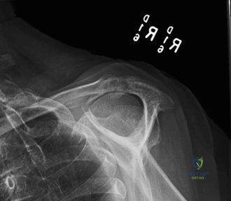
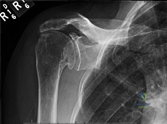
Rotator Cuff Arthropathy (RCA) is diagnosed by chronic shoulder pain, progressive pseudoparalysis, and specific clinical signs. Key findings include supraspinatus/infraspinatus atrophy, significant active ROM loss with preserved passive ROM, positive external rotation lag and drop arm tests. Radiographs confirm superior humeral head migration, acromiohumeral interval narrowing, and glenohumeral joint space narrowing, often with acromial erosion.
A 72-year-old male presents with a 3-year history of progressive shoulder pain and functional loss. He reports an inability to lift his arm above shoulder height despite his best effort. On examination, he demonstrates a 'shrug' sign during attempted abduction. Passive range of motion is significantly greater than active range of motion. Looking at the provided imaging, describe the key findings and provide your differential diagnosis.
Candidate: The X-ray shows superior migration of the humeral head, narrowing of the acromiohumeral interval, and osteophytes on the glenoid and humeral head. This is suggestive of rotator cuff arthropathy. The differential diagnosis includes adhesive capsulitis, primary glenohumeral osteoarthritis, and cervical radiculopathy.
Failing to mention the "pseudoparalysis" context, ignoring the specific radiographic signs like "acetabularization," or failing to organize the differential based on the clinical presentation of active/passive ROM discrepancy.
Structure your answer: 1) Radiographic Findings: Superior humeral head migration, acromiohumeral interval <7mm, acetabularization of the undersurface of the acromion, and glenohumeral joint space narrowing. This is Hamada Grade 4A/B. 2) Clinical Integration: The active-passive ROM discrepancy and "shrug sign" confirm rotator cuff deficiency leading to pseudoparalysis. 3) Differential: Narrow to: Adhesive capsulitis (restricted passive ROM), Primary OA (concentric wear, intact cuff), and Inflammatory arthropathy (r/o with inflammatory markers).
You have decided to proceed with a Reverse Total Shoulder Arthroplasty (RTSA). During your pre-operative planning using the CT scan, you identify significant posterior glenoid erosion and retroversion. How do you classify this, and how does it influence your surgical plan?
Candidate: I would classify this as a Walch Type B2 or B3 glenoid. It affects my plan because I need to address the bone loss to ensure the baseplate is stable. I would use reaming, or potentially bone grafting if the defect is severe, to restore the version.
Focusing only on the implant and forgetting the biomechanical principles of RTSA, such as the need for inferior positioning to prevent scapular notching or failing to mention the importance of screw purchase in the scapular pillar.
State the classification (Walch B2/B3). Explain the "3-pillar approach": 1) Bone Stock Management: Using structural bone graft or eccentric reaming to correct retroversion. 2) Baseplate Fixation: Ensuring central peg and peripheral screw purchase in the scapular spine and pillar. 3) Avoidance of Complications: Emphasize the importance of inferior and lateralized glenosphere positioning to increase deltoid tension and decrease the risk of scapular notching.
Post-operatively, the patient is concerned about the risk of dislocation. What are the biomechanical reasons for the stability of a reverse shoulder, and what specific movements should the patient avoid in the early rehabilitation phase?
Candidate: The reverse shoulder is stable because it moves the center of rotation medially and inferiorly, which tensions the deltoid. Patients should avoid combined adduction, internal rotation, and extension because this is the primary dislocation position.
Failing to explicitly link the deltoid function to the stability of the joint or not recognizing that the "Reverse" design relies on joint compression rather than the ligamentous constraint of an anatomic shoulder.
Explain: 1) Biomechanical Advantage: The RTSA medializes and distalizes the center of rotation, which significantly increases the deltoid lever arm and tension, creating a compressive force that stabilizes the joint. 2) Dislocation Mechanism: Avoid the "clunk" position: Combined Adduction, Internal Rotation, and Hyperextension. 3) Rehabilitation: Emphasize protecting the soft tissues for 6 weeks, specifically avoiding active internal rotation behind the back or reaching for the contralateral axilla.
