العربية
Part of the Master Guide

Orthopedic Ob Reconstru Review | Dr Hutaif Hip & Knee R -...

Adult Reconstructive Of The Hip And Review | Dr Hutaif - ...

23 Apr 2026 107 min read 155 Views
Illustration of knee selfassessment online - Dr. Mohammed Hutaif

Key Takeaway

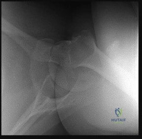
This interactive board review contains 100 randomly selected orthopedic surgery questions with clinical images, immediate feedback, and detailed references.

Adult Reconstructive Of The Hip And Review | Dr Hutaif - ...

Comprehensive 100-Question Exam


00:00

Start Quiz

Question 1

A 68-year-old female with a history of systemic lupus erythematosus (SLE) on chronic high-dose corticosteroids undergoes a total hip arthroplasty (THA) for avascular necrosis. Three months postoperatively, she presents with insidious onset of groin pain, mild swelling, and a low-grade fever (38.1°C). Her erythrocyte sedimentation rate (ESR) is 75 mm/hr and C-reactive protein (CRP) is 60 mg/L. Joint aspiration yields turbid fluid. Synovial fluid analysis shows a white blood cell (WBC) count of 18,000 cells/µL with 78% neutrophils and a single positive alpha-defensin test. Gram stain is negative. Which of the following is the most appropriate next step in confirming the diagnosis of periprosthetic joint infection (PJI)?


Explanation

The patient's clinical presentation, elevated inflammatory markers (ESR, CRP), and synovial fluid analysis (WBC count, neutrophil percentage) strongly suggest PJI, even with a negative Gram stain. The positive alpha-defensin test is a highly sensitive and specific marker for PJI. While antibiotics might be considered after cultures are obtained, initiating them immediately would jeopardize the culture results. Repeating aspiration for fungal/mycobacterial cultures is not the initial most definitive step, as the current data points strongly to bacterial PJI. Radionuclide scans are often used in equivocal cases but are less specific than joint aspiration and biopsy in chronic infections. The most definitive diagnostic step, especially when PJI is strongly suspected, is surgical exploration with intraoperative frozen section and collection of multiple periprosthetic tissue samples for microbiology culture. Frozen section analysis can provide rapid intraoperative confirmation of infection, and multiple tissue cultures improve sensitivity for identifying the causative organism. Given the immunosuppressed status due to SLE and corticosteroids, the presentation might be subtle, making robust diagnostic methods critical.

Question 2

A 72-year-old male presents with aseptic loosening of a cemented femoral component following THA performed 15 years prior. Radiographs show significant proximal femoral bone loss classified as Paprosky Type IIIB, with severe cortical thinning and a large defect involving the greater trochanter. There is evidence of a stress riser distal to the stem tip. The patient has good bone stock distally. Which of the following is the most appropriate reconstructive strategy for the femoral side?


Explanation

Paprosky Type IIIB femoral defects are characterized by extensive proximal bone loss, severe cortical thinning, and often a stress riser, making standard primary or short stems inadequate. While allograft-prosthesis composites are an option for massive defects, they carry risks of nonunion and infection. Proximal femoral replacement is typically reserved for even more severe defects, such as tumor resection or failed massive allografts. An extensively porous-coated uncemented stem with diaphyseal fixation is the most appropriate choice for Paprosky IIIB defects, as it bypasses the poor proximal bone and achieves stable fixation in the healthy distal diaphysis. Modular distally fixing stems are also a good option, but 'proximal femoral replacement' as an option here might be an overcall. The key is to achieve stable diaphyseal fixation and bypass the proximal defect. Cemented stems are less ideal in this setting due to bone loss and potential for recurrent aseptic loosening.

Question 3

A 35-year-old female with severe unilateral hip dysplasia (Crowe Type IV) is scheduled for a primary THA. She has significant limb length discrepancy and a false acetabulum. Which of the following pre-operative planning steps is the most crucial to ensure a successful outcome and minimize complications?


Explanation

Crowe Type IV dysplasia involves a high-riding femoral head and a dysplastic, shallow true acetabulum, often requiring a subtrochanteric osteotomy and/or extensive soft tissue release to bring the hip down. This scenario presents significant challenges in achieving limb length equality, restoring hip mechanics, and ensuring stable fixation. Detailed templating for both acetabular and femoral components is paramount. This includes planning for the placement of the acetabular component in the true anatomical acetabulum (often requiring medialization or grafting), determining the extent of potential limb lengthening (which is limited by sciatic nerve stretch tolerance), and planning for a subtrochanteric osteotomy if necessary to manage limb length and achieve proper hip center. While other options like MRI or CT are helpful, and tranexamic acid is standard, none are as critical as detailed templating for managing the specific complexities of Crowe IV dysplasia. Pre-stressing the limb with traction may give an estimate but doesn't replace meticulous templating for component sizing, placement, and osteotomy planning.

Question 4

A 55-year-old male undergoes revision THA for recurrent instability. The surgeon implants a modular dual mobility acetabular component. Six months postoperatively, he presents with another dislocation. Radiographs show all components are well-fixed and in good position. Which of the following is the most likely cause of this recurrent dislocation?


Explanation

Modular dual mobility systems are designed to reduce dislocation risk by providing two articulations: a large diameter articulation between a highly cross-linked polyethylene liner and the metal shell, and a standard articulation between the femoral head and the inner polyethylene liner. The 'jump distance' is significantly increased, making traditional dislocation mechanisms less likely. However, a common mode of failure unique to dual mobility designs (especially modular ones) is impingement of the femoral neck on the outer polyethylene liner, leading to lever-out of the construct or even dissociation of the head from the inner liner, or the inner liner from the shell. Given that components are well-fixed and in good position, and considering the design, neck-liner impingement (option C) is a well-described cause of recurrent dislocation in dual mobility THAs, leading to lever-out or intra-prosthetic dislocation. Bearing wear is less likely so early. Liner dissociation from the shell is a possibility but neck impingement is a more common initial mechanism leading to issues. Intra-prosthetic dislocation (IPD) is the result, but impingement is often the cause. Femoral component malrotation would typically be addressed as a cause for primary instability.

Question 5

A 70-year-old osteoporotic female presents with a Vancouver B3 periprosthetic femoral fracture around a well-fixed, extensively porous-coated, cementless femoral stem. The fracture extends proximally into the greater trochanter and distally well beyond the stem tip, with significant comminution and poor bone quality. What is the most appropriate treatment strategy?


Explanation

Vancouver B3 fractures are characterized by a loose stem or poor bone quality around a well-fixed stem, making stable fixation of the fracture fragments challenging. Given the extensive comminution, poor bone quality, and the fracture extending beyond the stem tip, the existing stem is compromised even if considered 'well-fixed' initially. ORIF alone (options A and B) is insufficient due to the lack of adequate bone stock for stable fixation and the likely inability to achieve durable construct. Excision arthroplasty is a salvage procedure, not primary treatment for a B3. The presence of significant bone loss and poor quality mandates stem removal and revision to a new stem that can bypass the fracture and achieve stable fixation in healthy bone distally. A cemented long-stem component or a modular distally fixing uncemented stem are both viable options. However, given the osteoporotic bone quality, a cemented long-stem revision often provides more predictable and immediate stability in compromised bone. While a modular distally fixing uncemented stem is also a strong option, the question highlights 'osteoporotic female' and 'poor bone quality', which sometimes sways towards cemented options for immediate stability. Cemented long-stem revision is a well-established strategy for Vancouver B3 fractures where robust fixation is required. Allografting may be needed for severe bone loss, but the primary strategy is stem revision.

Question 6

A 62-year-old male with a Metal-on-Metal (MoM) THA implanted 8 years ago presents with worsening groin and lateral hip pain, an audible 'squeak,' and elevated serum cobalt and chromium levels. MRI arthrogram reveals a large periprosthetic fluid collection (pseudotumor) and extensive synovial thickening consistent with an adverse reaction to metal debris (ARMD). The components appear radiographically well-fixed. What is the most definitive management strategy for this patient?


Explanation

The presence of elevated metal ions, pseudotumor, ARMD on MRI, and persistent symptoms in an MoM THA, even with radiographically well-fixed components, is indicative of progressive adverse tissue reaction. This reaction can lead to significant local tissue damage, osteolysis, and potentially systemic effects. While conservative management, injections, or debridement might provide temporary relief, they do not address the ongoing metal wear and debris generation. The most definitive management is revision THA with exchange of the MoM bearing surface to a non-MoM coupling, such as ceramic-on-polyethylene or ceramic-on-ceramic. This eliminates the source of metal wear debris, preventing further tissue damage and resolving the symptoms. Simply performing debridement and synovectomy without component exchange will not resolve the underlying issue. Regular follow-up is for asymptomatic or mildly symptomatic patients with lower ion levels.

Question 7

A 48-year-old active male presents for primary THA due to advanced bilateral avascular necrosis. During the procedure, while inserting an uncemented femoral stem, a longitudinal fracture of the calcar region is observed intraoperatively. The stem appears to be rotationally stable and well-seated distally. What is the most appropriate management of this intraoperative complication?


Explanation

An intraoperative calcar fracture during uncemented femoral stem insertion (often classified as a Vancouver A fracture or specific type A fracture) requires careful management. If the stem is rotationally stable and well-seated distally, and the fracture is contained without significant displacement or propagation, applying cerclage wires (option C) to stabilize the calcar region and maintain reduction is often the appropriate management, allowing the stem to achieve stable fixation and the fracture to heal. Removing the stem to ream larger or convert to cemented without first attempting to stabilize the fracture is not ideal if the stem is stable. Non-weight bearing alone is not sufficient to stabilize the fracture. Osteotomy is an extreme measure not indicated for a simple calcar fracture. Therefore, stable fixation of the stem and internal fixation of the fracture (e.g., cerclage wires) is the correct approach to manage this complication while maintaining the primary uncemented reconstruction. If the stem were unstable, then revision to a larger stem or a cemented stem would be considered. However, the question states 'rotationally stable and well-seated distally'.

Question 8

A 75-year-old female with chronic renal failure on hemodialysis presents with end-stage hip osteoarthritis and severe osteoporosis. She is scheduled for a THA. Which of the following is the most significant perioperative concern specific to this patient population?


Explanation

Patients with chronic renal failure, especially those on hemodialysis, present unique and complex challenges for THA. While all the listed options are potential concerns, challenges with fluid and electrolyte management and coagulopathy are most significant and specific to this population. Renal patients often have hyperkalemia, fluid overload, and metabolic acidosis, which need careful monitoring and management. Furthermore, they frequently have acquired platelet dysfunction and are often on anticoagulants for dialysis, leading to an increased risk of perioperative bleeding. While bone quality (osteoporosis and renal osteodystrophy) is a concern for fixation, and infection risk is elevated due to immunosuppression, the immediate life-threatening challenges often revolve around their metabolic and hematological derangements related to kidney function and dialysis. The risk of DVT can be high, but the combination of fluid/electrolyte and coagulopathy issues is particularly critical for managing these patients safely.

Question 9

A 58-year-old male with a history of ankylosing spondylitis (AS) is undergoing a bilateral THA for end-stage arthritis. He previously developed significant heterotopic ossification (HO) after a spinal fusion. What is the most effective prophylactic regimen to prevent severe HO in this patient?


Explanation

Patients with ankylosing spondylitis, especially those with a history of HO after other surgeries, are at extremely high risk for severe heterotopic ossification after THA. While NSAIDs (like indomethacin) are a standard prophylaxis for moderate-risk patients, they may not be sufficient for very high-risk individuals. Radiation therapy is highly effective for HO prophylaxis. The most effective protocol for high-risk patients, such as those with AS, is a single dose of 700 cGy delivered to the hip, typically within 24-72 hours postoperatively. Preoperative radiation is also effective but may delay surgery and might have slightly different logistical challenges. However, the timing (postoperative, within 24-72 hours) is critical for optimal efficacy. Corticosteroids and vitamin K antagonists are not standard or effective for HO prophylaxis. Therefore, postoperative radiation therapy is the most robust and evidence-based approach for this high-risk patient.

Question 10

A 60-year-old female undergoes a primary THA via a direct anterior approach. Postoperatively, she complains of numbness and burning pain in the lateral thigh. Sensory examination confirms diminished sensation in the distribution of the lateral femoral cutaneous nerve (LFCN). What is the most appropriate initial management for this iatrogenic complication?


Explanation

LFCN neuropathy (meralgia paresthetica) is a known complication of the direct anterior approach due to traction or direct injury to the nerve. While surgical exploration is an option for persistent or severe symptoms, the initial management for most iatrogenic nerve injuries is conservative, focusing on symptom control and allowing for spontaneous recovery. Medications like gabapentin or pregabalin (option B) are commonly used for neuropathic pain. Observation is crucial as many cases resolve spontaneously over weeks to months. NCS/EMG might be useful if symptoms persist or worsen significantly after an initial conservative period to assess the extent of damage but is not the immediate step. Local anesthetic injection can be diagnostic and therapeutic but typically follows medical management. Immediate surgical exploration is generally reserved for severe, progressive deficits or symptoms that fail extensive conservative management, or if there's suspicion of a transection. Therefore, starting with neuropathic pain medication and observation is the most appropriate initial management.

Question 11

A 32-year-old male, a competitive amateur triathlete, presents with bilateral end-stage avascular necrosis of the femoral heads. He requires THA and is concerned about the long-term durability and activity limitations. Considering his age, high activity level, and desire for longevity, which bearing surface combination would be most appropriate to recommend?


Explanation

For a young, active patient requiring THA, minimizing wear and maximizing longevity are critical. MoM bearings have largely been abandoned due to concerns about metal ion release, pseudotumor formation, and ARMD. Dual mobility, while excellent for instability, historically used conventional polyethylene (though highly cross-linked variants exist now) and the primary concern for this patient is wear, not instability. Conventional CoP has superior wear characteristics compared to conventional MoP. However, CoC bearings (option C) offer the lowest wear rates and excellent scratch resistance, making them ideal for young, active patients who demand maximum durability and minimal debris generation. While ceramic fractures are a rare but serious complication, the benefits of ultralow wear in a young, active patient generally outweigh this risk for many surgeons. MoP with highly cross-linked polyethylene is also a very good option, but CoC generally has even lower wear, making it the 'most appropriate' for this specific scenario of a highly active young patient prioritizing longevity and low wear. The options provided don't specify highly cross-linked polyethylene for the CoP option, making CoC a stronger choice here, as the question asks 'most appropriate' and lists CoC as a standalone option.

Question 12

A 67-year-old morbidly obese male (BMI 52 kg/m²) is undergoing primary THA for severe osteoarthritis. What is the most significant specific technical challenge related to his obesity that the surgeon must anticipate and prepare for?


Explanation

Morbid obesity significantly increases the complexity and risks of THA. While all listed options are relevant concerns in obese patients, the 'most significant specific technical challenge' during the actual surgery is often related to patient positioning and achieving adequate surgical exposure. Excess adipose tissue makes standard landmarks difficult to palpate, increases the depth of the surgical field, obscures anatomical structures, and makes retraction challenging. This can lead to longer operative times, increased blood loss, difficulty with component placement, and potential iatrogenic injury. Increased infection risk, DVT, and pain control are all important, but they are general perioperative concerns rather than direct technical challenges of the surgical act itself. Heterotopic ossification is not specifically related to obesity, though overall inflammation might be. Thus, managing the surgical exposure is a predominant intraoperative technical hurdle.

Question 13

During a revision THA for aseptic loosening of the acetabular component, it is discovered that the uncemented acetabular shell is well-fixed and difficult to remove without significant bone loss. The polyethylene liner is severely worn, and osteolysis is present around the shell-bone interface but not progressing beneath the shell. The femoral component is well-fixed and in good position. What is the most appropriate management of the acetabular component?


Explanation

This scenario describes aseptic loosening of the polyethylene liner within a well-fixed uncemented shell, with associated osteolysis. While simply exchanging the liner (option B) or cementing a new liner (option C) into an existing shell is an option for isolated polyethylene wear without significant osteolysis or shell malposition, the presence of progressive osteolysis around the shell-bone interface implies that the current shell-bone interface is compromised, even if the shell initially feels well-fixed. Leaving the existing shell will perpetuate the problem and make future revisions more challenging. Therefore, the most appropriate management is to remove the existing shell, address the osteolysis (debridement, grafting), and implant a new, stable uncemented acetabular shell to ensure long-term stability and prevent further bone loss. This allows for restoration of the correct hip center and better control of acetabular mechanics. Converting to a dual mobility component without removing the problematic shell is not ideal given the osteolysis. Girdlestone is a salvage procedure.

Question 14

A 50-year-old male presents with persistent thigh pain 5 years after an uncemented THA. Radiographs show no signs of component loosening, stable osteointegration, but significant proximal femoral stress shielding and distal cortical hypertrophy around the stem tip. Which of the following is the most likely diagnosis?


Explanation

The patient's symptoms (thigh pain) combined with radiographic findings of proximal stress shielding and distal cortical hypertrophy in the absence of loosening or infection are classic for 'thigh pain' associated with uncemented, extensively porous-coated femoral stems. This phenomenon is attributed to the mismatch in stiffness between the stiff implant and the bone, leading to altered load transfer. The proximal bone is 'shielded' from stress, leading to resorption (stress shielding), while the distal femur experiences increased load, leading to cortical hypertrophy. This altered biomechanics can cause persistent pain. Aseptic loosening would show radiographic signs such as lucencies. PJI would typically have inflammatory markers and systemic symptoms. HO would be visible as bone formation around the implant. Malposition could cause pain but not typically this specific radiographic pattern of remodeling in the absence of loosening. Therefore, stress shielding and subsequent bone remodeling are the most likely cause.

Question 15

A 68-year-old female presents with a painful THA 10 years after implantation. Radiographs demonstrate focal osteolytic lesions around the acetabular component and within the acetabular bone, but the femoral component appears well-fixed without evidence of loosening or osteolysis. Inflammatory markers (ESR, CRP) are within normal limits, and joint aspiration culture is negative. What is the most appropriate management strategy for the acetabular side?


Explanation

This patient presents with aseptic osteolysis around the acetabular component, a common long-term complication of THA, often due to polyethylene wear debris. Given the isolated acetabular osteolysis with a well-fixed femoral component and negative infection workup, the most appropriate management is an isolated acetabular revision. This involves exchanging the worn polyethylene liner, debriding the osteolytic lesions, and bone grafting these lesions to prevent further bone loss and restore structural integrity. If the metal shell is well-fixed and in good position, it can be retained (a liner exchange procedure). However, often, if osteolysis is significant, the shell itself might be contributing or could become loose, necessitating a full shell revision. The option 'Acetabular revision with exchange of the polyethylene liner, debridement of osteolytic lesions, and bone grafting' (option C) broadly covers the necessary steps. Observing with serial radiographs is appropriate for small, non-progressive lesions but not for symptomatic, progressive osteolysis. Exchanging both components is unnecessary if the femoral component is well-fixed. Debridement with a spacer or antibiotics is for infection. Therefore, addressing the wear and bone loss specifically at the acetabulum is key.

Question 16

A 70-year-old male presents with a persistent Trendelenburg gait and pain over the greater trochanter 1 year after THA. MRI demonstrates discontinuity of the abductor tendons (gluteus medius and minimus) from the greater trochanter. What is the most appropriate surgical intervention?


Explanation

Persistent Trendelenburg gait and pain over the greater trochanter, particularly with MRI evidence of abductor tendon discontinuity, strongly indicate abductor deficiency. This can be due to avulsion, non-healing of a trochanteric osteotomy, or direct injury during surgery (e.g., in a lateral approach). While physical therapy and injections might offer temporary symptomatic relief for bursitis, they do not address the underlying anatomical defect of tendon discontinuity. Revision THA with a larger femoral head primarily addresses instability, not abductor function. Excision of the greater trochanter is a drastic measure not indicated here. The most appropriate and definitive surgical intervention for abductor tendon discontinuity is direct surgical repair of the avulsed tendons to the greater trochanter. This aims to restore the continuity and function of the abductor mechanism, improving gait and reducing pain. Various techniques exist, including direct repair, advancement, or augmentation with allograft/autograft depending on the tissue quality.

Question 17

A 55-year-old female undergoes revision THA for recurrent dislocation. Intraoperatively, she is found to have a significant acetabular bone defect (Paprosky Type IIIB) with pelvic discontinuity. The surgeon plans to use a custom triflange acetabular component. What is the primary indication for using such a component in this scenario?


Explanation

Custom triflange acetabular components are highly specialized implants used in complex revision THA cases, particularly for massive acetabular bone loss (Paprosky Type IIIB, sometimes Type III with pelvic discontinuity) where standard cages or conventional uncemented shells cannot provide stable fixation. The primary indication for these components is their ability to achieve stable, peripheral fixation to intact pelvic bone (e.g., ischium, ilium, pubis) bypassing the central acetabular defect and discontinuity. They are custom-designed from preoperative CT scans to perfectly match the patient's unique pelvic anatomy, providing a durable solution in otherwise unreconstructible acetabular defects. While they aim for biological ingrowth, their primary strength is mechanical stability in areas of severe bone loss, distributing load to healthy bone. The other options describe general benefits of THA components or are not the main indication for a triflange design.

Question 18

A 65-year-old male with a history of hypertension and diabetes undergoes elective primary THA. Approximately 18 hours postoperatively, he suddenly develops acute hypotension (BP 80/40 mmHg), tachycardia (HR 120 bpm), hypoxia (SpO2 88% on room air), and altered mental status. There is no evidence of significant blood loss from the wound. What is the most likely diagnosis?


Explanation

The patient's acute presentation with hypotension, tachycardia, hypoxia, and altered mental status in the immediate postoperative period after THA, without significant surgical site bleeding, is highly suggestive of Fat Embolism Syndrome (FES). FES typically occurs within 24-72 hours post-trauma or orthopedic surgery (especially long bone fractures or joint replacement) involving reaming or cementing. It involves the release of fat globules into the bloodstream, leading to pulmonary, cerebral, and systemic inflammatory responses. Pulmonary embolism often presents with acute dyspnea, pleuritic chest pain, and hypoxia, but the combination with hypotension and acute encephalopathy is more characteristic of FES. PJI would usually present later with fever and local signs. AMI would be more chest pain-centric, and anaphylaxis would usually present much sooner after drug administration. The constellation of respiratory, neurological, and circulatory symptoms points strongly to FES.

Question 19

During a primary THA, a significant leg length discrepancy (LLD) of 3 cm is noted intraoperatively with the ipsilateral limb being shorter. The surgeon has already reduced the hip and achieved stable component fixation. What is the most appropriate next step to address this LLD while minimizing complications?


Explanation

Intraoperative leg length discrepancy >2-2.5 cm can lead to complications such as sciatic nerve palsy, lower back pain, and gait abnormalities. While accepting LLD with a shoe lift is an option for smaller discrepancies, 3 cm is significant. A femoral shaft lengthening osteotomy (option B) is a major procedure generally reserved for very large discrepancies or reconstructive needs outside of typical THA. Decreasing the acetabular component size (option D) would likely compromise stability and fixation. Controlled release of adductors and psoas (option E) is used to prevent impingement or to facilitate reduction in cases of severe contracture, but it doesn't directly lengthen the limb. The most practical and common intraoperative solution to address a short limb after initial stable reduction is to increase the effective neck length of the femoral component. This can be achieved by using a longer modular neck, a different femoral head length (e.g., +4, +8mm offset), or, if necessary, revising to a stem that allows for more lengthening, provided it doesn't compromise stability or risk neurovascular injury. This increases the offset and length without disturbing the established fixation of the stem or acetabulum.

Question 20

A patient with a history of Charcot arthropathy of the hip due to syringomyelia requires THA for pain and instability. What is the most significant challenge and perioperative consideration unique to THA in patients with Charcot arthropathy?


Explanation

Charcot arthropathy is a destructive joint disease caused by nerve damage, leading to loss of sensation and proprioception, which can result in repetitive microtrauma, bone resorption, and joint instability. For THA in the setting of Charcot arthropathy, the most significant challenge and perioperative consideration is the difficulty in achieving stable implant fixation due to neurogenic osteolysis and bone fragility (option B). The abnormal bone quality, often with significant bone loss and sclerosis, makes it challenging to achieve primary stability and long-term ingrowth for uncemented components. Patients with Charcot joints are also at a very high risk of accelerated bone resorption and prosthetic loosening postoperatively due to their ongoing neuropathy and lack of protective pain sensation. While other options (HO, DVT, infection, pain management) can be concerns, the unique challenge of bone quality and achieving durable fixation is paramount in Charcot joints, often requiring cemented fixation, custom implants, or extensive bone grafting.

Question 21

A 72-year-old male with a history of hypertension and diabetes presents with persistent pain, erythema, and purulent drainage from his left hip incision 6 months after a primary cementless total hip arthroplasty. A previous debridement, antibiotics, and implant retention (DAIR) procedure 3 months ago failed. Synovial fluid analysis prior to the DAIR showed a leukocyte count of 65,000 cells/µL with 92% neutrophils and culture grew Staphylococcus aureus (MRSA). Given the failed DAIR and chronic nature of the infection, what is the most appropriate next step in management?


Explanation

The patient has a confirmed chronic periprosthetic joint infection (PJI) with MRSA that failed a DAIR procedure. For chronic PJI, especially with resistant organisms or a failed DAIR, a two-stage exchange arthroplasty is the gold standard for eradication. This involves explantation of all components, thorough debridement, placement of an antibiotic-loaded cement spacer, and a period of systemic antibiotics, followed by reimplantation after infection markers normalize and cultures are negative. A one-stage exchange is an option in select cases (e.g., susceptible organism, good soft tissues, non-resistant infection), but for a failed DAIR with MRSA, two-stage is generally preferred due to higher success rates. Repeated DAIR for failed chronic PJI is unlikely to succeed. Chronic suppressive therapy may be considered for patients who are not surgical candidates but does not eradicate the infection. Resection arthroplasty is reserved for patients with severe comorbidities, extensive bone loss, or failed two-stage revisions, as it results in significant functional impairment.

Question 22

A 68-year-old female presents with severe left hip pain and inability to bear weight after a fall. She underwent a cementless total hip arthroplasty 10 years prior. Radiographs reveal a periprosthetic fracture of the femur classified as Vancouver B3. The femoral stem appears loose and has subsided significantly within the femur. What is the most appropriate definitive surgical management?


Explanation

A Vancouver B3 periprosthetic fracture indicates a loose femoral stem with significant bone loss in the proximal femur. In such cases, the existing stem must be removed, and a new, more stable fixation obtained. ORIF around the existing loose stem is inappropriate. Cemented stems are generally used for elderly, low-demand patients, or in specific bone defects, but a long, modular cementless stem is often preferred for B3 fractures to bypass the fracture and achieve distal fixation, especially in cases with significant bone loss. An extended trochanteric osteotomy provides excellent exposure for stem removal and facilitates placement of a revision stem. Explantation for a spacer is for infection, not fracture. Pelvic reconstruction plates are for acetabular fractures, not femoral. Therefore, revision of the femoral stem with an extended trochanteric osteotomy and a long, modular cementless stem is the most appropriate definitive surgical management.

Question 23

A 55-year-old active male undergoes primary total hip arthroplasty (THA) for severe osteoarthritis. Postoperatively, he experiences recurrent anterior dislocations despite adequate component positioning (cup inclination 40°, anteversion 15°). Further evaluation reveals a 'flatback' deformity in the lumbar spine, with a diminished lumbar lordosis and a sacral slope of 25° in standing, decreasing to 10° in sitting. Which of the following adjustments would be most appropriate during a revision surgery to address his recurrent dislocations related to his spinal-pelvic mechanics?


Explanation

Patients with stiff spinal deformities, particularly 'flatback' (diminished lumbar lordosis, low sacral slope), are at higher risk for hip dislocation after THA. In standing, these patients often have a posterior pelvic tilt, but in sitting, they demonstrate a significant anterior pelvic tilt. This anterior pelvic tilt in sitting effectively reduces the functional anteversion of the acetabular component, increasing the risk of anterior dislocation. To compensate for this, the acetabular component needs to be placed in a position that provides increased effective anteversion in the sitting position, which means positioning it with more anteversion than traditional guidelines, or with a more anterior tilt. A dual-mobility construct also offers increased jump distance and stability. Increasing anteversion and inclination excessively (Option A) can lead to posterior impingement or instability. Decreasing anteversion and inclination (Option B) would worsen the problem. A constrained liner is an option for recurrent dislocation but may not fully address the underlying spinal-pelvic kinematics causing the problem and comes with its own risks. A larger femoral head increases jump distance but might not be sufficient for severe spinopelvic malalignment. Therefore, placing the acetabular component in a more anteriorly tilted position (increased functional anteversion in sitting) or using a dual-mobility construct directly addresses the kinematic challenges posed by the 'flatback' deformity.

Question 24

A 58-year-old male with a history of hip developmental dysplasia presents with severe acetabular bone loss (Paprosky Type IIIB) requiring revision total hip arthroplasty. The patient has a healthy, active lifestyle and good bone quality in the remaining pelvis. What is the most appropriate reconstructive option for his acetabular defect?


Explanation

Paprosky Type IIIB acetabular defects are characterized by extensive peripheral rim loss and medial wall deficiency, often with dissociation between the anterior and posterior columns. While standard hemispherical cups (A, E) are insufficient due to lack of peripheral support, and impaction grafting (B) is useful for contained defects, for severe uncontained defects like Type IIIB, a custom triflange acetabular component (C) offers the best chance for stable fixation by conforming to the patient's unique bone defect and providing screw fixation into viable bone. Antiprotrusio cages (D) are generally used for more contained medial wall defects or when the columns are intact. Given the patient's activity level and good bone quality, a custom component offers a durable solution.

Question 25

A 45-year-old male presents with increasing groin pain and limp 8 years after a left hip resurfacing arthroplasty. Radiographs show significant osteolysis around the acetabular component and a cystic lesion in the femoral neck, concerning for loosening and potential metallosis. Blood metal ion levels (cobalt and chromium) are significantly elevated. What is the most appropriate surgical management?


Explanation

The patient's symptoms, radiographic findings of osteolysis and cystic lesions, and elevated metal ion levels are highly suggestive of adverse local tissue reaction (ALTR) or metallosis due to wear of the metal-on-metal resurfacing components. Both components likely contribute to the metal ion burden and are subject to failure. For symptomatic failure of hip resurfacing due to ALTR/metallosis, conversion to a conventional total hip arthroplasty (THA) is the gold standard. This involves removing both the femoral and acetabular resurfacing components and replacing them with standard THA components (typically ceramic-on-polyethylene or ceramic-on-ceramic) to eliminate the metal-on-metal bearing. Revising only one component (A or B) would not fully address the problem. Debridement and irrigation (D) without component removal is ineffective for ALTR. Conservative management (E) is not appropriate for symptomatic ALTR with significant osteolysis.

Question 26

A 70-year-old female with a history of multiple previous abdominal surgeries presents for a revision total hip arthroplasty due to aseptic loosening of her cementless acetabular component. Intraoperatively, after removal of the old cup, a large contained cavitary defect (Paprosky Type 2B) is noted in the posterior superior acetabulum. The remaining acetabular bone appears healthy. What is the best strategy for managing this bone defect to ensure stable fixation of the new acetabular component?


Explanation

For contained cavitary defects (Paprosky Type 2B), the strategy is to restore the geometry and provide support for the new acetabular component. Filling the defect with autogenous bone graft (e.g., reamings from reaming the healthy host bone) (B) is an excellent option as it provides biological fill and allows for ingrowth around the new cup. The new cementless cup can then be placed with good press-fit and supplemental screw fixation. Simply omitting bone grafting (A) might leave an unsupported area. Jumbo cups (C) are for uncontained defects. Cemented cups with cages (D) are generally reserved for more complex, uncontained defects or pelvic discontinuity. Allograft-prosthesis composites (E) are for massive defects. Therefore, filling the defect with autogenous bone graft and implanting a standard cementless cup is the most appropriate and common management for a contained cavitary defect.

Question 27

During a primary total hip arthroplasty via a posterolateral approach, a 65-year-old male develops sudden hypotension, tachycardia, and a large hematoma rapidly expanding in the proximal thigh and gluteal region immediately after acetabular reaming and cup insertion. Despite attempts at local compression, the hematoma continues to grow. Which of the following vascular structures is most likely injured?


Explanation

The superior gluteal artery (C) exits the pelvis through the greater sciatic notch, superior to the piriformis muscle, and supplies the gluteus medius and minimus. Its close proximity to the posterior-superior acetabulum makes it highly vulnerable to injury during acetabular reaming, screw placement, or retraction in a posterolateral approach, especially with vigorous superior retraction or violation of the true acetabular wall superiorly. Injury to this vessel can lead to rapid, significant retroperitoneal or gluteal hemorrhage. The inferior gluteal artery (D) exits inferior to piriformis and is less commonly injured during acetabular preparation itself. The femoral artery (A) and deep femoral artery (E) are anterior and medial, respectively, and are typically not at direct risk during a posterolateral approach to the acetabulum. The popliteal artery (B) is in the distal thigh/knee and irrelevant to hip surgery.

Question 28

A 75-year-old female with a history of diffuse idiopathic skeletal hyperostosis (DISH), Parkinson's disease, and previous right THA (now undergoing left THA) is identified as high risk for heterotopic ossification (HO). Which of the following is the most effective prophylactic measure against severe HO after THA?


Explanation

Patients with DISH, Parkinson's disease, and prior HO are at high risk for developing heterotopic ossification (HO) after THA. The two most effective prophylactic measures are NSAIDs and radiation therapy. For NSAIDs, a regimen of indomethacin 25 mg three times daily for 6 weeks is a common and effective protocol (B). A 1-week course (A) is generally insufficient for high-risk patients. For radiation, a single dose of 700-800 cGy (7-8 Gy) administered within 24-72 hours pre- or postoperatively to the affected hip is also highly effective (C). A dose of 2000 cGy (D) is too high and not standard for HO prophylaxis. Warfarin (E) is an anticoagulant and has no role in HO prophylaxis. Therefore, a 6-week course of NSAIDs is an appropriate and effective choice for high-risk patients.

Question 29

A 48-year-old male presents with severe proximal femoral bone loss (Paprosky Type IV) due to chronic aseptic loosening of his previous revision femoral stem. The entire proximal femur is a sclerotic tube with massive cavitary defects and cortical thinning. The patient is otherwise healthy and active. What is the most appropriate reconstructive option for the femur?


Explanation

Paprosky Type IV femoral defects involve extensive bone loss, often with a 'stovepipe' or sclerotic proximal femur, making traditional distal fixation challenging. In such cases, a custom femoral component (C) is often the best solution. It is designed preoperatively based on CT scans to precisely fit the remaining host bone and bypass the defect, achieving optimal distal fixation and rotational stability. While modular cementless revision stems (B) are excellent for Paprosky Type II and III defects, they may not provide adequate fixation or fill for Type IV. Allograft-prosthesis composites (D) are also an option for massive proximal femoral defects but carry risks of allograft nonunion, fracture, and infection. Long cemented stems (A) are less favored in active patients with extensive defects, and a standard primary stem (E) is clearly inadequate. Given the patient's activity level and the extent of the bone loss, a custom component offers a durable and precise solution.

Question 30

A 62-year-old immunocompromised patient undergoing a two-stage revision for periprosthetic joint infection (PJI) has persistently elevated inflammatory markers and positive cultures for Candida albicans from both the explanted tissue and the spacer during the first stage. What is the most appropriate next step in managing this fungal PJI?


Explanation

Fungal PJI is a severe and challenging complication. Standard antibacterial antibiotics are ineffective against fungal infections. If Candida albicans is cultured, the initial management involves thorough debridement, removal of all foreign material (including the existing spacer if it doesn't contain antifungal agents), and placement of a new spacer specifically loaded with antifungal agents (e.g., amphotericin B) in addition to systemic antifungal therapy (B). Proceeding to reimplantation (A) without addressing the fungal infection will lead to failure. Continuing antibacterial antibiotics (C) alone is ineffective. Resection arthroplasty (D) is a salvage option but not the first line when eradication might be possible. Intra-articular injections (E) are not a primary treatment for established fungal PJI. Therefore, the most appropriate next step is to address the fungal infection with targeted antifungal loaded components and systemic therapy.

Question 31

A 50-year-old male develops a foot drop immediately after primary total hip arthroplasty performed via a posterior approach. Clinical examination reveals weakness in ankle dorsiflexion and eversion, and sensory loss over the dorsum of the foot. What is the most likely injured nerve, and which factor is most commonly implicated in this type of injury?


Explanation

Foot drop (weakness in ankle dorsiflexion and eversion) with sensory loss over the dorsum of the foot is the classic presentation of sciatic nerve palsy, specifically the peroneal division, which is more susceptible to stretch injury. The sciatic nerve (C) is at risk during a posterior approach due to direct trauma, thermal injury, or, most commonly, excessive limb lengthening. Limb lengthening exceeding 4 cm is a significant risk factor for sciatic nerve injury. The femoral nerve (A) is anterior. The obturator nerve (B) is medial. The lateral femoral cutaneous nerve (D) causes numbness in the lateral thigh (meralgia paresthetica). While the peroneal nerve (E) is affected, it's typically due to injury to its parent, the sciatic nerve, at the hip level, rather than isolated direct trauma during closure.

Question 32

A 60-year-old male undergoes revision THA for painful metallosis and pseudotumor formation related to a previously implanted metal-on-metal articulation. During the revision, both the femoral head and acetabular liner are replaced with ceramic-on-highly cross-linked polyethylene components. What is the most critical step to prevent recurrence of metallosis in this patient?


Explanation

For revision THA due to metallosis and pseudotumor, the primary goal is to remove the source of the metal debris and excise the reactive tissue. Aggressive debridement of all pseudotumor tissue (A) and thorough lavage is critical to remove the inflammatory burden and any residual metal debris that could perpetuate the inflammatory response. While changing bearing surfaces eliminates the source, residual metallosis and pseudotumor must be excised. A larger femoral head (B) is for instability, not metallosis. Antibiotic-loaded cement (C) is for infection. Metal chelating agents (D) are not standard treatment for local tissue reactions from joint replacements. Long-term monitoring (E) is important but does not prevent recurrence; it merely detects it. Therefore, aggressive debridement is the most critical surgical step.

Question 33

A 68-year-old female presents with persistent groin pain 2 years after an uncomplicated primary cementless THA. Radiographs show well-fixed components with no signs of loosening, osteolysis, or heterotopic ossification. Inflammatory markers are normal, and aspiration of the joint is negative for infection. On physical exam, she has pain with resisted hip flexion and internal rotation. Which of the following is the most likely cause of her persistent pain?


Explanation

Given the patient's symptoms of persistent groin pain, pain with resisted hip flexion, and well-fixed components with no signs of infection or other obvious pathology, iliopsoas impingement (C) is a highly likely diagnosis. This occurs when the iliopsoas tendon rubs against an overhanging or anteriorly prominent acetabular component, particularly if the cup is placed with excessive anteversion or a larger than necessary component. Aseptic loosening (A) and PJI (B) are ruled out by imaging and workup. Trochanteric bursitis (D) would present with lateral hip pain. A pubic ramus stress fracture (E) would typically be associated with different pain characteristics and likely seen on imaging or bone scan.

Question 34

A 60-year-old male with a 20-year history of a right hip arthrodesis for post-traumatic arthritis presents with increasing contralateral hip pain and ipsilateral low back pain. He desires conversion of his hip arthrodesis to a total hip arthroplasty (THA). Which of the following is a recognized major challenge and potential complication specific to converting a hip arthrodesis to THA?


Explanation

Converting a hip arthrodesis to THA is a complex procedure with several unique challenges. Significant limb length discrepancy (LLD) is common, as the fused hip often limits growth and positioning. Correcting LLD can involve substantial femoral lengthening, which carries a high risk of sciatic or femoral nerve palsy (neurovascular injury) due to stretch (C). While DVT risk (A) and PJI risk (B) are generally higher in revision surgery, the specific challenge of nerve injury due to limb lengthening is paramount in arthrodesis conversion. Limited exposure (D) can be challenging but is overcome with appropriate extensile approaches. Avascular necrosis of the femoral head (E) is not a direct complication of this procedure, as the femoral head is typically replaced.

Question 35

A 55-year-old female with a history of cervical cancer treated with pelvic radiation 10 years prior requires a total hip arthroplasty for severe post-radiation osteonecrosis. What is the most significant anticipated complication specific to performing THA in a previously irradiated hip?


Explanation

Pelvic radiation causes damage to soft tissues (skin, subcutaneous tissue, muscle) and bone, leading to fibrosis, impaired vascularity, and reduced cellularity. This significantly compromises wound healing and increases the risk of infection (C). The weakened and sclerotic bone (A) can also lead to increased risk of intraoperative fracture and reduced long-term implant survival (E) due to poor osseointegration, but wound healing and infection are often the most immediate and challenging complications to manage. DVT risk (B) is general to surgery. Neuropathic pain (D) is a possible long-term effect of radiation but not the most significant surgical complication.

Question 36

For a 40-year-old active male undergoing primary THA for avascular necrosis, which bearing surface combination is generally considered to offer the best long-term durability and lowest wear rates, assuming no contraindications (e.g., allergy, renal disease)?


Explanation

For young, active patients requiring long-term durability, ceramic-on-ceramic (CoC) bearings (D) have historically demonstrated the lowest wear rates and negligible osteolysis in studies, making them an excellent choice for longevity. Highly cross-linked polyethylene (HXLPE) has significantly improved the wear performance of CoP bearings (B), making them a very popular and durable option, but CoC generally shows even lower wear. Metal-on-metal (C) has fallen out of favor due to concerns regarding metallosis, adverse local tissue reactions, and pseudotumor formation. Standard polyethylene (A, E) has higher wear rates compared to HXLPE and ceramics.

Question 37

A 35-year-old female with severe osteopetrosis requires a total hip arthroplasty for debilitating osteoarthritis. What is the primary surgical challenge encountered during femoral preparation in this patient population?


Explanation

Osteopetrosis is a rare genetic disorder characterized by abnormally dense, brittle bones due to defective osteoclast function. The primary surgical challenge in THA is the extreme bone density, which makes reaming and broaching the femoral canal extremely difficult. This hard, sclerotic bone increases the risk of intraoperative femoral fracture (B) due to the forces applied during preparation, and can lead to excessive heat generation. The intramedullary canal is typically narrowed, not excessively wide (A). While bone quality is poor in terms of brittleness, the density leads to different challenges than porous bone. Rapid osseointegration (D) and soft tissue issues (E) are not the primary concerns specific to osteopetrosis.

Question 38

A 70-year-old male undergoes revision THA for severe polyethylene wear and associated periacetabular osteolysis around a well-fixed cementless acetabular shell. The acetabular shell itself appears well-integrated and stable. What is the most appropriate management strategy for this scenario?


Explanation

For well-fixed cementless acetabular shells with isolated polyethylene wear and associated osteolysis, the preferred management is acetabular bone grafting for the osteolytic lesions and exchange of the polyethylene liner only (B). This procedure, known as isolated liner exchange, avoids the morbidity of removing a well-fixed shell and has good success rates if the shell is truly stable. Full revision of the shell (A) is unnecessary and adds risk. Cementing a new liner into an old one (C) is not standard practice for a cementless shell. Observation (D) is inappropriate given active osteolysis. A constrained liner (E) is for instability, not wear or osteolysis, and would not address the underlying bone loss.

Question 39

Which of the following statements most accurately reflects the current understanding of robotic-assisted total hip arthroplasty (THA) compared to conventional manual THA?


Explanation

While long-term clinical superiority of robotic-assisted THA over conventional THA (A) is still being investigated, current evidence strongly supports that robotic systems achieve more reproducible and accurate component positioning, especially for acetabular inclination and anteversion (C). This precision can potentially reduce complications like dislocation and impingement. Operative times may initially be longer during the learning curve (B, D) and blood loss is not consistently reduced. Robotic assistance often complements, rather than eliminates, conventional templating and sometimes requires intraoperative imaging for registration (E).

Question 40

A 70-year-old male with a history of Parkinson's disease undergoing primary THA via a direct anterior approach for severe osteoarthritis. Due to his underlying condition and a significant leg length discrepancy, the surgeon anticipates increased risk of neurological injury. Which of the following intraoperative neuromonitoring techniques would be most appropriate to mitigate this risk, specifically for the femoral nerve?


Explanation

For monitoring the femoral nerve specifically, electromyography (EMG) of the muscles innervated by the femoral nerve (e.g., vastus medialis, quadriceps) provides real-time feedback on nerve irritation or impingement during surgery. The question asks to mitigate risk, so real-time intraoperative monitoring is required. Given the DA approach, the femoral nerve is at risk from retraction, not typically from limb lengthening as much as the sciatic nerve. SSEPs (A) primarily monitor sensory pathways. MEPs (B) and TcMEPs (D) monitor motor pathways but are more commonly used for spinal cord monitoring and may not provide specific peripheral nerve localization or real-time feedback for femoral nerve stretching during hip procedures. NCS (E) is a diagnostic tool used postoperatively, not for intraoperative risk mitigation. Therefore, EMG of the relevant muscles is the most appropriate.

Question 41

A 65-year-old active female undergoes primary THA. She has a high-riding greater trochanter and significant hip abductor weakness despite no overt abductor tear. The surgeon performs a direct anterior approach. What is a potential unique advantage of a modified direct anterior approach, specifically related to abductor function, in this patient compared to a standard posterior or lateral approach?


Explanation

The direct anterior approach (DAA) is unique in that it is an intermuscular and internervous interval approach, typically done between the tensor fascia lata (innervated by superior gluteal nerve) and the sartorius (innervated by femoral nerve). This approach largely preserves the hip abductor mechanism (gluteus medius and minimus) and their insertions, as well as the external rotators (C). This can be particularly advantageous in patients with pre-existing abductor weakness, potentially leading to quicker recovery of abductor strength and reduced risk of postoperative limp or abductor tears, compared to approaches that involve detaching or splitting these muscles (e.g., transtrochanteric, direct lateral, or posterior approach with repair). Visualization of the sciatic nerve (A) is typically better with a posterior approach. Leg length assessment (B) can be achieved with various approaches, often with aids. External rotation (D) is typically performed for femoral preparation in a DAA. Superior exposure for complex revision (E) is often better achieved with extensile posterior or lateral approaches.

Question 42

A 58-year-old male presents for revision total hip arthroplasty (THA) due to aseptic loosening of his acetabular component. Intraoperative assessment reveals a Paprosky Type IIIB acetabular defect, characterized by significant segmental loss and superior migration beyond the tear drop. The anterior and posterior columns are compromised, but some host bone stock remains. What is the most appropriate reconstructive option for this acetabular defect?


Explanation

For Paprosky Type IIIB acetabular defects, which involve significant segmental loss and often column discontinuity, a standard hemispheric cup is insufficient. Impaction bone grafting is primarily for contained cavitary defects. While reinforcement cages with bulk allografts were historically used, they are associated with high non-union and infection rates. Custom triflange components are an option for very severe, irregular defects, but modern highly porous uncemented cups with adjunctive augments (trabecular metal or equivalent) and supplemental screws offer a robust and more frequently utilized solution. These augments restore bone stock and provide stable fixation, allowing for biologic ingrowth. The highly porous design maximizes surface area for integration and provides immediate stability even with compromised host bone. For Type IIIB, the combination of a highly porous cup and structural augments addresses both fixation and bone loss effectively.

Question 43

A 65-year-old patient with a well-fixed, cemented THA develops chronic groin pain, fatigue, and occasional low-grade fever two years post-surgery. A hip aspiration is performed, and multiple cultures consistently grow Cutibacterium acnes (formerly Propionibacterium acnes). Inflammatory markers (ESR, CRP) are mildly elevated. What is the most appropriate management strategy for this periprosthetic joint infection (PJI) based on the organism and presentation?


Explanation

Cutibacterium acnes is a low-virulence organism often associated with delayed-onset PJI. In cases of well-fixed components and a relatively acute presentation of chronic infection, DAIR (Debridement, Antibiotics, and Implant Retention) is a viable option, especially for low-virulence organisms. The key is thorough debridement, capsulectomy, exchange of modular components (femoral head and polyethylene liner), and prolonged (typically 3-6 months) organism-specific intravenous and then oral antibiotics. Two-stage revision is generally reserved for high-virulence infections or failed DAIR. One-stage revision might be considered but DAIR is often preferred for low-virulence organisms in well-fixed components to minimize surgical morbidity. Lifelong suppression is typically for patients who cannot undergo surgery, and explantation without reimplantation is a salvage procedure.

Question 44

A 72-year-old male with a 10-year-old uncemented THA sustains a fall, resulting in a periprosthetic femoral fracture. Radiographs show a Vancouver Type B3 fracture, characterized by a fracture around or distal to a loose femoral stem, with significant proximal femoral bone loss. What is the most appropriate surgical management for this fracture?


Explanation

Vancouver Type B3 fractures imply a loose stem and compromised proximal femoral bone stock. ORIF with plates and wires/cables (Option A) is typically indicated for stable stems (Type B1). A short-stem prosthesis (Option B) would not provide adequate bypass of the fracture or stability given the bone loss. Girdlestone arthroplasty (Option D) is a salvage procedure for severe infection or medical comorbidities precluding reconstruction. The most appropriate treatment for a Vancouver B3 fracture is revision of the femoral component. This requires a long, extensively porous-coated or cemented stem (depending on surgeon preference and bone quality) that bypasses the fracture by at least two cortical diameters (typically 5-10 cm) to ensure stable fixation in healthy bone. Cerclage wires or cables are often used adjunctively to stabilize the fracture fragments to the new stem.

Question 45

A 48-year-old female with Crowe Type IV developmental dysplasia of the hip (DDH) undergoes total hip arthroplasty. The surgeon plans to bring the acetabulum to the true anatomical hip center to restore biomechanics and leg length. What is a specific major intraoperative challenge or potential postoperative complication associated with this strategy in Crowe Type IV DDH?


Explanation

In Crowe Type IV DDH, the femoral head is significantly displaced superiorly, leading to a chronically shortened limb. Reconstructing the hip at the true anatomical center can require substantial lengthening of the limb, often exceeding 4 cm. This significant lengthening can put the sciatic nerve under extreme tension, leading to a high risk of sciatic nerve palsy, which can be devastating. Intraoperative neuromonitoring, sequential lengthening, and careful soft tissue releases (e.g., adductor tenotomy, psoas release, femoral shortening osteotomy) are often employed to mitigate this risk. Femoral nerve palsy is less common with posterior approaches but can occur with anterior retraction. Heterotopic ossification is a general risk but not specific to limb lengthening. Acetabular stability is a concern but addresses by various grafting and component selection, not the primary concern of nerve injury with lengthening.

Question 46

A 60-year-old patient with rheumatoid arthritis presents with severe bilateral protrusio acetabuli, graded as Paprosky Type IIIA defects, with significant loss of the medial wall. Which of the following reconstructive strategies is most appropriate for the acetabulum?


Explanation

Protrusio acetabuli involves medial displacement of the femoral head and acetabulum. For severe protrusio (Paprosky IIIA), characterized by significant medial wall bone loss, merely placing a standard cup or resecting bone will not adequately restore hip mechanics or provide durable fixation. A small cemented cup without grafting is insufficient for significant bone loss. The ideal approach often involves restoring the medial wall with a structural bone graft (autograft or allograft) to support the acetabular component, followed by medialization of the cup into a more anatomical position and often supported by an anti-protrusio cage or reinforcement plate to prevent further medial migration and achieve stability. Custom triflange implants (Option E) are typically reserved for much more complex and irregular defects than a Paprosky IIIA protrusio, though they could technically address it, they are often an overkill and more expensive option for this scenario. Medializing the cup into the true hip center with graft support is the reconstructive principle.

Question 47

A 55-year-old female, 3 years post-THA with a cobalt-chromium femoral head and titanium acetabular shell, develops a chronic, diffuse eczematous rash and persistent, non-infectious hip pain. Patch testing reveals a significant hypersensitivity reaction to cobalt and chromium. All other work-up for infection and loosening is negative. What is the most appropriate next step in management?


Explanation

Given the positive patch tests for cobalt and chromium, and the clinical symptoms of dermatitis and persistent non-infectious hip pain, metal hypersensitivity is the most likely diagnosis. While conservative measures (A, D) might alleviate some symptoms temporarily, they do not address the underlying issue. A diagnostic aspiration (B) would be redundant if prior workup for infection was negative and symptoms are consistent with hypersensitivity. A bone scan (E) is generally not specific enough to diagnose hypersensitivity and typically looks for loosening or infection, which have already been ruled out. The definitive treatment for symptomatic metal hypersensitivity, when conservative measures fail, is revision arthroplasty to remove the offending metal components and replace them with hypoallergenic materials such as titanium or ceramic bearing surfaces.

Question 48

A 78-year-old male with a history of Parkinson's disease and two prior dislocations after a primary total hip arthroplasty (THA) is scheduled for revision surgery. The surgeon plans to use a dual mobility acetabular component. What is the primary biomechanical advantage offered by a dual mobility component in preventing recurrent dislocation?


Explanation

Dual mobility components incorporate two articulations: a small diameter femoral head articulates within a mobile polyethylene liner, which then articulates with a larger metal acetabular shell. This design significantly increases the 'jump distance' — the distance the femoral head must travel out of the acetabulum before dislocating. This larger jump distance provides a much greater barrier to dislocation compared to conventional THA, making it particularly beneficial in patients with high risk of instability, such as those with neuromuscular disorders, abductor insufficiency, or a history of recurrent dislocations. While dual mobility can be porous-coated (D) and may eventually reduce wear over long terms by larger articulation (A), the primary and immediate mechanical advantage for instability is the increased jump distance. It does not inherently increase soft tissue tension (E), and a smaller femoral head alone (B) would increase dislocation risk in conventional THA.

Question 49

A 55-year-old female, one year after a total hip arthroplasty (THA) with a greater trochanteric osteotomy (GTO) for severe hip dysplasia, presents with persistent Trendelenburg gait, lateral hip pain, and weakness of hip abduction. Radiographs show a clear fibrous non-union of the osteotomized greater trochanter. What is the most appropriate management strategy?


Explanation

A symptomatic fibrous non-union of a greater trochanteric osteotomy leads to persistent abductor weakness and pain due to the lack of a stable lever arm for the abductor muscles. Prolonged non-weight bearing (A) or physical therapy alone (B) will not address a structural non-union. Hardware removal (D) would further destabilize the trochanter. While a constrained acetabular component (E) might help with stability, it does not address the abductor deficiency or pain from the non-union. The most appropriate management is surgical repair. This typically involves revision internal fixation (e.g., using cables, screws, or plates) to achieve compression and stability, often augmented with bone graft to promote osteotomy union. This aims to restore the continuity of the abductor mechanism and improve function.

Question 50

During a direct anterior approach for total hip arthroplasty, after placing the retractors, the surgeon notes a sudden, brisk, pulsatile hemorrhage deep and medial to the rectus femoris and lateral to the psoas. What is the most likely injured vessel?


Explanation

In the direct anterior approach, the surgical interval is between the tensor fascia lata (superior gluteal nerve) and the sartorius/rectus femoris (femoral nerve). The femoral neurovascular bundle lies medial to the rectus femoris. Retraction of the rectus femoris and psoas medially can place the external iliac artery (which becomes the common femoral artery distal to the inguinal ligament) at risk, particularly with medial retractors or during acetabular reaming. The deep femoral artery and superficial femoral artery are more distal branches. The lateral circumflex femoral artery is a branch of the deep femoral, typically seen more laterally. The obturator artery is deep within the pelvis and less likely to be injured during standard anterior approach exposure unless there is significant medial breach of the acetabulum. Therefore, the external iliac artery (or its immediate continuation, the common femoral artery) is the most vulnerable vessel in this position.

Question 51

A 70-year-old male presents with persistent pain, instability, and recurrent drainage from his hip, 5 years after undergoing a Girdlestone resection arthroplasty for a previous infected THA. He is medically fit for further surgery and desires improved function. What is the most appropriate definitive surgical management to attempt a functional reconstruction?


Explanation

A Girdlestone arthroplasty is a salvage procedure that results in a flail, painful, and often unstable hip with significant limb shortening and severe bone loss, particularly in the proximal femur. While lifelong antibiotics (A) may suppress infection, they do not address the functional deficit. A repeat Girdlestone (B) provides no functional improvement. Hip fusion (C) is a possibility, but given modern reconstructive options, a Girdlestone is often an intermediate step for a planned staged reimplantation. For a patient who desires improved function and is medically fit, a staged reimplantation with a proximal femoral allograft-prosthesis composite (APC) is often the preferred and most effective option. This involves radical debridement, antibiotic treatment, and then at a later stage, reconstruction using a large structural allograft combined with prosthetic components to restore bone stock, length, and stability. Single-stage revision (E) with a standard stem is generally not feasible due to the massive bone loss following Girdlestone.

Question 52

A 50-year-old male with severe ankylosing spondylitis and a fused, kyphotic spine ('chin-on-chest' deformity) requires bilateral total hip arthroplasties. What is the single most critical consideration during intraoperative positioning and component placement for successful THA in this patient to optimize postoperative function and prevent dislocation?


Explanation

Patients with severe ankylosing spondylitis and a fused kyphotic spine have a fixed sagittal imbalance, which significantly alters their functional pelvic tilt. In their typical standing or sitting posture, the pelvis is often in a functionally retroverted position relative to anatomical landmarks. If components are implanted based on standard anatomical landmarks without considering this altered functional pelvic tilt, the patient may be at high risk for impingement and dislocation postoperatively. For example, a 'standard' anteverted cup in an anatomically neutral pelvis could become highly anteverted functionally when the spine is fused in kyphosis. Therefore, it is critical to anticipate the patient's functional pelvic tilt and adjust acetabular component anteversion and inclination accordingly. Performing a spinal osteotomy (E) before THA is a major procedure and not always necessary or feasible. Other options are less critical than understanding functional pelvic tilt.

Question 53

A 65-year-old patient with a long history of poorly controlled diabetes presents with rapidly progressive, painless destruction of the right hip joint, significant instability, and profound bone loss evident on radiographs, consistent with a Charcot arthropathy. He is otherwise medically optimized for surgery. What is the most appropriate surgical management for end-stage neuropathic arthropathy of the hip?


Explanation

End-stage Charcot arthropathy of the hip is characterized by severe joint destruction, significant bone loss, and profound instability. Standard uncemented or even cemented THA (E) often fails due to the poor bone quality, fragmentation, and persistent instability. Arthrodesis (B) is technically very challenging in the presence of severe bone loss and may not be feasible. Girdlestone (C) is a salvage procedure, but in a medically fit patient with severe instability and bone loss, it may be indicated when other options are not viable. However, the most appropriate reconstructive option to restore function and stability is often a constrained total hip arthroplasty (D). Constrained liners provide mechanical stability, compensating for the severe capsular and bone destruction and chronic instability inherent in Charcot joints, reducing the risk of dislocation. Cementation is often preferred due to compromised bone stock. Conservative management (A) is rarely effective for end-stage disease.

Question 54

A 35-year-old active male requires a total hip arthroplasty for post-traumatic arthritis. He has high activity demands and a long life expectancy. He values longevity and minimizing the risk of revision. Which bearing surface combination is generally considered most appropriate for this patient, offering the lowest wear rates and optimal longevity?


Explanation

For young, active patients with long life expectancies, minimizing wear and maximizing longevity are paramount. Metal-on-polyethylene (A) has historically high wear rates and osteolysis risk. Metal-on-metal (C) was once considered for young patients due to low wear, but concerns about metal ion release, pseudotumors, and hypersensitivity have largely led to its abandonment. Polyethylene-on-polyethylene (E) is not a standard bearing. Ceramic-on-polyethylene with highly cross-linked polyethylene (B) offers excellent wear characteristics, but ceramic-on-ceramic (D) currently boasts the lowest reported in vivo wear rates, making it a very attractive option for young, active patients, despite potential risks like squeaking or ceramic fracture (which are increasingly rare with modern ceramics). The decision often weighs CoC's lowest wear against the slightly higher fracture risk and noise, versus CoP with highly cross-linked poly which also has excellent wear and negligible fracture risk.

Question 55

A 40-year-old male sustained a displaced femoral neck fracture, which was treated with cannulated screws. One year post-op, he develops persistent groin pain and radiographic evidence of femoral head collapse and avascular necrosis (AVN). He has no signs of infection. What is the most appropriate definitive surgical intervention for this active patient?


Explanation

For a 40-year-old active male with painful femoral head collapse due to avascular necrosis after failed femoral neck fracture fixation, the femoral head is no longer viable. Repeat internal fixation (A) or core decompression (E) would be ineffective as the head is already necrotic and collapsed. Hemiarthroplasty (B) is generally considered less ideal for active younger patients due to the risk of acetabular cartilage erosion and the potential need for revision to THA in the future. Given his age and activity level, a total hip arthroplasty (C) is the most appropriate definitive intervention, as it replaces both the femoral head and the acetabular socket, providing durable pain relief and restoration of function. Girdlestone (D) is a salvage procedure and not suitable for this patient.

Question 56

A 68-year-old female presents with groin pain and hip instability 12 years after a primary THA. Radiographs show a well-fixed femoral stem but extensive periacetabular osteolysis and a large contained Paprosky Type IIB acetabular defect caused by polyethylene wear, with the acetabular component still in place. There are no signs of infection. What is the most appropriate management strategy for the acetabulum?


Explanation

Extensive periacetabular osteolysis with a Paprosky Type IIB defect indicates significant bone loss that compromises the stability and integrity of the acetabular component. A liner exchange only (A) would address the wear source but would not treat the underlying osteolysis or restore the bone defect, leading to continued progression and potential catastrophic failure. Observation (B) is inappropriate for progressive osteolysis. Revision of both components (D) is unnecessary if the femoral component is well-fixed. Explantation (E) is a salvage procedure. The most appropriate management is revision of the acetabular component (C). This involves removing the old acetabular shell, debriding the osteolytic lesions, grafting the bone defect (often with morselized allograft), and implanting a new acetabular component, typically an uncemented porous-coated shell, to allow for bone ingrowth and long-term stability.

Question 57

During a primary total hip arthroplasty via a posterior approach, the patient is noted to have a preoperative leg length discrepancy of 2.5 cm, with the operative leg being shorter. The surgeon plans to restore leg length to within 5 mm of the contralateral side. What is a crucial intraoperative maneuver or consideration to minimize the risk of sciatic nerve injury during this limb lengthening?


Explanation

Restoring significant leg length discrepancy (e.g., >2-3 cm) during THA carries a notable risk of sciatic nerve palsy due to excessive tension. While a femoral shortening osteotomy (not an option here) is a definitive way to prevent excessive lengthening, in this scenario, options to mitigate risk are crucial. Intraoperative neuromonitoring (B) is a critical tool to detect impending nerve compromise. It provides real-time feedback on nerve function, allowing the surgeon to adjust lengthening or perform additional soft tissue releases if nerve signals diminish. Staged lengthening (A) is for very extreme cases or where nerve function cannot be monitored or salvaged. Extreme external rotation (C) may temporarily reduce tension but is not a primary protective maneuver. Steroids (D) are not proven prophylactic. Avoiding soft tissue releases (E) increases nerve tension. Therefore, neuromonitoring is key for safe lengthening.

Question 58

A patient undergoes revision THA through a direct lateral approach. Postoperatively, they develop severe abductor weakness, and radiographs reveal a complete detachment of the greater trochanter, including the reattached abductor muscles. What is the most appropriate management for this acute complete trochanteric detachment?


Explanation

A complete detachment of the greater trochanter with the abductors, especially in the acute setting, is a severe complication that results in significant abductor insufficiency, Trendelenburg gait, and often pain. This requires operative management. Observation (A) or physical therapy alone (B) will not allow the bony fragment to heal or restore abductor function. Surgical repair (C) is the most appropriate management. This typically involves re-attaching the greater trochanter to the proximal femur using strong internal fixation, such as cerclage wires, cables, or plates, often with an emphasis on recreating a stable tension band. Early and stable repair is crucial for functional recovery. Referral for chronic pain (D) would be premature, and Girdlestone (E) is a salvage procedure for profound failure, not an acute traumatic detachment amenable to repair.

Question 59

A 60-year-old female experiences persistent, non-specific pain in the buttock and posterior thigh 6 months after an uncomplicated primary THA performed via a posterior approach. Radiographs are normal, inflammatory markers (ESR, CRP) are within normal limits, and a nuclear medicine scan shows no evidence of loosening or infection. Physical examination reveals tenderness over the piriformis muscle and pain with resisted external rotation and abduction. What is the most likely diagnosis?


Explanation

Given the normal radiographs, inflammatory markers, and nuclear medicine scan, aseptic loosening (A), PJI (B - though a low-grade infection cannot be entirely ruled out without aspiration, the exam points elsewhere), and osteolysis (D) are less likely. Iliopsoas impingement (E) typically presents as anterior groin pain, often worse with hip flexion. The symptoms of buttock and posterior thigh pain, tenderness over the piriformis, and pain with resisted external rotation and abduction, particularly after a posterior approach (which can involve manipulation of the piriformis), are highly suggestive of piriformis syndrome (C). This is a common cause of persistent pain after THA, related to irritation or inflammation of the piriformis muscle or sciatic nerve entrapment by the muscle, and is a non-arthroplasty-related issue.

Question 60

A 55-year-old morbidly obese patient (BMI 45 kg/m²) undergoes a primary total hip arthroplasty. What is a commonly cited increased risk specific to the immediate postoperative period in morbidly obese patients undergoing THA compared to non-obese patients?


Explanation

Morbid obesity significantly increases the risk of several complications after THA, but certain risks are particularly elevated in the immediate postoperative period. While DVT risk (C) is generally increased in obese patients, surgical site infection (SSI) (D) is arguably the most significantly and consistently cited increased risk specific to the immediate postoperative period for THA in morbidly obese individuals. The increased subcutaneous tissue thickness, poor vascularity of adipose tissue, difficulty with wound care, and impaired immune function contribute to higher rates of superficial and deep SSI. Periprosthetic fracture (A) and aseptic loosening (B) are typically longer-term complications, and heterotopic ossification (E) is a general risk but not disproportionately higher in obesity compared to SSI.

Question 61

A 70-year-old female presents with progressive groin pain 3 years after primary uncemented THA. Radiographs show superior migration of the uncemented acetabular component by 5 mm, without gross instability or signs of infection. The femoral component is well-fixed. The acetabular defect is classified as Paprosky Type IIA. What is the most appropriate surgical management for the acetabulum?


Explanation

Superior migration of an uncemented acetabular component, even if mild (Paprosky Type IIA, involving a superior rim defect), indicates failure of ingrowth and mechanical loosening. Observation (A) is inappropriate for a failed component. A liner exchange (B) only addresses the articulation, not the failed fixation of the shell to the bone. Cementing a new liner into a loose metal shell (D) would not provide durable fixation. Explantation (E) is a salvage procedure. The most appropriate treatment for a loose uncemented acetabular component with a Paprosky IIA defect is revision of the acetabular component (C). This involves removing the loose shell, debriding any fibrous tissue, bone grafting the superior defect, and implanting a new, often larger, uncemented porous-coated cup that gains purchase in healthy bone and fills the defect. Supplemental screws are commonly used to enhance primary stability and promote ingrowth.

Question 62

A 72-year-old male presents with persistent groin pain and instability four years after undergoing revision total hip arthroplasty (THA) for aseptic loosening of a cemented femoral stem. Radiographs reveal a Paprosky Type IIIB femoral defect with a well-fixed, extensively porous-coated acetabular component. He has a positive Girdlestone sign on physical exam. What is the most appropriate next step in surgical management for this patient?


Explanation

The patient presents with a Paprosky Type IIIB femoral defect, which signifies significant metaphyseal and diaphyseal bone loss requiring extensive reconstruction. While a tapered fluted modular stem (Option A) can be used for Type IIIA defects, Type IIIB often necessitates more substantial support like an allograft-prosthesis composite (APC). An APC (Option D) provides structural support and bone stock replacement, allowing for biological incorporation and stable fixation of the new stem, which is crucial for such a large defect. The 'Girdlestone sign' suggests significant instability and likely a functional Girdlestone, but converting to a permanent Girdlestone (Option C) is generally reserved for frail, non-ambulatory patients or those with insurmountable infection. A two-stage revision for PJI (Option B) is indicated if infection is suspected; however, the clinical scenario primarily describes aseptic loosening and bone loss, with no explicit signs of infection (fever, erythema, elevated inflammatory markers) given. A constrained liner (Option E) addresses instability but does not resolve the underlying issue of severe femoral bone loss and loosening, and a debridement without stem revision is insufficient for a Type IIIB defect.

Question 63

A 65-year-old female with a history of recurrent dislocations after a primary total hip arthroplasty (THA) undergoes revision with a constrained acetabular liner. Three months post-revision, she presents with severe acute groin pain and inability to bear weight. Radiographs show no obvious dislocation but reveal a fracture of the acetabular rim surrounding the constrained liner. What is the most likely diagnosis and appropriate initial management?


Explanation

The scenario describes a patient with acute severe groin pain and inability to bear weight after revision THA with a constrained liner, with radiographs showing a fracture of the acetabular rim. This is highly suggestive of a pelvic stress fracture or a fracture through the constrained construct itself, which can occur due to the increased forces transmitted to the bone by a constrained liner, especially in patients with compromised bone quality. Initial management (Option E) would involve immobilization to prevent further displacement and a thorough evaluation, potentially including CT scan, to assess the fracture pattern and determine the need for surgical stabilization. While infection (Option A) should always be considered, the immediate finding of a fracture on imaging makes it less likely to be the primary diagnosis. Impingement (Option B) or polyethylene wear (Option D) typically cause more gradual symptoms or different radiographic findings, and liner dissociation (Option C) would typically present with frank dislocation or gross instability rather than a fracture of the surrounding bone.

Question 64

A 45-year-old male with a history of long-standing ankylosing spondylitis presents for bilateral total hip arthroplasty due to severe pain and bilateral hip ankylosis in a flexion-adduction-internal rotation deformity. What is the most significant perioperative challenge specific to this patient population undergoing THA?


Explanation

Patients with ankylosing spondylitis undergoing total hip arthroplasty (THA) have a significantly increased risk of heterotopic ossification (HO) compared to the general THA population, often requiring prophylactic measures like NSAIDs, radiation therapy, or both. While difficulty with patient positioning and surgical exposure (Option B) can be challenging due to severe deformities and spinal rigidity, it's a technical challenge addressed by experienced surgeons. The incidence of PJI (Option C) and DVT/PE (Option D) are general risks of THA but not specifically higher in AS patients compared to HO. Achieving full range of motion (Option E) is a goal, but HO is a major impediment to this, making its prevention critical. The increased HO risk is a very specific and well-documented perioperative challenge in this patient group.

Question 65

A 32-year-old female presents with groin pain and stiffness following a metal-on-metal (MoM) hip resurfacing arthroplasty performed five years prior. Serum cobalt and chromium levels are elevated, and advanced imaging (MARS-MRI) reveals a large periprosthetic pseudotumor. She is asymptomatic apart from mild pain. What is the most appropriate management strategy?


Explanation

Despite being largely asymptomatic, the presence of elevated metal ion levels and a large periprosthetic pseudotumor identified on MARS-MRI after a metal-on-metal (MoM) hip resurfacing (or THA) is an indication for revision surgery. These pseudotumors can be progressive, lead to osteolysis, and cause significant tissue damage, even in the absence of severe pain. Revision THA (Option C) with removal of both MoM components and exchange to a non-MoM bearing (e.g., ceramic-on-polyethylene or metal-on-polyethylene) is the recommended treatment to address the source of metal ion release and prevent further adverse local tissue reactions (ALTR). Observation (Option A) is not appropriate given the documented pseudotumor and elevated ions. Aspiration and corticosteroid injection (Option B) or debridement with implant retention (Option D) do not address the fundamental issue of ongoing metal wear and release. Medical chelation (Option E) is not an established or effective treatment for periprosthetic metal ion toxicity.

Question 66

A 58-year-old male undergoes revision THA for recurrent dislocation. Intraoperatively, after removal of the previously well-fixed cementless acetabular component, a large cavitary defect with an intact rim and deficient medial wall is encountered, consistent with Paprosky Type IIIA acetabular bone loss. The femoral stem is stable. What is the most appropriate reconstruction strategy for the acetabulum?


Explanation

For a Paprosky Type IIIA defect, characterized by a cavitary defect with an intact rim and deficient medial wall, a common and effective reconstruction strategy is to re-ream to a larger size and place an oversized conventional cementless hemispheric cup (Option A) with adjunctive screw fixation. This allows for stable fixation in the remaining host bone. Impaction bone grafting (Option B) can be used to fill the defect and promote bone ingrowth but an oversized conventional cup can often achieve stability. Custom triflange components (Option C) are typically reserved for more severe, uncontained defects or pelvic discontinuity (Type IV). Modular cages or reconstruction rings (Option D) are often used in conjunction with impaction grafting for Type IIIB or IIIc defects, or Type IV. Bulk allograft reconstruction (Option E) might be considered for more extensive defects or when the rim is also compromised, but for a contained cavitary defect with an intact rim (Type IIIA), an oversized conventional cup is often sufficient.

Question 67

During a complex revision THA for a Vancouver Type B3 periprosthetic femoral fracture, the surgeon encounters a large defect in the proximal femur involving both the greater and lesser trochanters. The remaining host bone is insufficient for stable stem fixation. What is the most biomechanically sound reconstruction technique in this scenario?


Explanation

For a Vancouver Type B3 fracture with extensive bone loss and insufficient host bone for stable stem fixation, the most biomechanically sound reconstruction is often an extensively porous-coated uncemented stem (Option B). This stem design achieves diaphyseal fixation distally, bypassing the fracture and areas of bone loss by at least two cortical diameters, providing robust long-term stability. While APC (Option C) is an option for massive proximal femoral defects, it is more complex, has higher complication rates, and is not always necessary for B3 fractures that can be managed with diaphyseal engaging stems. Cortical strut allografts (Option D) are adjunctive and provide cortical reinforcement, but they do not provide primary fixation of the stem itself. A cemented tapered stem (Option A) may not provide sufficient fixation in significant bone loss scenarios, and an extended trochanteric osteotomy is a surgical approach, not a primary fixation method for the stem itself. A calcar-replacing stem (Option E) primarily addresses calcar deficiency and is not typically sufficient for large defects involving the trochanters and compromising stem stability.

Question 68

A 70-year-old female presents with persistent pain and a limp three years after revision THA for aseptic loosening. Imaging reveals a well-fixed acetabular component and a stable, extensively porous-coated femoral stem. A bone scan shows mild uptake around the tip of the femoral stem but is otherwise unremarkable. Lab work (ESR, CRP) is normal. She has a history of opioid use and significant psychosocial distress. What is the most appropriate next step in her management?


Explanation

This patient presents with chronic pain after revision THA with a well-fixed acetabular and stable femoral component, normal inflammatory markers, and a history of psychosocial distress. This scenario is highly suggestive of 'painful hip arthroplasty with no obvious cause' or 'unexplained chronic pain' after THA. In such cases, and after ruling out mechanical issues, the focus shifts to non-operative management. Referral for pain management, psychological evaluation, and physical therapy (Option B) is the most appropriate next step. These measures aim to address the multifactorial nature of chronic pain and improve functional outcomes without resorting to unnecessary surgery. Explantation (Option A) is a drastic measure, especially with normal labs and stable components, and should be considered only after exhausting non-operative options and if a clear diagnosis still eludes. An infectious workup (Option C) is warranted if there's any suspicion of PJI, but normal ESR/CRP make it less likely, and should typically be done before considering explantation. Diagnostic arthroscopy (Option D) is rarely indicated in THA. NSAIDs (Option E) may offer symptomatic relief but do not address the underlying psychosocial and chronic pain issues.

Question 69

A 78-year-old male with a history of hypertension and atrial fibrillation on warfarin presents for a scheduled revision THA due to recurrent dislocations of his primary THA. His INR is 2.8. What is the most appropriate management of his anticoagulation in the perioperative period?


Explanation

For patients on warfarin undergoing major orthopedic surgery like revision THA, bridging with LMWH (Option C) is typically recommended for those at high risk of thromboembolism (like atrial fibrillation) when warfarin is held. However, this question implies an INR of 2.8 for a scheduled surgery, meaning warfarin is still active. Therefore, the most appropriate management is to hold warfarin, bridge with therapeutic doses of LMWH (Option C) once the INR has fallen into a safe range for surgery (usually below 1.5, although this varies), and then restart warfarin postoperatively. Simply canceling the surgery (Option A) isn't the management of anticoagulation. Administering Vitamin K (Option B) is typically reserved for urgent situations or excessively high INRs, not routine preoperative management. Holding warfarin without bridging (Option D) increases the risk of thromboembolic events in high-risk patients. Switching to aspirin (Option E) is insufficient for a patient with atrial fibrillation on warfarin for stroke prevention.

Question 70

A 55-year-old patient with a severe valgus neck-shaft angle (coxa valga) and femoral head hypoplasia secondary to Legg-Calve-Perthes disease in childhood presents with end-stage arthritis requiring THA. What specific technical consideration is paramount during femoral preparation in this case?


Explanation

Patients with Legg-Calve-Perthes disease often have significant proximal femoral deformities, including coxa valga, femoral head hypoplasia, and a narrowed, often anteverted femoral canal. These morphological abnormalities make femoral preparation challenging during THA. The narrowed canal, combined with the often dense bone, significantly increases the risk of intraoperative femoral fracture (Option D) during reaming and broaching. Therefore, using an undersized broach, careful progressive reaming, and potentially considering custom stems or non-standard stem designs are crucial. While addressing offset (Option E) and limb length discrepancy (Option C, with shortening osteotomy) are important considerations in THA, the immediate and most critical technical concern during femoral preparation in this specific context is preventing fracture. Reaming to avoid varus malpositioning (Option B) is important in any THA, but the specific anatomy of Perthes makes fracture a higher risk. Shorter stems (Option A) are not directly related to the unique challenges of Perthes morphology.

Question 71

During a primary THA via a direct anterior approach, the surgeon encounters significant difficulty in achieving adequate exposure of the acetabulum due to obesity and muscular build. After release of the rectus femoris and capsular structures, visualization remains suboptimal, leading to concerns about accurate cup placement. Which of the following is the most appropriate next step?


Explanation

When faced with inadequate acetabular exposure during a direct anterior approach due to patient anatomy (obesity, muscular build), the most appropriate and safest next step to ensure accurate cup placement is often to convert to an approach that provides better visualization (Option B), such as a posterolateral or direct lateral approach. This prioritizes patient safety and optimal implant positioning over adhering strictly to the initial approach. While fluoroscopy (Option A) and navigation (Option E) can aid in positioning, they do not fundamentally solve the issue of inadequate visualization, which can still lead to soft tissue impingement, unrecognized pathology, or difficulties with reaming. A traction table (Option C) can improve femoral exposure but is less effective for acetabular exposure, and it's generally used from the start of the case. A limited trochanteric osteotomy (Option D) is not part of the direct anterior approach and is typically reserved for complex revision cases or specific primary situations via different approaches, not for simple exposure issues in a primary DAA.

Question 72

A 68-year-old female presents with severe groin pain and a leg length discrepancy following an uncemented THA performed 10 years ago. Radiographs show a well-fixed acetabular component, but the femoral stem has subsided significantly, with extensive osteolysis around the stem extending distally. There are no signs of infection. The Paprosky femoral defect is classified as Type IIIB. What is the most appropriate surgical strategy for femoral reconstruction?


Explanation

A Paprosky Type IIIB femoral defect signifies significant metaphyseal and diaphyseal bone loss, requiring a stem that can achieve stable diaphyseal fixation distally. An extensively porous-coated stem (Option A) that bypasses the defect and obtains fixation in healthy bone distally is a common and effective solution for Type IIIB defects. It relies on scratch fit and subsequent bone ingrowth for stability. Impaction bone grafting (Option B) is typically used for Type IIA/B defects, or in conjunction with stems for Type III defects, but not as the sole reconstruction method for significant diaphyseal loss. Modular tapered fluted stems (Option C) are also excellent for Type IIIB defects but often used in conjunction with screws or for more complex defects; without specific mention of modularity or other features, the extensively porous-coated stem is a strong general option. A proximal femoral allograft-prosthesis composite (Option D) is generally reserved for massive bone loss (Type IV) or cases where diaphyseal fixation is not achievable. A calcar-replacing stem (Option E) addresses only proximal medial bone loss and is insufficient for extensive diaphyseal osteolysis.

Question 73

Which of the following scenarios in a total hip arthroplasty (THA) patient is most indicative of early, acute periprosthetic joint infection (PJI) rather than aseptic loosening or other complications?


Explanation

Acute periprosthetic joint infection (PJI) typically presents with an early, sudden onset of severe pain, warmth, erythema, and often purulent drainage from the surgical site, usually within weeks of surgery (Option B). This clinical picture, especially with purulent drainage, is highly suggestive of acute infection. Gradual onset of pain with normal inflammatory markers (Option A) is more consistent with aseptic loosening or other mechanical issues. Chronic thigh pain with lucency at the cement-bone interface years after THA (Option C) is classical for aseptic loosening. Recurrent dislocation (Option D) and polyethylene wear (Option E) are mechanical complications, not indicative of infection unless superinfected.

Question 74

A 40-year-old male with a history of sickle cell disease and avascular necrosis (AVN) of the femoral head undergoes THA. One year post-op, he develops persistent pain, elevated inflammatory markers, and a lucent line around the femoral stem on radiographs. Aspiration confirms PJI with coagulase-negative Staphylococcus. What is the most significant long-term complication risk in this patient population following revision for PJI?


Explanation

Patients with sickle cell disease (SCD) undergoing THA are at a significantly higher risk for developing periprosthetic joint infection (PJI) and, more importantly, have a higher rate of recurrent PJI (Option A) even after successful initial treatment. This is due to their immunocompromised state, chronic osteomyelitis, and impaired microcirculation. While SCD patients are at increased risk for DVT/PE (Option C) and sickle cell crisis (Option D) during surgery, these are acute perioperative risks. Development of new AVN (Option B) is not a direct complication of PJI treatment. Poor bone healing (Option E) can occur but the most prominent long-term concern after revision for PJI in SCD patients is the high risk of infection recurrence, which can lead to multiple further revisions or limb loss.

Question 75

A 62-year-old male presents with chronic hip pain and progressive leg length discrepancy after a ceramic-on-ceramic (CoC) THA performed 8 years ago. Radiographs show no component loosening or migration, but a 'squeaking' sound is audible with hip motion. What is the most likely cause of his symptoms and potential complication?


Explanation

The combination of chronic hip pain, leg length discrepancy (which can be a subtle sign of collapse), and a 'squeaking' sound in a ceramic-on-ceramic THA, despite no radiographic signs of gross loosening or migration, is highly indicative of a ceramic liner fracture (Option C). While ceramic liners are durable, they are brittle and can fracture due to trauma, edge loading, or material defects. Aseptic loosening (Option A) would usually show radiographic lucency. Acetabular component malposition (Option B) can cause impingement and squeaking but wouldn't typically cause a sudden change in leg length or the specific collapse implied by chronic pain with no obvious loosening. Heterotopic ossification (Option D) can cause stiffness but typically presents with radiographic evidence. Trunnionosis (Option E) is a problem specific to metal-on-metal or ceramic-on-metal bearing surfaces, or when a ceramic head articulates with a metal trunnion, and would not typically present with ceramic squeaking without a significant metal-on-metal component.

Question 76

In a revision THA for pelvic discontinuity, which surgical approach and fixation strategy is generally preferred to maximize stability and minimize complications?


Explanation

Pelvic discontinuity (Paprosky Type IV defect) represents a complete circumferential separation of the acetabulum from the rest of the hemipelvis, requiring robust fixation of both columns. The most stable and preferred strategy is often a combined anterior and posterior approach (circumferential fixation) (Option E) to achieve stable fixation of the pelvic fracture and reconstruct the acetabulum with a reconstruction cage or custom component. This allows for direct visualization and repair of both columns of the pelvis. A posterolateral approach (Option A) alone may not provide adequate access for complete anterior column fixation. A direct anterior approach (Option B) would not allow posterior column fixation. A jumbo cup (Option C) or a dual-mobility cup (Option D) are not sufficient to address the underlying fracture and lack the structural support needed for pelvic discontinuity; cages and rings are typically used in conjunction with these, but the key is the fracture fixation strategy.

Question 77

A 75-year-old male with a history of previous pelvic radiation therapy for prostate cancer presents with a periprosthetic acetabular fracture (modified Paprosky Type IIB, stable) occurring 6 months after THA. What is the primary concern for surgical management in this patient?


Explanation

Patients who have undergone pelvic radiation therapy present a unique challenge in total hip arthroplasty due to radiation-induced osteonecrosis and severely compromised bone quality. For a periprosthetic acetabular fracture or revision surgery, the primary concern (Option B) is the difficulty in achieving biological fixation and bony ingrowth due to poor bone stock and impaired healing potential. This significantly increases the risk of aseptic loosening and non-union of the fracture. While increased infection risk (Option A) and intraoperative blood loss (Option D) are concerns, the most significant and specific challenge related to radiation is the compromised bone quality and healing capacity. Nerve injury (Option C) and visceral injury (Option E) are general risks in revision THA but are not uniquely exacerbated by prior radiation to the same extent as bone healing. The Paprosky Type IIB fracture, although stable, will still require robust fixation and the compromised bone quality will make it difficult.

Question 78

A 48-year-old female undergoes a THA for severe osteonecrosis of the femoral head. Postoperatively, she develops a painful sciatic nerve palsy. Which of the following is the most likely intraoperative cause of this complication?


Explanation

Sciatic nerve palsy is a known, albeit rare, complication of THA. In the absence of direct trauma from retractors or malpositioning, the most common cause is excessive limb lengthening (Option A), particularly when lengthening exceeds 4 cm. The sciatic nerve, especially if previously scarred or stretched, can be put under significant tension with lengthening, leading to neuropraxia or permanent injury. Direct trauma from retractors (Option B) is possible but less likely if careful technique is used. Heat necrosis from cement (Option C) is more likely to cause femoral nerve palsy in the anterior approach or local tissue damage, but less commonly sciatic nerve palsy, which is more posterior. Malpositioning of the acetabular component (Option D) can cause impingement but typically presents with pain and instability, not nerve palsy unless it causes direct compression. DVT (Option E) can cause leg swelling and pain but not directly a nerve palsy unless it leads to compartment syndrome, which is rare in the context of nerve palsy post-THA.

Question 79

Which factor is considered the strongest independent predictor of recurrent dislocation after primary total hip arthroplasty?


Explanation

While all options listed can contribute to the risk of dislocation, neuromuscular diseases (Option E), such as Parkinson's disease, stroke with hemiparesis, or multiple sclerosis, are consistently identified as the strongest independent predictors of recurrent dislocation after primary total hip arthroplasty. These conditions compromise muscle control, coordination, and proprioception, making patients highly susceptible to instability despite technically perfect component placement. Surgical approach (Option A) has been debated, but modern techniques and repair of posterior capsule/short external rotators have minimized differences. Patient age (Option B), previous hip surgery (Option C), and female gender (Option D) are also risk factors but typically secondary to neuromuscular impairment or specific patient activity levels/ligamentous laxity.

Question 80

A 35-year-old male with a history of chronic glucocorticoid use for systemic lupus erythematosus presents with bilateral femoral head osteonecrosis and collapses, requiring THA. What specific complication risk is heightened in this patient population following THA, requiring careful preoperative planning and postoperative monitoring?


Explanation

Patients with chronic glucocorticoid use, such as those with systemic lupus erythematosus, are at significant risk of developing adrenal insufficiency. If their steroid regimen is not properly managed perioperatively, they can experience an adrenal crisis (Option C), which is a life-threatening complication characterized by severe hypotension, shock, and electrolyte imbalances. Therefore, careful preoperative endocrinological evaluation and stress-dose steroid administration are crucial. While DVT (Option A), PJI (Option B), aseptic loosening (Option D), and HO (Option E) are general risks of THA, the risk of adrenal crisis is uniquely and significantly heightened in patients on chronic steroids and requires specific preventative measures.

Question 81

What is the primary role of a modular junction failure in a modern total hip arthroplasty (THA) system?


Explanation

The primary role of a modular junction (e.g., a taper junction between the femoral stem and femoral head) in modern THA systems is to allow customization of femoral head offset and leg length (Option B). This modularity allows the surgeon to independently adjust these critical parameters to optimize soft tissue tension, joint stability, and biomechanics for each individual patient. While it allows selection of different stem and head sizes independently (Option D), this is part of the broader customization goal. It's not primarily for rotational stability (Option A), facilitating various bearing surfaces (Option C) (though different heads can have different bearings), or reducing stress shielding (Option E) (which is more related to stem design and material).

Question 82

A 72-year-old female sustains a minor fall 10 years after a primary total hip arthroplasty. Radiographs reveal a periprosthetic femur fracture extending just distal to the tip of the femoral stem. The stem is loose, but there is excellent proximal and distal bone stock. According to the Vancouver classification, which of the following is the most appropriate surgical treatment?


Explanation

This is a Vancouver B2 fracture (fracture around a loose stem with adequate bone stock). The gold standard treatment is revision to a cementless long stem (extensively porous-coated or fluted tapered) to bypass the fracture by at least two cortical diameters.

Question 83

A 62-year-old male with a metal-on-polyethylene THA placed 7 years ago presents with new-onset anterior groin pain. Serum cobalt is elevated at 8.5 ppb, while serum chromium is normal. A MARS-MRI demonstrates a large cystic fluid collection around the hip joint. What is the most likely etiology of this patient's condition?


Explanation

Elevated cobalt levels with normal chromium in the setting of a metal-on-polyethylene bearing strongly suggests mechanically assisted crevice corrosion at the head-neck taper (trunnionosis). This generates an adverse local tissue reaction (ALTR) mimicking a pseudotumor.

Question 84

A 68-year-old female undergoes an acetabular revision for aseptic loosening. Preoperative radiographs demonstrate superior cup migration of 3.5 cm, significant ischial osteolysis, and an intact Kohler line. Based on the Paprosky classification, which of the following is the most appropriate acetabular reconstruction strategy?


Explanation

Superior migration greater than 3 cm with ischial osteolysis and an intact teardrop/Kohler line defines a Paprosky IIIA defect. A highly porous hemispherical cup supported by trabecular metal augments is the preferred reconstructive option to restore the joint center.

Question 85

A 65-year-old male presents with acute onset of severe hip pain and fever 3 weeks after an uncomplicated primary THA. Aspiration yields 45,000 WBCs/mcL with 92% neutrophils. Radiographs show well-fixed components. What is the most appropriate definitive surgical management?


Explanation

DAIR with modular component exchange is indicated for acute postoperative periprosthetic joint infections (typically within 4 weeks of surgery) with well-fixed implants. This approach minimizes morbidity while effectively eradicating early biofilm.

Question 86

A 42-year-old male with a ceramic-on-ceramic THA complains of a reproducible squeaking noise during deep hip flexion and walking. Radiographs are unremarkable. Which of the following is the most common underlying cause of this phenomenon?


Explanation

Squeaking in ceramic-on-ceramic THA is most commonly associated with edge loading caused by component malposition, such as excessive cup anteversion, steep inclination, or loss of fluid film lubrication (stripe wear).

Question 87

During a primary THA, the surgeon inadvertently decreases the patient's femoral offset by 10 mm. Which of the following is the most likely clinical consequence of this technical error?


Explanation

Decreasing femoral offset reduces the moment arm of the abductor muscles, leading to weakness and a Trendelenburg gait. It also decreases soft tissue tension, thereby significantly increasing the risk of instability and postoperative dislocation.

Question 88

Following a primary THA via a posterior approach, a patient exhibits a foot drop and inability to extend the great toe, but plantar flexion is preserved. Which specific nerve division is most likely injured?


Explanation

The common peroneal division of the sciatic nerve is uniquely vulnerable to stretch injury during THA, particularly when lengthening the limb. Injury presents with weakness in ankle dorsiflexion and great toe extension, resulting in a clinical foot drop.

Question 89

A 74-year-old female with a prior long segment lumbar fusion (T10-pelvis) for scoliosis is scheduled for a THA. How does her altered spinopelvic biomechanics influence acetabular component positioning?


Explanation

A fused lumbar spine cannot flex (loss of posterior pelvic tilt/rollback) to accommodate hip flexion, placing the patient at a high risk for posterior dislocation. The acetabular component should be placed in increased anteversion and inclination to compensate for this stiffness.

Question 90

A 55-year-old female presents with persistent anterior groin pain 1 year post-THA. Pain is elicited with an active straight leg raise. Cross-sectional imaging reveals the acetabular component overhangs the anterior bone edge by 12 mm. What is the most appropriate definitive management?


Explanation

While iliopsoas impingement is often treated with tenotomy, significant anterior cup overhang (generally >8 mm) provides a mechanical block that will cause tenotomy to fail. Acetabular component revision is the definitive treatment in this scenario.

Question 91

What is the primary advantage of utilizing highly cross-linked polyethylene (HXLPE) compared to conventional ultra-high-molecular-weight polyethylene (UHMWPE) in total hip arthroplasty?


Explanation

The cross-linking process significantly reduces volumetric wear, minimizing the generation of wear debris and macrophage-mediated osteolysis. However, this process comes at the cost of reduced fracture toughness and fatigue strength.

Question 92

A 68-year-old male with a history of external beam pelvic irradiation for prostate cancer requires a THA for secondary hip osteoarthritis. What is the most significant concern regarding implant fixation in this patient?


Explanation

Pelvic radiation impairs local osteoblast function and vascularity, leading to high rates of aseptic loosening in standard cementless acetabular cups due to poor biological ingrowth. A cemented cup with a cage or a highly porous trabecular metal construct is preferred.

None

Dr. Mohammed Hutaif Clinic
Medically Verified Content by
Prof. Dr. Mohammed Hutaif Clinic
Consultant Orthopedic & Spine Surgeon
Chapter Index