
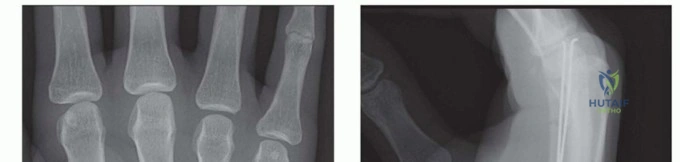
We are discussing the biomechanical evolution of long bone fracture fixation. Consider the two primary methods of load transmission: Intramedullary Nails (IMN) and External Fixation (ExFix). Briefly define the mechanical differences in how these devices handle axial loading and describe the concept of "load-sharing" versus "load-bearing".
Candidate: IMNs are internal devices that sit in the medullary canal, providing load-sharing, whereas external fixators are placed outside the body and generally act as a load-bearing frame.
A poor answer fails to define the "why." Candidates often say "it shares the load" without explaining that the IMN is positioned close to the neutral axis of the bone, which reduces the bending moment. Furthermore, they fail to mention that "load-bearing" devices (like ExFix or bridging plates) often risk stress shielding and non-union if they bypass the fracture site entirely without biological stimulation.
The perfect answer classifies the constructs by their proximity to the neutral axis: 1. IMN (Load-Sharing): Situated centrally (near the neutral axis), allowing the bone-implant construct to share axial forces, which reduces the bending moment and promotes secondary bone healing through physiologic loading and controlled micromotion. 2. ExFix (Load-Bearing): Situated eccentrically (outside the bone envelope). The device must bear the entire mechanical load, protecting the fracture. While excellent for initial stabilization (Damage Control Orthopedics), excessive rigidity without functional loading can lead to stress shielding and delayed union. Mentioning Wolff’s Law in the context of load-sharing adds significant depth.
You have a 35-year-old polytrauma patient with an open tibial shaft fracture. You decide to utilize a temporary external fixator as part of Damage Control Orthopedics. What specific design principles must you employ to maximize the frame's rigidity?
Candidate: I would use a multiplanar frame, increase the number of pins, and make sure the pins are spread far apart from the fracture site. I would also place the bars as close to the skin as possible.
The candidate lists factors but fails to explain their mechanical impact. A failure is neglecting the "frame-to-bone distance"—a critical concept where increased distance exponentially decreases stiffness—or failing to mention pin diameter, which has a cubic relationship with stiffness.
To maximize frame rigidity, I focus on the following: 1. Pin Diameter: Stiffness increases to the 4th power of the radius. 2. Frame Geometry: Multiplanar frames are significantly stiffer than uniplanar constructs. 3. Pin Spread: Increasing the distance between pins in the same fragment improves stability. 4. Frame-to-Bone Distance: Minimizing this distance is crucial; the further the bar is from the bone, the greater the bending moment and the less rigid the construct. 5. Connecting Rods: Using stiffer materials (like stainless steel vs carbon fiber) and shortening the length of the connecting rods increases overall construct stiffness.
Regarding the intramedullary nail, explain the difference between static and dynamic locking, and indicate when you might convert a static nail to a dynamic one.
Candidate: Static locking means screws are placed in both proximal and distal fragments, locking the length. Dynamic locking means removing a screw to allow the bone to shorten or move slightly. I do this if the fracture isn't healing.
The candidate lacks clinical precision. Simply saying "if it isn't healing" is too vague. They must distinguish between "delayed union" and "non-union" and recognize that dynamic locking is contraindicated in unstable fracture patterns (like comminuted fractures) where shortening would be catastrophic.
Static Locking: Uses screws in both fragments to control length, rotation, and alignment. This is mandatory for unstable, comminuted, or segmental fractures.
Dynamic Locking: Involves removing a proximal or distal locking screw to allow axial micromotion and load transfer across the fracture site, stimulating callus formation.
Conversion: I would only consider dynamization in a stable (transverse) fracture pattern that shows signs of delayed union (e.g., at 6-8 weeks) where there is evidence of callus but insufficient bridging. It must NEVER be performed in unstable, comminuted patterns as it risks uncontrollable shortening or malalignment.
Detailed Chapters & Topics
Dive deeper into specialized chapters regarding nails-and-external-fixators