Knee Dislocation: Epidemiology, Anatomy, and Initial Management
Key Takeaway
Knee dislocation (KD) is a severe, often limb-threatening injury involving tibiofemoral articulation disruption. Characterized by multi-ligamentous damage, it poses a high risk of neurovascular compromise. Initial management prioritizes limb salvage through prompt recognition, neurovascular assessment, and immediate reduction, addressing critical structures like the popliteal artery and common peroneal nerve.
A 28-year-old male presents to the Emergency Department following a high-energy motor vehicle accident. He has a grossly deformed knee. You are the registrar on call. What is your immediate priority, and how do you systematically conduct your primary assessment?
Candidate: My absolute priority is to rule out limb-threatening injuries, specifically vascular compromise. I would perform an ATLS primary survey. For the knee, I would perform a rapid neurovascular exam, check the Ankle-Brachial Index (ABI), and proceed to immediate closed reduction under sedation, followed by re-assessment of neurovascular status and immobilization in a splint.
Candidates often forget to mention the "ABI" specifically or fail to mention the need for an emergency reduction before obtaining formal imaging. Some also neglect to mention checking for compartment syndrome post-reduction, which is a critical oversight.
Start by prioritizing the "Limb-at-Risk." Structure the answer: 1. ATLS protocols for systemic stability. 2. Neurovascular assessment (documenting pulses and common peroneal/tibial nerve function). 3. Immediate ABI calculation (noting < 0.9 mandates further investigation like CTA). 4. Urgent closed reduction (emphasizing immediate restoration of limb alignment to decompress the popliteal artery). 5. Re-check neurovascular status and apply a protective splint in slight flexion.
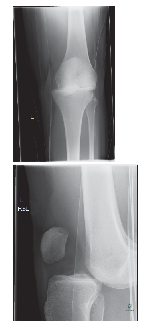
The patient has been reduced. Imaging is performed. Please describe the injury seen in this radiograph and how it influences your management strategy.
Candidate: The radiograph shows a multi-ligamentous knee injury pattern. I would use the Schenck classification to grade the instability. This requires MRI to define the specific ligaments involved (ACL, PCL, PLC, or MCL). Management will be surgical reconstruction, likely involving allografts, and the timing will depend on the resolution of soft tissue swelling.
Failing to emphasize the Posterolateral Corner (PLC). Candidates often focus only on the cruciate ligaments, missing the fact that if the PLC is ignored, the ACL/PCL reconstruction will fail.
Structure by: 1. Classification (Schenck system). 2. Diagnostic steps (MRI is essential for pre-operative planning). 3. Surgical decision-making (Address the PCL first, then ACL, then collateral/PLC structures). 4. Mentioning that the PLC is a critical stabilizer for rotational instability and must be reconstructed anatomically to prevent failure of the cruciate reconstructions.
Discuss the complications of a high-energy knee dislocation. Which one has the highest impact on potential limb salvage and why?
Candidate: The most critical complication is popliteal artery injury, as it directly threatens limb viability. Other complications include common peroneal nerve palsy, stiffness/arthrofibrosis, and post-traumatic arthritis. Vascular injury has the highest impact because delay in diagnosis beyond 8 hours exponentially increases the amputation rate.
Listing complications but failing to provide the "Why" (e.g., the anatomical tethering of the popliteal artery at the adductor hiatus and soleal arch) or forgetting to mention the timeline for nerve recovery.
Categorize into Vascular, Neurological, and Functional complications. Explain the anatomical basis for the vascular injury (tethering points). State clearly that popliteal artery injury carries a high amputation risk if not addressed within 8 hours. Mention that nerve injury (Common Peroneal) is often a stretch injury and requires patient counseling regarding the 3-6 month window for potential recovery.
