Osteomyelitis of the Metacarpals and Phalanges: Principles of Diagnosis and Surgical Management
Key Takeaway
Osteomyelitis of the metacarpals and phalanges presents a complex reconstructive challenge, often arising from contiguous soft tissue infections, open fractures, or systemic immunocompromise. Successful management demands aggressive surgical debridement, targeted antimicrobial therapy, and early mobilization to prevent debilitating stiffness. This guide details the diagnostic algorithms, surgical approaches for joint drainage, and definitive management strategies, including sequestrectomy and amputation, to optimize functional outcomes in the infected hand.
Comprehensive Introduction and Patho-Epidemiology
Osteomyelitis of the metacarpals and phalanges represents a catastrophic, potentially limb-threatening, and highly function-threatening condition that demands aggressive medical and surgical intervention. Unlike long bone osteomyelitis, where the robust soft tissue envelope provides a forgiving buffer, infections in the hand are uniquely challenging due to the intimate proximity of osseous structures to critical soft tissues. The flexor and extensor tendons, delicate neurovascular bundles, and complex articular capsules are separated from the periosteum by mere millimeters. Consequently, the primary goal of treatment transcends the mere eradication of infection; the orthopedic surgeon must prioritize the preservation of a functional, sensate, and mobile digit, a task that frequently tests the limits of surgical salvage.
In the adult hand, osteomyelitis is rarely an isolated or hematogenous event. It is overwhelmingly the sequela of contiguous spread from neighboring soft tissue infections, such as neglected felons, suppurative flexor tenosynovitis, or deep fascial space infections. Furthermore, high-energy trauma resulting in open fractures, traumatic amputations, or the open surgical treatment of closed fractures with internal fixation introduces direct inoculation pathways. Systemic factors play an undeniable and critical role in the patho-epidemiology; patients with peripheral vascular disease, poorly controlled diabetes mellitus, end-stage renal disease, and immunodeficiency states are at a markedly increased risk for both the development of osteomyelitis and the failure of subsequent eradication efforts.
The microbiological profile of hand osteomyelitis is heavily influenced by the mechanism of injury and the patient's host status. Staphylococcus aureus remains the most commonly isolated organism across all etiologies, encompassing both methicillin-susceptible (MSSA) and methicillin-resistant (MRSA) strains. In cases of contiguous spread from "diabetic hand syndrome" or in patients with severe peripheral vascular disease, polymicrobial infections involving Gram-negative rods (such as Pseudomonas aeruginosa) and obligate anaerobes are frequently encountered. Bite wounds introduce unique flora; human bites (clenched fist injuries) frequently inoculate Eikenella corrodens, while feline or canine bites introduce Pasteurella multocida, both of which can rapidly progress to fulminant osteomyelitis if the joint capsule or periosteum is breached.
The pathophysiologic cascade of bone destruction begins with the introduction of these pathogens into the osseous architecture, inciting a profound acute inflammatory response. The rigid, unyielding nature of the cortical bone dictates that inflammatory exudate rapidly increases intramedullary pressure. This pressure mechanically occludes the delicate intraosseous microcirculation, leading to localized ischemia, hypoxia, and subsequent bone necrosis. If the process lingers without adequate surgical decompression, the necrotic bone separates from viable surrounding tissue, forming a sequestrum. Simultaneously, the elevated periosteum attempts to wall off the infection by laying down new, reactive bone known as an involucrum. The presence of a sequestrum is the definitive hallmark of chronic osteomyelitis and represents an avascular nidus of infection that is entirely impervious to systemic antibiotics, mandating radical surgical extirpation.
Detailed Surgical Anatomy and Biomechanics
A profound understanding of the complex osteology and vascular supply of the metacarpals and phalanges is a prerequisite for the surgical management of hand osteomyelitis. The metacarpals and phalanges are miniature long bones, possessing a diaphysis, metaphysis, and epiphysis. The nutrient arteries to the metacarpals generally enter the volar aspect of the diaphysis and are directed distally, except for the first metacarpal, where the nutrient artery is directed proximally. The phalanges receive their primary osseous blood supply from the digital arteries via branches that enter the volar and lateral aspects of the metaphyses. This delicate vascular network is easily compromised by intramedullary edema, periosteal stripping during trauma, or iatrogenic injury during surgical exposure, accelerating the progression of ischemic bone necrosis.
The soft tissue envelope of the digits is extraordinarily thin and highly specialized, leaving the underlying bone vulnerable to direct inoculation from seemingly minor lacerations or puncture wounds. Dorsally, the extensor mechanism lies directly over the periosteum of the phalanges, with virtually no intervening subcutaneous fat. Volarly, the flexor tendons glide within a rigid fibro-osseous sheath that is intimately attached to the volar periosteum. This anatomical arrangement explains why suppurative flexor tenosynovitis can rapidly destroy the underlying volar cortical bone if not emergently decompressed. Furthermore, the digital neurovascular bundles are tethered by Cleland’s ligaments (dorsal to the bundle) and Grayson’s ligaments (volar to the bundle), which must be carefully navigated and preserved during any midaxial or volar surgical approach to the infected bone.
The articular anatomy of the metacarpophalangeal (MCP) and interphalangeal (IP) joints facilitates the rapid contiguous spread of infection. The joint capsules are reinforced laterally by the proper collateral ligaments and the accessory collateral ligaments. The accessory collateral ligaments insert volarly into the thick, fibrocartilaginous volar plate. Infections originating in the flexor tendon sheath can easily rupture through the thin proximal membranous portion of the volar plate, directly entering the joint space and subsequently invading the intra-articular epiphyseal bone. Conversely, primary septic arthritis can rapidly destroy the articular cartilage and penetrate the subchondral bone, resulting in epiphyseal osteomyelitis that compromises the mechanical integrity of the joint.
Specialized anatomical consideration must be given to the distal phalanx and the digital pulp. The distal pulp is divided into multiple enclosed compartments by dense fibrous septa connecting the volar skin to the periosteum of the distal phalanx. When an abscess (felon) forms in this closed space, the pressure rises exponentially. Because the diaphysis of the distal phalanx receives its blood supply from vessels traversing these rigid septa, the increased pressure rapidly leads to ischemic necrosis and subsequent osteomyelitis of the diaphyseal tuft. Notably, in the pediatric population, the epiphysis of the distal phalanx receives a separate, proximal blood supply that does not traverse the pulp space. Therefore, even in the presence of severe diaphyseal erosion from a felon, the pediatric epiphysis often survives, possessing a remarkable capacity to regenerate once the soft tissue abscess is adequately decompressed.
Exhaustive Indications and Contraindications
The decision-making algorithm for the management of metacarpal and phalangeal osteomyelitis hinges on differentiating between acute, subacute, and chronic phases of the disease, as well as assessing the patient's overall physiologic reserve. Operative intervention is the cornerstone of treatment for the vast majority of cases, as the unique anatomy of the hand rarely allows for the successful eradication of established bone infection through systemic antimicrobial therapy alone. The threshold for surgical intervention must remain low to prevent the irreversible destruction of articular surfaces, the necrosis of critical tendon excursion pathways, and the systemic dissemination of the pathogen.
Absolute indications for surgical intervention include the presence of a macroscopic soft tissue abscess, radiographic evidence of a sequestrum, the presence of infected orthopedic hardware, or the failure of acute osteomyelitis to respond to targeted intravenous antibiotics within 24 to 48 hours. Furthermore, if the pathogen cannot be reliably isolated via closed needle aspiration, open surgical biopsy and debridement become mandatory to obtain accurate deep tissue cultures. Empiric antibiotic therapy without a definitive microbiological diagnosis in the setting of subacute or chronic osteomyelitis is condemned, as it frequently leads to the selection of highly resistant organisms and the masking of ongoing osseous destruction.
Relative indications and contraindications require a nuanced assessment of the patient's functional demands and systemic health. In patients with profound peripheral vascular disease or end-stage diabetic microangiopathy, the vascular inflow may be insufficient to support the healing of a radical debridement and subsequent reconstructive efforts. In such scenarios, extensive salvage procedures are relatively contraindicated, as they are likely to result in protracted morbidity, chronic non-healing wounds, and eventual amputation under less favorable conditions. Similarly, severe systemic illness, profound immunosuppression, or documented patient non-compliance with complex postoperative rehabilitation protocols may push the surgeon toward an early, definitive amputation rather than a multi-staged salvage attempt.
The most challenging paradigm in hand osteomyelitis is the decision between digit salvage and amputation. While modern reconstructive techniques, including diaphysectomy, antibiotic spacers, and secondary bone grafting, make the anatomical salvage of almost any digit technically feasible, the functional outcome is frequently poor. A stiff, insensate, and painful finger acts as a mechanical obstruction, severely degrading the global function of the hand. Therefore, a non-salvageable digit—defined functionally rather than purely anatomically—is a strong indication for ray amputation. A well-executed amputation followed by early, aggressive rehabilitation often yields a vastly superior functional outcome compared to a prolonged, multi-staged salvage attempt that results in a stiff, non-functional ray.
Table of Indications and Contraindications
| Category | Operative Salvage (Debridement/Reconstruction) | Amputation (Ray or Transphalangeal) |
|---|---|---|
| Absolute Indications | Acute osteomyelitis failing 48h IV antibiotics; Presence of sequestrum/involucrum; Infected hardware; Drainable intraosseous abscess. | Irreversible ischemia/gangrene of the digit; Life-threatening sepsis originating from the digit; Unresectable malignant transformation (Marjolin's ulcer). |
| Relative Indications | Localized chronic osteomyelitis with preserved neurovascular bundles; Compliant patient; Intact flexor/extensor mechanisms. | Severe stiffness of the affected digit precluding function; Multilevel joint destruction; Loss of protective sensation; Intractable pain. |
| Absolute Contraindications | Non-salvageable vascular supply (e.g., failed revascularization); Patient medically unfit for anesthesia. | Salvageable thumb with intact basal joint function (thumb length must be preserved at all costs); Patient refusal. |
| Relative Contraindications | Severe peripheral vascular disease; End-stage renal disease; Severe malnutrition (albumin < 2.5 g/dL); Active IV drug abuse. | Single-digit involvement in a high-demand manual laborer where early return to work is paramount (salvage may take months). |
Pre-Operative Planning, Templating, and Patient Positioning
Thorough pre-operative planning is essential to navigate the complex anatomical constraints of the hand and to anticipate the requirement for dead space management and skeletal stabilization. The clinical evaluation must be supplemented by a rigorous imaging protocol. Standard posteroanterior, lateral, and oblique radiographs are the first-line modality, though early acute osteomyelitis may show no abnormalities for the first 10 to 14 days. Subsequent findings include focal osteopenia, periosteal reaction, and eventually, cortical destruction. When radiographs are equivocal or when evaluating the extent of marrow edema and contiguous soft tissue involvement, Magnetic Resonance Imaging (MRI) with gadolinium contrast is the modern gold standard. MRI precisely delineates the boundary between necrotic and viable bone, guiding the extent of the planned surgical resection.
Microbiological planning is equally critical. The surgeon must coordinate with infectious disease specialists and the microbiology laboratory prior to the procedure. Superficial swab cultures of draining sinuses are notoriously unreliable, frequently growing colonizing skin flora (such as coagulase-negative staphylococci or diphtheroids) rather than the true osseous pathogen. Deep tissue biopsies and bone aspirates obtained during the operative debridement are mandatory. Empiric antibiotics should be strictly withheld until these deep cultures are obtained, unless the patient is exhibiting signs of systemic sepsis or hemodynamic instability.
Surgical templating involves mapping out the anticipated incisions, the extent of bone resection, and the strategy for dead space management. The surgeon must decide between a midaxial approach, which safely avoids the volar neurovascular bundles and the flexor apparatus, or a volar zig-zag (Bruner) incision if concomitant flexor tenosynovitis requires decompression. If a segmental diaphysectomy is anticipated, the surgeon must be prepared to implement the Masquelet technique. This requires pre-operative templating of the skeletal defect to ensure adequate polymethyl methacrylate (PMMA) cement is available, along with appropriate heat-stable antibiotics (typically tobramycin, vancomycin, or gentamicin) for intra-operative mixing. Furthermore, the method of skeletal stabilization—whether rigid Kirschner wires, miniature plates, or a mini-external fixator—must be selected and available in the surgical suite.
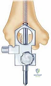
Patient positioning and tourniquet management require specific attention to prevent the iatrogenic proximal spread of infection. The patient is typically positioned supine with the affected extremity extended on a radiolucent hand table. Regional anesthesia (such as an axillary or supraclavicular brachial plexus block) is preferred, as it provides excellent intra-operative anesthesia and prolonged post-operative sympathectomy, which enhances distal perfusion. Crucially, the extremity must not be exsanguinated with an Esmarch bandage, as the mechanical compression can force purulent material proximally into uninvolved fascial spaces or the systemic circulation. Instead, the arm is simply elevated for 3 to 5 minutes to allow for gravity exsanguination before the pneumatic tourniquet is inflated to 250 mmHg.
Step-by-Step Surgical Approach and Fixation Technique
The surgical management of metacarpal and phalangeal osteomyelitis demands meticulous technique to eradicate the infection while preserving the delicate surrounding soft tissue envelope. When non-operative measures fail, or in the presence of a sequestrum, open drainage of pus and radical debridement of necrotic material are imperative. The procedure begins with the establishment of a safe and extensible surgical exposure. For infections of the phalanges, the midaxial approach is the workhorse incision, providing excellent access to the diaphysis and adjacent joints while minimizing the risk of flexor tendon adhesions and neurovascular injury.
The Midaxial Surgical Approach
The incision is planned on either the radial or ulnar side of the involved digit. The midaxial line is identified by connecting the apices of the flexion creases when the digit is fully flexed. An incision along this line places the surgical approach dorsal to the neurovascular bundle. The surgeon carefully dissects through the subcutaneous tissues, identifying the neurovascular bundle and gently retracting it volarly. To access the underlying bone and joint capsule, the transverse retinacular ligament is identified and sectioned. This critical step allows the lateral band of the extensor mechanism to be mobilized and retracted dorsally, creating a wide, safe window to the periosteum and the lateral aspect of the joint capsule.
Radical Debridement and Sequestrectomy
Once the bone is exposed, the periosteum is incised and elevated only as much as necessary to expose the infected area, preserving the vascular supply to the remaining viable bone. The core principle of chronic osteomyelitis surgery is radical resection: all infected, necrotic, and avascular bone must be excised. This process, known as sequestrectomy or diaphysectomy, is performed using a combination of rongeurs, curettes, and high-speed burrs under constant saline irrigation. The surgeon must resect bone until punctate cortical and cancellous bleeding is observed—the classic "paprika sign." This sign confirms that the remaining osseous bed is viable, well-vascularized, and capable of mounting an immune response and integrating a future bone graft. Multiple deep tissue and bone specimens are sent for aerobic, anaerobic, mycobacterial, and fungal cultures.
Dead Space Management and the Masquelet Technique
Following radical resection, a significant osseous void is created. This dead space must be meticulously managed to prevent hematoma formation, which serves as an ideal culture medium for recurrent infection. The standard of care involves the insertion of an antibiotic-impregnated polymethyl methacrylate (PMMA) cement spacer. The PMMA is mixed intra-operatively with heat-stable antibiotics (e.g., 2 to 4 grams of vancomycin and 3.6 grams of tobramycin per 40-gram bag of cement). The cement is molded into a cylinder that matches the dimensions of the resected diaphysis and is inserted into the defect while still in the doughy phase. This spacer delivers a massive, localized concentration of antibiotics directly to the infected bed without systemic toxicity. Over the subsequent 6 to 8 weeks, the spacer induces the formation of a biologically active pseudosynovial membrane (the first stage of the Masquelet technique), which secretes osteoinductive factors essential for the second stage of reconstruction.
Skeletal Stabilization
The resected digit must be rigidly stabilized to maintain length, alignment, and rotation, and to prevent soft tissue contracture during the eradication phase. Depending on the extent of bone loss and the quality of the remaining soft tissues, stabilization is achieved using either longitudinal Kirschner wires driven across the adjacent joints, or a mini-external fixator applied dorsally or midaxially. External fixation is particularly advantageous when there is significant soft tissue loss, as it allows for daily wound care and avoids placing hardware directly through the infected bed. The surgical wounds are copiously irrigated. If the infection was acute and the debridement is deemed completely clean, loose primary closure may be attempted. However, in chronic or grossly purulent cases, the wounds are left open to heal by secondary intention or to be closed secondarily once the infection is definitively controlled.
Complications, Incidence Rates, and Salvage Management
The surgical treatment of hand osteomyelitis is fraught with complications, stemming both from the destructive nature of the disease and the iatrogenic trauma of radical surgical intervention. The surgeon must counsel the patient extensively regarding these risks, setting realistic expectations for the final functional outcome. The most ubiquitous and debilitating complication is severe, intractable stiffness of the involved digit, which frequently extends to involve the entire hand. This stiffness is multifactorial, resulting from prolonged immobilization, severe edema, the formation of dense tendon adhesions within the fibro-osseous canals, and the development of Complex Regional Pain Syndrome (CRPS) or sympathetic dystrophy.
Recurrent or persistent infection is another major complication, particularly in cases of chronic osteomyelitis where biofilms have formed on necrotic bone or retained hardware. Inadequate initial debridement, failure to recognize contiguous joint involvement, or the selection of inappropriate systemic antibiotics based on superficial cultures are the primary drivers of recurrence. When infection persists despite radical diaphysectomy and antibiotic spacer placement, the surgeon must suspect a compromised host immune system or an unrecognized area of avascular tissue. Non-union following the second-stage bone grafting procedure is also a significant risk, particularly if the induced pseudosynovial membrane was damaged during spacer removal or if the patient continues to smoke, which severely compromises microvascular ingrowth.
Iatrogenic injuries during the initial debridement can catastrophically alter the salvage potential of the digit. Inadvertent laceration of the digital neurovascular bundles during a midaxial approach results in a dysvascular or insensate digit, which is highly prone to recurrent ulceration and infection. Similarly, excessive disruption of the extensor mechanism or the volar plate can lead to severe boutonnière or swan neck deformities, rendering the digit mechanically useless even if the infection is successfully eradicated.
When complications prove insurmountable, or when a salvaged digit remains stiff, painful, and non-functional, salvage management pivots to amputation. A stiff, insensate finger acts as a mechanical post that obstructs the function of the adjacent normal digits, severely degrading overall grip strength and dexterity. Ray amputation—the removal of the digit and its corresponding metacarpal—is frequently the most functional salvage procedure. By removing the metacarpal, the adjacent digits can approximate, closing the web space and restoring a functional, albeit narrower, grasp. In cases involving the thumb, however, length must be preserved at all costs; arthrodesis of the MCP or IP joints, combined with advanced soft tissue coverage (e.g., neurovascular island flaps), is preferred over amputation whenever technically feasible.
Table: Complications, Incidence Rates, and Management Strategies
| Complication | Estimated Incidence | Primary Etiology | Management / Salvage Strategy |
|---|---|---|---|
| Intractable Stiffness | 40% - 60% | Edema, tendon adhesions, prolonged immobilization. | Aggressive hand therapy, tenolysis (post-eradication), amputation if functionally obstructive. |
| Recurrent Infection | 15% - 25% | Retained sequestrum, biofilm, inadequate debridement. | Repeat radical debridement, exchange of PMMA spacer, modification of IV antibiotics. |
| Bone Graft Non-union | 10% - 20% | Poor vascular bed, smoking, premature hardware removal. | Revision bone grafting (iliac crest), rigid internal fixation, use of bone morphogenetic proteins (BMPs). |
| CRPS / Sympathetic Dystrophy | 5% - 10% | Altered central/peripheral pain processing post-trauma. | Stellate ganglion blocks, gabapentinoids, aggressive desensitization therapy. |
| Iatrogenic Nerve Injury | < 5% | Poor surgical exposure, altered anatomy from severe edema. | Microsurgical nerve repair/grafting if clean; otherwise, terminal neuroma management or amputation. |
Phased Post-Operative Rehabilitation Protocols
The post-operative protocol following the surgical management of hand osteomyelitis is as critical to the final functional outcome as the surgical intervention itself. A perfectly executed debridement and reconstruction will inevitably result in a stiff, useless hand if the post-operative rehabilitation is neglected. The rehabilitation process is divided into distinct, progressive phases, requiring close collaboration between the orthopedic surgeon, the infectious disease specialist, and a certified hand therapist.
Phase I encompasses the immediate post-operative period (Days 1 to 7). The primary objective during this phase is the aggressive mitigation of edema, which is the primary driver of secondary stiffness and tendon adhesions. The hand is strictly elevated above the level of the heart continuously. If the wounds were left open for drainage, bulky, non-adherent dressings are applied and changed daily or twice daily. Hydrotherapy, utilizing exercise periods in a sterile whirlpool bath, is highly recommended for open wounds. This provides gentle mechanical debridement of fibrinous exudate, promotes local microvascular blood flow, and facilitates the very earliest stages of passive motion. The hand must be splinted in the intrinsic-plus position (the "position of safety") between therapy sessions: the wrist extended 20-30 degrees, the MCP joints flexed 70-90 degrees, and the IP joints in full extension to prevent collateral ligament contracture.
Phase II marks the initiation of early mobilization and soft tissue healing (Weeks 2 to 6). As the acute inflammatory phase subsides and the infection is controlled by local and systemic antibiotics, active and active-assisted range-of-motion exercises are instituted for all uninvolved digits, and carefully introduced to the involved digit if skeletal stability permits (e.g., if an external fixator is spanning the defect but leaving adjacent joints free). The goal is to maintain tendon glide and prevent the organization of fibrinous exudate into dense, unyielding scar tissue. If the wound bed demonstrates healthy, beefy-red granulation tissue without purulence, it may be closed secondarily, or covered with a split-thickness skin graft.
Phase III involves strengthening and preparation for secondary reconstruction (Weeks 6 to 12). For patients undergoing single-stage treatment (e.g., simple debridement without massive bone loss), progressive resistance exercises and functional task training are initiated. For patients managed with a PMMA spacer (the Masquelet technique), this phase culminates in the return to the operating room. Once systemic inflammatory markers (CRP, ESR) have normalized and clinical signs of infection are absent, the spacer is removed, carefully preserving the induced pseudosynovial membrane, and the defect is packed with autologous cancellous bone graft.
Phase IV is the post-reconstruction integration phase (Months 3 to 6+). Following bone grafting, the digit is protected until radiographic evidence of graft consolidation is observed. Rehabilitation then focuses on maximizing the range of motion of the reconstructed digit, managing scar tissue through massage and silicone gel sheeting, and integrating the digit into global hand function. The patient must be counseled that maximal medical improvement may take up to a year, and that some degree of permanent impairment is the rule rather than the exception.
Summary of Landmark Literature and Clinical Guidelines
The evolution of the surgical management of metacarpal and phalangeal osteomyelitis has been profoundly shaped by several landmark concepts and clinical guidelines. Historically, the treatment of chronic hand osteomyelitis was heavily biased toward early amputation due to the unacceptably high rates of recurrent infection and the dismal functional outcomes of early salvage attempts. However, the adaptation of long-bone osteomyelitis principles to the miniature skeleton of the hand has revolutionized limb salvage capabilities.
A pivotal advancement in the literature is the adaptation of the Cierny-Mader classification system to the hand. While originally designed for long bones, its principles—categorizing osteomyelitis not just by anatomy (medullary, superficial, localized, diffuse) but critically by the physiologic status of the host (A, B, or C hosts)—have become the bedrock of clinical guidelines. Literature consistently demonstrates that attempting complex, multi-staged salvage in a "C-host" (a patient with profound systemic or local compromise, such as severe diabetic microangiopathy) results in complication rates approaching 100%, strongly guiding modern surgeons toward early amputation in this specific demographic.
The application of the Masquelet technique (induced membrane technique) to the small bones of the hand represents another landmark shift in the literature. Originally described by Alain Masquelet for massive diaphyseal defects in the tibia and femur, numerous recent case series and retrospective reviews have validated its efficacy in the metacarpals and phalanges. The literature confirms that the PMMA spacer not only provides high local concentrations of antibiotics but also induces a membrane rich in vascular endothelial growth factor (VEGF) and bone morphogenetic protein-2 (BMP-2). This membrane prevents the resorption of the subsequent cancellous bone graft and prevents soft tissue interposition, leading to union rates exceeding 85% in salvaged digits.
Pharmacokinetic studies regarding antibiotic elution from PMMA cement have also generated critical clinical guidelines. Landmark in vitro and in vivo studies have established that the elution profile of antibiotics from cement is highly dependent on the surface-area-to-volume ratio and the specific antibiotics used. Guidelines now strictly mandate the use of heat-stable antibiotics in powder form (liquid antibiotics severely compromise the mechanical integrity and curing process of the cement). Tobramycin and vancomycin remain the gold standard combination, providing broad-spectrum coverage against the most common hand pathogens, including MRSA and Pseudomonas, with elution levels remaining well above the minimum inhibitory concentration (MIC) for up to 6 weeks locally, without elevating systemic serum levels.
Finally, comparative outcome literature evaluating amputation versus salvage continues to guide surgical philosophy. Long-term functional studies utilizing metrics such as the Disabilities of the Arm, Shoulder and Hand (DASH) questionnaire consistently reveal a paradox: patients who undergo early, definitive ray amputation for severe, destructive osteomyelitis often report higher satisfaction and better global hand function scores at two years compared to patients who undergo successful, but prolonged, anatomical salvage of a stiff digit. These landmark findings underscore the fundamental principle of hand surgery: the ultimate goal is not the preservation of anatomy, but the restoration of function.
