Operative Management of Complex Hand Infections: A Master Surgical Guide
Key Takeaway
Complex hand infections demand swift, aggressive surgical intervention to prevent catastrophic functional loss. This comprehensive guide details the evidence-based operative management of pyogenic flexor tenosynovitis, necrotizing fasciitis, deep space abscesses, and atypical mycobacterial infections. It provides orthopedic surgeons with step-by-step surgical approaches, biomechanical considerations, and postoperative rehabilitation protocols to optimize patient outcomes and preserve hand mechanics.
Comprehensive Introduction and Patho-Epidemiology
The human hand is an intricate biomechanical marvel, characterized by a dense concentration of specialized tissues—tendons, neurovascular bundles, and synovial sheaths—confined within tight, unyielding fascial compartments. When bacterial, fungal, or atypical pathogens breach these compartments through trauma, hematogenous spread, or contiguous extension, the resulting inflammatory cascade can rapidly lead to ischemia, tissue necrosis, and irreversible functional impairment. The seminal literature, ranging from Allen B. Kanavel’s foundational anatomical studies in the early 20th century to contemporary analyses by Gonzalez, Jebson, and others, underscores a singular, unifying surgical principle: time is tissue. The operative management of complex hand infections requires a profound understanding of compartmental anatomy, precise surgical execution, and aggressive, protocol-driven postoperative rehabilitation.
The patho-epidemiology of hand infections is deeply intertwined with patient demographics, occupational hazards, and systemic comorbidities. In the acute care setting, hand infections account for a significant proportion of surgical emergencies, with Staphylococcus aureus (particularly Methicillin-Resistant S. aureus, MRSA) and Streptococcus species representing the most commonly isolated organisms. However, the microbiological landscape is highly variable. Clenched-fist injuries (human bites) frequently yield polymicrobial flora, including Eikenella corrodens, while animal bites are classically associated with Pasteurella multocida. Furthermore, the rising prevalence of immunocompromising conditions—such as poorly controlled diabetes mellitus, end-stage renal disease, human immunodeficiency virus (HIV), and iatrogenic immunosuppression for rheumatologic conditions—has dramatically altered the clinical presentation of these infections. In such hosts, the classic cardinal signs of inflammation may be dangerously blunted, leading to delayed presentation and catastrophic outcomes.
At the cellular and microvascular level, the pathophysiology of closed-space hand infections is driven by the rapid accumulation of purulent exudate within non-distensible anatomical boundaries. In conditions such as pyogenic flexor tenosynovitis (PFT), the intrasynovial pressure quickly exceeds capillary perfusion pressure. This mechanical tamponade obliterates the delicate vincular blood supply to the flexor tendons. Concurrently, the release of bacterial exotoxins and host proteolytic enzymes (e.g., matrix metalloproteinases) degrades the collagenous architecture of the tendon and its gliding surface. If surgical decompression and targeted antimicrobial therapy are not instituted emergently, the tendon undergoes liquefactive necrosis, resulting in spontaneous rupture, profound stiffness, and ultimate loss of digital function.
Understanding this rapid progression is paramount for the orthopedic surgeon. The transition from a localized cellulitis to a fulminant necrotizing soft-tissue infection (NSTI) or deep space abscess can occur within hours. Therefore, the threshold for operative intervention must remain appropriately low. The surgeon must be prepared to execute a spectrum of procedures, ranging from minimally invasive closed catheter irrigation to radical debridement, fasciotomy, and, in life-threatening scenarios, proximal amputation. This masterclass delineates the evidence-based surgical protocols for managing the most challenging and limb-threatening hand infections encountered in clinical practice.
Detailed Surgical Anatomy and Biomechanics
A masterful approach to hand infections demands an encyclopedic knowledge of its complex spatial anatomy. The hand is compartmentalized by a series of fascial septa that dictate the predictable pathways of purulent dissemination. The flexor tendon sheaths are closed, double-walled synovial cylinders that facilitate frictionless tendon excursion. Anatomically, these sheaths extend from the metacarpal neck (just proximal to the A1 pulley) to their distal insertion at the base of the distal phalanx. The structural integrity of the sheath is reinforced by a highly specialized retinacular system comprising five annular (A1-A5) and three cruciform (C1-C3) pulleys. The A2 and A4 pulleys, located over the proximal and middle phalanges respectively, are the critical biomechanical fulcrums. Excision or iatrogenic destruction of these pulleys during surgical debridement results in severe bowstringing, drastically reducing the mechanical advantage, excursion, and active flexion of the digit.
The synovial sheaths of the thumb and small finger exhibit unique anatomical extensions that hold profound clinical implications. The flexor pollicis longus (FPL) sheath continues proximally through the carpal tunnel to form the radial bursa. Similarly, the flexor sheath of the small finger expands proximally to form the ulnar bursa. In approximately 50% to 80% of individuals, the radial and ulnar bursae communicate within the carpal tunnel. Consequently, an infection originating in the small finger can track proximally into the ulnar bursa, cross the wrist, and aggressively descend into the radial bursa of the thumb—a catastrophic presentation classically termed a "horseshoe abscess." Furthermore, the potential space situated deep to the flexor tendons and volar to the pronator quadratus in the distal forearm—known as Parona's space—serves as a proximal reservoir for purulence escaping the bursae, necessitating deep forearm exploration.
Beyond the synovial sheaths, the hand contains discrete deep fascial spaces that can harbor massive, occult purulence. The thenar space is located volar to the adductor pollicis muscle and dorsal to the flexor tendons of the index finger; its ulnar border is defined by the oblique septum attaching to the third metacarpal. Infection within the thenar space forces the thumb into a characteristic abducted and extended posture due to the massive accumulation of pus in the first web space. The midpalmar space lies deep to the flexor tendons of the middle, ring, and small fingers, resting volar to the interosseous fascia and the second and third volar interossei. The hypothenar space, encompassing the hypothenar musculature, is tightly bound by the hypothenar fascia and rarely communicates with the other spaces.
Biomechanically, the presence of infection within these spaces drastically alters the kinematics of the hand. The accumulation of fluid increases the resting tension of the intrinsic musculature and the extrinsic flexors. The collateral ligaments of the metacarpophalangeal (MCP) joints, which are uniquely eccentric, become lax when the joint is extended. If an infected hand is allowed to rest in a position of comfort (typically wrist flexion, MCP extension, and interphalangeal flexion), the collateral ligaments will rapidly contract and fibrose. This biomechanical reality dictates not only the urgency of surgical decompression but also the absolute necessity of postoperative splinting in the "Intrinsic Plus" or safe position, which maintains the MCP collateral ligaments at their maximal length.
Exhaustive Indications and Contraindications
The decision to proceed with operative intervention in hand infections relies on a synthesis of clinical examination, chronicity, host immune status, and the specific anatomical compartment involved. Non-operative management—consisting of elevation, immobilization, and intravenous antibiotics—is strictly reserved for early, uncomplicated cellulitis or superficial paronychia without fluctuance. However, once an infection breaches a closed anatomical space, surgical decompression becomes the definitive standard of care.
For Pyogenic Flexor Tenosynovitis (PFT), the Michon classification system guides surgical decision-making. Michon Stage I (increased serous exudate, visually inflamed but intact synovium) and Stage II (purulent exudate, thickened synovium) are strong indications for minimally invasive closed catheter irrigation. Michon Stage III (frank necrosis of the tendon, pulleys, or sheath) mandates extensive open debridement. Contraindications to closed irrigation include delayed presentation (symptoms > 48 hours), the presence of digital ischemia, or severe immunocompromise where the risk of retained purulence outweighs the benefits of a limited incision.
In the context of Necrotizing Soft-Tissue Infections (NSTI), there are zero absolute contraindications to immediate surgical exploration. The presence of hemodynamic instability or profound septic shock is an indication for accelerated resuscitation concurrent with emergent radical debridement, not a reason to delay surgery. The Laboratory Risk Indicator for Necrotizing Fasciitis (LRINEC) score may assist in risk stratification, but clinical suspicion of NSTI (pain out of proportion, crepitus, blistering, rapid progression) overrides all laboratory parameters.
| Pathology / Condition | Primary Operative Indications | Absolute or Relative Contraindications | Preferred Surgical Modality |
|---|---|---|---|
| Pyogenic Flexor Tenosynovitis (Early: Michon I/II) | Kanavel's signs present, symptom duration < 48 hours, intact tendon on ultrasound/MRI. | Frank tendon necrosis, overlying skin compromise, digital ischemia. | Closed continuous or intermittent catheter irrigation via A1/A5 windows. |
| Pyogenic Flexor Tenosynovitis (Late: Michon III) | Delayed presentation, purulent drainage from wounds, palpable fluctuance, tendon necrosis. | None. Limb-threatening emergency. | Extensive open debridement via mid-axial or Brunner incisions. |
| Deep Fascial Space Abscess | Palpable fluctuance, systemic signs of sepsis, failure of 24h IV antibiotics, severe pain. | Uncomplicated cellulitis without discrete fluid collection on imaging. | Open incision and drainage (dorsal/volar approaches depending on space). |
| Necrotizing Soft-Tissue Infection (NSTI) | Crepitus, skin necrosis, dishwater pus, rapid proximal spread, septic shock. | None. Immediate life-saving intervention required. | Radical fascial excision, open wound management, mandatory second look. |
| Human Bite / Clenched Fist Injury | Penetration of MCP joint capsule, suspected retained tooth fragment, purulent drainage. | Superficial abrasion not violating the dermis or joint capsule. | Formal arthrotomy, joint irrigation, open management (NO primary closure). |
Pre-Operative Planning, Templating, and Patient Positioning
Thorough pre-operative planning is the cornerstone of successful surgical management in complex hand infections. The clinical evaluation must meticulously document the exact anatomical boundaries of erythema, swelling, and tenderness. Laboratory investigations should routinely include a complete blood count (CBC), comprehensive metabolic panel (CMP), erythrocyte sedimentation rate (ESR), C-reactive protein (CRP), and serum lactate. These markers not only aid in diagnosis but serve as critical baselines for monitoring the efficacy of surgical and antimicrobial therapy.
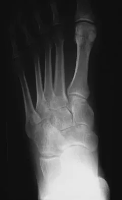
Imaging plays a vital, albeit adjunctive, role. Plain radiographs in multiple orthogonal planes are mandatory to rule out radiopaque foreign bodies (e.g., retained teeth in fight bites, metallic shards), subcutaneous gas indicative of gas-forming organisms or NSTI, and established osteomyelitis. While magnetic resonance imaging (MRI) provides unparalleled resolution of soft-tissue compartments, synovial hypertrophy, and occult deep space abscesses, its procurement must never delay emergent surgical intervention in cases of acute PFT or NSTI. High-resolution bedside ultrasonography has emerged as a rapid, highly sensitive tool for identifying fluid collections within the tendon sheath or deep fascial spaces, allowing the surgeon to precisely map the planned incisions.
Patient positioning and anesthesia require careful coordination with the anesthesia team. The patient is positioned supine with the affected upper extremity extended on a radiolucent hand table. General anesthesia or a dense regional block (e.g., axillary or supraclavicular brachial plexus block) is mandatory. Local anesthesia, such as the Wide-Awake Local Anesthesia No Tourniquet (WALANT) technique, is generally contraindicated in severe, complex hand infections. The injection of local anesthetic volumes into already compromised, tightly bound compartments can exacerbate microvascular ischemia. Furthermore, local infiltration risks proximal dissemination of the purulent payload along fascial planes.
Tourniquet management in the infected hand differs fundamentally from elective hand surgery. Exsanguination of the limb must be achieved solely by prolonged elevation (typically 3 to 5 minutes). The use of an Esmarch bandage or tight compressive wrapping over the infected site is strictly prohibited, as the mechanical pressure will force bacteria and purulent material proximally into uncontaminated compartments, potentially converting a localized digital infection into a catastrophic forearm or systemic sepsis. Once elevated, the pneumatic tourniquet is inflated to 250 mm Hg (or 100 mm Hg above systolic pressure). The surgeon must operate efficiently, as tourniquet times exceeding 2 hours will compound the ischemic insult to the already compromised tissues.
Step-by-Step Surgical Approach and Fixation Technique
Pyogenic Flexor Tenosynovitis: Closed Catheter Irrigation
For early-stage PFT (Michon Stage I and II), closed catheter irrigation is the gold standard, balancing adequate decompression with minimal surgical trauma.
1. Proximal Exposure: A transverse or oblique incision is made in the distal palmar crease directly over the A1 pulley of the affected digit. Blunt dissection with tenotomy scissors is utilized to spread the palmar fascia longitudinally, protecting the adjacent neurovascular bundles. The proximal edge of the flexor tendon sheath is identified.
2. Distal Exposure: A mid-axial incision is made along the non-dominant border of the middle or distal phalanx. The incision is placed dorsal to the neurovascular bundle, exposing the distal A4 or A5 pulley.
3. Sheath Fenestration: A small, 2-3 mm transverse window is sharply created in the sheath just proximal to the A1 pulley, and another distal to the A4 pulley. Immediate egress of purulent fluid is typically observed. Swabs and fluid aspirates are immediately sent for aerobic, anaerobic, mycobacterial, and fungal cultures.
4. Catheter Insertion: A 16-gauge pediatric feeding tube, or a specialized multi-holed irrigation catheter, is introduced into the proximal window. It is advanced 1 to 2 cm distally into the sheath and secured to the palmar skin with a 4-0 nylon suture to prevent dislodgement.
5. Irrigation Protocol: The sheath is flushed with 500 mL of sterile normal saline using a large syringe. The surgeon must visually confirm clear, unobstructed egress of fluid from the distal window. If the fluid does not flow freely, the sheath may be loculated, necessitating conversion to an open debridement.
6. Closure: The distal wound is left completely open to allow for continuous drainage. The proximal wound may be loosely approximated around the catheter. Postoperatively, the catheter is flushed with 10-20 mL of saline every 2 to 4 hours for 48 hours before removal.
Deep Fascial Space Infections: Surgical Decompression
The surgical approach to deep space infections must provide wide exposure while meticulously protecting motor and sensory nerves.
* The Thenar Space: Decompression can be achieved via a volar or dorsal approach. The volar approach utilizes a longitudinal incision parallel to, but slightly radial to, the thenar crease. Meticulous blunt dissection is paramount to avoid iatrogenic injury to the recurrent motor branch of the median nerve, which enters the thenar musculature proximally. Alternatively, a dorsal longitudinal incision in the first web space allows for excellent drainage of the adductor pollicis region without risking the median nerve.
* The Midpalmar Space: A transverse incision within the mid-palmar crease provides direct access. Alternatively, a longitudinal incision in the web space between the middle and ring fingers can be utilized. Dissection is carried dorsally between the flexor tendons and the lumbrical muscles, piercing the midpalmar fascia to evacuate the abscess.
* Wound Management: Following copious irrigation, the spaces are packed loosely with iodoform gauze or fitted with a small Penrose drain. The skin is never primarily closed.
Necrotizing Soft-Tissue Infections (NSTI): Radical Debridement
The operative management of NSTI requires an aggressive, oncologic-style resection of all necrotic tissue.
1. Incision Planning: Extensive longitudinal incisions are made along the entire zone of suspected involvement. While crossing flexion creases at 90 degrees should be avoided to prevent future contractures, the immediate priority is absolute exposure; future scar cosmesis is a secondary concern.
2. Fascial Assessment: The "finger sweep" test is employed. Healthy fascia is glistening and firmly adherent to the overlying subcutaneous fat. Necrotic fascia is dull, grayish-green, and separates from the fat with minimal digital resistance.
3. Radical Excision: All non-viable skin, subcutaneous fat, fascia, and necrotic muscle are sharply excised until briskly bleeding, healthy tissue is encountered. "Marginal" or questionable tissue must be resected.
4. Postoperative Care: The massive wounds are left entirely open. Application of a negative pressure wound therapy (NPWT) device is highly effective for managing exudate and promoting angiogenesis.
5. Mandatory Second Look: A planned return to the operating room within 24 to 48 hours is absolute dogma for re-evaluation and further debridement.
Osteomyelitis and Septic Arthritis (Clenched Fist Injuries)
Human bites to the MCP joint are notorious for sealing virulent oral flora deep within the joint capsule.
1. Incision and Exposure: The traumatic laceration is extended longitudinally, both proximally and distally.
2. Dynamic Assessment: The MCP joint must be flexed to 90 degrees to recreate the exact position of the hand at the moment of impact. This maneuver aligns the capsular and tendinous lacerations, exposing the true depth of the penetration.
3. Arthrotomy and Debridement: A formal arthrotomy is performed. The joint is copiously irrigated with several liters of normal saline. Any indented osteochondral fragments or retained tooth matter must be meticulously debrided.
4. Wound Care: The wound is left open to heal by secondary intention. Primary closure of a human bite wound is universally contraindicated and constitutes surgical malpractice.
Atypical and Mycobacterial Infections: Radical Tenosynovectomy
Infections caused by Mycobacterium marinum present as chronic, boggy tenosynovitis and require radical surgical clearance.
1. Exposure: An extended Brunner (zigzag) incision is utilized along the volar aspect of the affected digit, extending proximally into the palm or wrist to expose the entire extent of the synovial disease.
2. Synovectomy: The hypertrophic, granulomatous synovium (often containing fibrinous "rice bodies") is meticulously excised from the flexor tendons. The tendons must be completely skeletonized.
3. Pulley Preservation: Extreme surgical discipline is required to preserve the A2 and A4 pulleys. If the granulomatous tissue is densely adherent beneath these pulleys, a small curette or a pediatric right-angle clamp is used to gently clear the space without compromising the pulley's structural integrity.
Complications, Incidence Rates, and Salvage Management
Despite aggressive surgical intervention and targeted antimicrobial therapy, complex hand infections carry a high morbidity profile. The dense anatomical crowding of the hand means that the inflammatory response alone, even after the pathogen is eradicated, can lead to devastating fibrotic complications. The most common complication following pyogenic flexor tenosynovitis is profound digital stiffness, resulting from restrictive adhesions forming between the flexor tendons and the surrounding sheath.
More catastrophic complications arise from delayed presentation or inadequate initial debridement. Ischemic necrosis of the flexor tendons can lead to spontaneous rupture, requiring complex, two-stage tendon reconstruction using silicone Hunter rods once the infection is definitively cleared. In cases of fulminant NSTI or overwhelming deep space infections, progressive microvascular thrombosis can lead to irreversible digital or limb ischemia. In these scenarios, the surgeon must pivot from limb salvage to life salvage, executing precise amputations to halt the systemic inflammatory response syndrome (SIRS) and prevent fatal sepsis.
| Complication | Estimated Incidence | Pathophysiology / Risk Factors | Salvage Management / Intervention |
|---|---|---|---|
| Digital Stiffness / Adhesions | 40% - 60% (Post-PFT) | Fibroblastic proliferation and scarring between tendon and sheath; prolonged immobilization. | Aggressive early hand therapy, dynamic splinting. Late: Surgical tenolysis (minimum 6 months post-op). |
| Flexor Tendon Rupture | 5% - 10% | Liquefactive necrosis from prolonged increased intrasynovial pressure and bacterial proteases. | Two-stage tendon reconstruction (Hunter rod placement followed by free tendon graft). |
| Bowstringing | 2% - 5% | Iatrogenic destruction or necrotic rupture of the A2 and/or A4 pulleys during debridement. | Pulley reconstruction using palmaris longus or extensor retinaculum grafts after infection clears. |
| Chronic Osteomyelitis | 10% - 15% (Post-bite) | Inadequate initial debridement of bone; retained foreign body; avascular bone segments. | Radical sequestrectomy, dead space management with PMMA beads, vascularized soft-tissue coverage (e.g., ADM flap). |
| Systemic Sepsis / Death | 10% - 20% (In NSTI) | Rapid systemic dissemination of exotoxins; delayed surgical intervention; immunocompromised host. | Emergent radical debridement, broad-spectrum IV antibiotics, ICU support. Proximal amputation (wrist/forearm) if progressive. |
Phased Post-Operative Rehabilitation Protocols
The ultimate functional outcome of a surgically managed hand infection is inextricably linked to the rigor and execution of the postoperative rehabilitation protocol. The surgeon and the certified hand therapist must work in lockstep to achieve the dual, often competing, goals of the postoperative phase: eradicating the residual infection while aggressively preventing debilitating adhesions and joint contractures.
Phase I: Acute Immobilization and Edema Control (Days 0 to 3)
Immediately postoperatively, the hand is enveloped in a bulky, non-compressive dressing and immobilized in a custom orthosis in the "Intrinsic Plus" (Safe) Position. The wrist is extended 20 to 30 degrees, the MCP joints are flexed to 70 to 90 degrees, the interphalangeal (IP) joints are held in full extension, and the thumb is abducted and opposed. This specific posture is non-negotiable; it maintains the collateral ligaments of the MCP joints at their maximal length to prevent extension contractures, and it prevents the volar plates of the IP joints from adhering and causing flexion contractures. Strict elevation above the level of the heart is mandatory to facilitate venous and lymphatic drainage. Interstitial edema acts as a biological glue, rich in fibrinogen, which rapidly converts to restrictive fibrous adhesions if not aggressively managed.
Phase II: Early Active Motion and Tendon Gliding (Days 3 to 14)
Once the acute inflammatory phase begins to subside—typically 48 to 72 hours post-debridement, provided there is no evidence of worsening infection—the splint is removed for strictly supervised therapy sessions. The cornerstone of this phase is the initiation of specific tendon gliding exercises. Patients are instructed to perform active and active-assisted range of motion exercises, cycling through straight fist, hook fist, and full fist postures. These exercises promote maximum differential glide between the flexor digitorum superficialis (FDS) and flexor digitorum profundus (FDP) tendons, preventing them from scarring to each other or to the surrounding sheath. If closed catheter irrigation was utilized, the catheter is removed at 48 hours, and motion is initiated immediately thereafter.
Phase III: Strengthening, Scar Management, and Antimicrobial Tailoring (Weeks 2 to 12)
As the surgical wounds heal—either by secondary intention or delayed primary closure—the focus shifts to restoring grip strength and managing scar tissue. Retrograde massage, compressive garments (e.g., Isotoner gloves), and silicone gel sheeting are employed to soften the surgical scars and reduce hypersensitivity.
Concurrently, antimicrobial therapy is carefully managed. Empiric broad-spectrum intravenous antibiotics (e.g., Vancomycin combined with Piperacillin-Tazobactam or a third-generation cephalosporin) are initiated immediately after intraoperative cultures are obtained. Once definitive culture and sensitivity results are available, therapy is narrowed. For uncomplicated PFT or space infections, a 7 to 14-day course of targeted oral antibiotics is typically sufficient following clinical improvement. However, for atypical infections such as Mycobacterium marinum, a prolonged multidrug regimen (e.g., Clarithromycin, Ethambutol, and Rifampin) for 3 to 6 months is often required in conjunction with the initial radical surgical debridement.
Summary of Landmark Literature and Clinical Guidelines
The surgical mastery of hand infections is heavily dependent on an appreciation of the landmark literature that has shaped modern protocols. Allen B. Kanavel’s seminal text, Infections of the Hand (1912), remains the bedrock of our anatomical understanding. Kanavel meticulously detailed the fascial spaces, the synovial sheaths, and the predictable pathways of infectious spread, introducing the four cardinal signs of flexor tenosynovitis that remain the diagnostic gold standard over a century later.
In the modern era, Michon’s classification of pyogenic flexor tenosynovitis (1974) revolutionized the surgical approach, providing a clear algorithm that delineates when minimally invasive catheter irrigation is appropriate versus when open radical debridement is mandatory. This classification was further refined by Pang et al., who validated the efficacy of closed continuous irrigation in reducing morbidity in early-stage disease.
For necrotizing soft-tissue infections, the work of Wong et al. (2004) in developing the Laboratory Risk Indicator for Necrotizing Fasciitis (LRINEC) score provided a crucial, objective tool for differentiating NSTI from severe cellulitis, though clinical judgment remains paramount. Furthermore, contemporary clinical guidelines from the American Society for Surgery of the Hand (ASSH) and the Orthopaedic Trauma Association (OTA) consistently emphasize the absolute contraindication of primary closure in human bite wounds and the necessity of formal arthrotomy for clenched-fist injuries involving the MCP joint. Mastery of these guidelines, combined with precise surgical execution and rigorous rehabilitation, ensures the highest probability of restoring function and salvaging the limb in the face of complex hand infections.
End of Chapter.
