The Swashbuckler Approach to the Distal Femur: A Comprehensive Surgical Guide
Key Takeaway
The Swashbuckler approach, popularized by Starr et al., is a highly utilitarian surgical technique for complex distal femur fractures. By utilizing a lateral parapatellar arthrotomy and elevating the vastus lateralis without violating the quadriceps tendon, it provides unparalleled exposure of the femoral condyles. This approach is particularly indicated for AO/OTA Type C intra-articular fractures requiring precise anatomical reduction and locking plate osteosynthesis.
Comprehensive Introduction and Patho-Epidemiology
The management of complex, comminuted, and intra-articular fractures of the distal femur—specifically those classified as AO/OTA Type 33-B (partial articular) and 33-C (complete articular)—presents a formidable and enduring challenge to the orthopedic surgeon. Achieving absolute anatomical reduction of the articular block while simultaneously preserving the delicate, vascularized soft-tissue envelope of the metadiaphysis remains the immutable cornerstone of successful osteosynthesis. Historically, standard lateral approaches provided grossly inadequate visualization of the medial femoral condyle and the deep intercondylar notch. This limitation frequently forced surgeons into utilizing highly morbid extensile exposures, such as the quadriceps-splitting or quadriceps-peeling approaches, or resorting to dual medial and lateral incisions, both of which drastically increased the risk of devascularization, profound arthrofibrosis, and extensor mechanism dysfunction.
To circumvent the profound limitations of traditional exposures, Starr, Jones, and Reinert introduced and popularized the Swashbuckler approach in 1999. Conceived as a highly modified, extensile anterior approach, the Swashbuckler technique ingeniously combines a lateral parapatellar arthrotomy with the meticulous elevation of the vastus lateralis muscle off the lateral intermuscular septum. By reflecting the entire intact extensor mechanism medially—without incising or splitting the quadriceps tendon—the operating surgeon gains unparalleled, panoramic visualization of the entire distal femoral articular surface. This "open book" exposure allows for the precise manipulation and direct visualization necessary to reconstruct highly comminuted articular blocks, including elusive coronal plane shear fragments (Hoffa fractures), while minimizing iatrogenic trauma to the extensor mechanism.
The patho-epidemiology of distal femur fractures dictates the necessity of such a versatile approach. These injuries characteristically demonstrate a bimodal demographic distribution. In the younger, physiologically robust population, these fractures are typically the sequelae of high-energy trauma, such as motor vehicle collisions or falls from significant heights, resulting in severe comminution, profound soft-tissue injury, and multi-system polytrauma. Conversely, in the elderly population, distal femur fractures frequently occur following low-energy mechanisms, such as ground-level falls, superimposed upon severely osteoporotic bone. The exponential rise in total knee arthroplasties (TKA) has also precipitated a dramatic increase in complex periprosthetic distal femur fractures. In these osteoporotic and periprosthetic scenarios, the bone stock is highly compromised, making secure fixation exceptionally difficult and elevating the importance of direct, unimpeded articular visualization.
In the contemporary era of orthopedic traumatology, the Swashbuckler approach is inextricably linked with the principles of Minimally Invasive Plate Osteosynthesis (MIPO) and the deployment of advanced locking plate technologies. While the approach allows for the absolute stability required for articular reconstruction, it seamlessly transitions into a biological, relative-stability construct for the metadiaphysis. Once the articular block is anatomically reconstituted and secured with interfragmentary lag screws, it is effectively converted into an extra-articular fracture. The Swashbuckler exposure then facilitates the submuscular insertion of a locking condylar plate proximally along the lateral femoral shaft, bridging the zone of comminution. This masterclass in surgical strategy perfectly balances the mechanical imperative of articular congruity with the biological imperative of fracture hematoma preservation.
Detailed Surgical Anatomy and Biomechanics
The successful execution of the Swashbuckler approach demands an intimate and exhaustive understanding of the surgical anatomy of the lateral thigh and the extensor mechanism of the knee. The approach exploits the intermuscular plane between the vastus lateralis and the lateral intermuscular septum (LIMS). The vastus lateralis, the largest component of the quadriceps femoris, is enveloped superficially by the unyielding fascia lata and the iliotibial band (ITB). Deeply, it originates from the greater trochanter, the lateral lip of the linea aspera, and the LIMS. The LIMS acts as a robust fascial partition separating the anterior compartment (extensors, innervated by the femoral nerve) from the posterior compartment (flexors, innervated by the sciatic nerve). By dissecting the vastus lateralis off the LIMS and elevating it anteriorly, the surgeon exposes the entire lateral and anterior aspects of the distal femoral shaft without violating the muscle belly itself.
A critical neurovascular consideration during this deep dissection is the presence of the perforating branches of the profunda femoris artery. As the profunda femoris descends in the posterior compartment, it gives off three to four perforating arteries that pierce the adductor magnus and subsequently the lateral intermuscular septum to supply the vastus lateralis. These vessels are typically encountered dynamically as the surgeon elevates the vastus lateralis from distal to proximal. They are short, under high arterial pressure, and tethered closely to the femur. Meticulous identification, isolation, and secure ligation or electrocoagulation of these perforators are absolutely mandatory. Inadvertent avulsion or inadequate hemostasis of these vessels can result in catastrophic acute intraoperative hemorrhage, massive postoperative hematoma, or the rapid development of thigh compartment syndrome.
The anatomy of the extensor mechanism is central to the joint-sparing nature of the Swashbuckler approach. Traditional extensile approaches, such as the rectus snip or V-Y quadricepsplasty, directly violate the quadriceps tendon, leading to inevitable scarring, weakness, and significant extensor lag. The Swashbuckler approach entirely avoids the quadriceps tendon. Distally, the dissection transitions into a lateral parapatellar arthrotomy, incising the lateral retinaculum and the joint capsule parallel to the lateral border of the patella and the patellar tendon. Because the vastus lateralis has been mobilized proximally, the entire extensor mechanism (quadriceps tendon, patella, and patellar tendon) can be gently subluxated and retracted medially. This provides an unobstructed, 180-degree view of the trochlea, the intercondylar notch, and both the medial and lateral femoral condyles, facilitating the anatomical reduction of complex intra-articular fracture lines.
From a biomechanical perspective, the distal femur is subjected to immense physiological loads, including massive axial compression, bending moments, and torsional forces generated by the mechanical axis of the lower extremity and the pull of powerful muscle groups. The gastrocnemius muscles, originating on the posterior aspect of the medial and lateral condyles, exert a profound deforming force, typically pulling the distal articular fragment into severe flexion (recurvatum deformity). Fracture fixation must counteract these forces. Following anatomical articular reduction (achieving absolute stability via interfragmentary compression), fixation is typically achieved using a lateral Locking Condylar Plate (LCP). Locking plates function as fixed-angle constructs, which are biomechanically superior to conventional plates in osteoporotic bone. The distal locking screws provide a rigid scaffold that resists varus/valgus collapse of the condyles, while the proximal shaft screws, utilized in a bridging fashion, act as an extramedullary splint. This construct provides relative stability across the comminuted metaphysis, permitting the micromotion necessary to stimulate robust secondary bone healing via endochondral ossification.
Exhaustive Indications and Contraindications
The Swashbuckler approach is an incredibly versatile surgical exposure, but its utilization must be carefully tailored to the specific pathoanatomy of the fracture and the physiological status of the patient. The primary utility of this approach lies in its unparalleled ability to expose the entire distal femoral articular surface, making it the exposure of choice for fractures where precise, direct visualization of the joint is non-negotiable. It is particularly valuable when preoperative imaging reveals complex, multi-planar articular comminution that cannot be adequately reduced via closed means or through limited "mini-open" windows. The surgeon must weigh the benefits of this extensile exposure against the inherent risks of increased surgical time, potential blood loss, and the creation of a large surgical dead space.
The most definitive indications for the Swashbuckler approach involve high-grade, intra-articular distal femur fractures. According to the AO/OTA classification, Type 33-C fractures (complete articular fractures with metaphyseal dissociation) are the classic indication. Within this category, C2 (simple articular, multifragmentary metaphyseal) and C3 (multifragmentary articular, multifragmentary metaphyseal) fractures demand the panoramic visualization provided by the Swashbuckler to meticulously reconstruct the articular puzzle before attaching it to the femoral shaft. Furthermore, Type 33-B fractures (partial articular), particularly those involving coronal plane shear fractures (Hoffa fragments) of either the lateral or medial condyle, are prime indications. Hoffa fractures are notoriously difficult to visualize and reduce through standard lateral approaches, whereas the Swashbuckler allows for direct visualization of the posterior condyles and facilitates precise anterior-to-posterior lag screw placement.
Beyond acute trauma, the Swashbuckler approach is highly indicated in the realm of complex reconstructive orthopedics. Distal femoral nonunions and malunions frequently require extensive surgical exposure for callus takedown, intra-articular osteotomies, and the correction of multi-planar deformities (varus/valgus, flexion/extension, and rotational malalignment). The approach provides the necessary access to perform these intricate structural corrections and to apply massive structural bone grafts if required. Additionally, in the context of periprosthetic fractures above a total knee arthroplasty (TKA)—specifically Vancouver Type B and C equivalents in the distal femur—the approach allows the surgeon to directly inspect the implant-bone interface, verify the stability of the femoral component, and achieve secure fixation around the existing hardware.
Despite its vast utility, the Swashbuckler approach is not universally applicable and possesses strict relative and absolute contraindications. The approach relies heavily on the medial mobilization of the extensor mechanism; therefore, severe trauma to the medial soft tissue envelope is a significant relative contraindication. If the medial tissues are crushed, degloved, or highly compromised, forcibly subluxating the patella medially may exacerbate tissue ischemia and precipitate catastrophic soft tissue necrosis. Furthermore, isolated medial pathology, such as an isolated medial condyle fracture (AO/OTA 33-B2) or a medial Hoffa fracture without lateral involvement, is better served by a direct medial approach, avoiding the unnecessary morbidity of a massive lateral dissection. Active local infection, such as overlying cellulitis or frank intra-articular sepsis, represents an absolute contraindication to utilizing this extensile approach for elective reconstruction, as it risks disseminating the infection throughout the entire lateral thigh and knee joint.
| Clinical Parameter | Indications for Swashbuckler Approach | Contraindications for Swashbuckler Approach | Rationale / Clinical Note |
|---|---|---|---|
| Fracture Classification | AO/OTA 33-C2, 33-C3 (Complete articular, highly comminuted). | AO/OTA 33-A (Extra-articular) without need for joint inspection. | Extra-articular fractures can often be managed with entirely percutaneous or MIPO techniques without arthrotomy. |
| Partial Articular Patterns | AO/OTA 33-B3 (Coronal shear / Hoffa fractures), especially lateral or bicondylar. | AO/OTA 33-B2 (Isolated medial condyle fractures). | Isolated medial pathology is accessed more safely and directly via a medial subvastus or medial parapatellar approach. |
| Soft Tissue Envelope | Intact or mildly bruised medial and lateral soft tissues. | Severe medial degloving injury, Morel-Lavallée lesion medially. | Medial subluxation of the patella requires a pliable and vascularized medial envelope to prevent ischemic necrosis. |
| Reconstructive Scenarios | Intra-articular malunions, nonunions requiring complex osteotomy. | Active intra-articular sepsis, overlying lateral cellulitis. | Extensile approaches in the presence of infection risk massive dissemination and deep space abscess formation. |
| Periprosthetic Status | Supracondylar fractures above TKA requiring component inspection. | Loose TKA requiring immediate revision arthroplasty (relative). | If the primary goal is revision arthroplasty, a standard midline anterior approach is typically preferred by arthroplasty surgeons. |
Pre-Operative Planning, Templating, and Patient Positioning
The success of complex distal femur reconstruction via the Swashbuckler approach is largely determined before the patient ever enters the operating theater. Exhaustive preoperative planning is absolutely critical. The imaging protocol must begin with high-quality, orthogonal plain radiographs (anteroposterior and lateral) of the entire femur, including the hip and knee joints, to rule out ipsilateral femoral neck fractures or segmental shaft injuries. Traction radiographs can be exceptionally helpful in overcoming muscle spasm to delineate the major fracture fragments and estimate the degree of metaphyseal comminution. However, plain radiography is vastly insufficient for understanding the true three-dimensional complexity of the articular block. A fine-cut Computed Tomography (CT) scan (1mm slices) with 2D multi-planar reformats (sagittal and coronal) and 3D surface-rendered reconstructions is mandatory. The CT scan is the only reliable modality for identifying occult coronal shear fragments (Hoffa fractures), assessing the exact geometry of the intercondylar notch comminution, and mapping the precise trajectory of the fracture lines.
Preoperative templating, utilizing either advanced digital orthopedic software or traditional analog acetate overlays, is an indispensable step. The surgeon must utilize the templating process to formulate a tactical blueprint for the surgery. This includes determining the optimal sequence of reduction, selecting the appropriate implant, and planning the trajectory of every single screw. When selecting a Lateral Locking Condylar Plate, the surgeon must calculate the required plate length to ensure adequate bridging of the comminuted metaphysis. The biomechanical rule of thumb dictates a plate span ratio (plate length to fracture length) of at least 2:1 to 3:1 in comminuted fractures, and a screw density (number of screws divided by the number of plate holes) of less than 0.5 to prevent construct over-stiffness. Furthermore, the surgeon must meticulously plan the trajectory of the independent interfragmentary lag screws used to reconstruct the articular block, ensuring they do not occupy the spatial corridors required for the distal locking screws of the condylar plate.
Patient positioning in the operating room must facilitate unimpeded fluoroscopic imaging and allow for dynamic manipulation of the limb. The patient is placed supine on a fully radiolucent operating table. A standard fracture table is generally contraindicated, as it severely restricts the ability to flex the knee and manipulate the limb dynamically. The ipsilateral hip may be slightly bumped to correct natural external rotation, bringing the patella to face directly anteriorly. A sterile bump, a rolled blanket, or a specialized radiolucent triangle must be placed under the ipsilateral knee. Maintaining the knee in 30 to 60 degrees of flexion is a critical reduction maneuver; it relaxes the powerful deforming force of the gastrocnemius muscle, which originates on the posterior condyles and invariably pulls the distal articular fragment into a recurvatum (extension) deformity. This flexed position also places the quadriceps mechanism under slight tension, which paradoxically can aid in the medial translation of the patella once the lateral retinacular release is performed.
The use of a surgical tourniquet during the Swashbuckler approach is a subject of intense debate among orthopedic traumatologists. While a tourniquet provides a bloodless field during the initial exposure, its inflation on the proximal thigh carries significant biomechanical disadvantages. Inflating a tourniquet effectively tethers the quadriceps muscle bulk to the femoral diaphysis, severely restricting the excursion of the extensor mechanism. This tethering makes the critical maneuver of medially retracting and subluxating the patella significantly more difficult, thereby limiting the distal articular exposure that the Swashbuckler approach is designed to provide. Therefore, the prevailing clinical recommendation is to place a sterile tourniquet high on the thigh as a fail-safe measure, but to avoid inflating it unless absolutely necessary for catastrophic hemorrhage control. Careful, meticulous hemostasis using electrocautery during the dissection is vastly preferred over routine tourniquet use.
Step-by-Step Surgical Approach and Fixation Technique
The surgical execution of the Swashbuckler approach demands meticulous soft-tissue handling and a systematic progression from superficial exposure to deep articular reconstruction. The skin incision is longitudinal, beginning proximally over the lateral aspect of the distal femur. The exact proximal starting point is dictated by the degree of metaphyseal comminution and the anticipated length of the locking plate. The incision extends distally, remaining over the lateral aspect of the femur, before curving gently anteriorly to parallel the lateral border of the patella, ultimately terminating at or just lateral to the tibial tubercle. Superficial dissection is carried sharply down through the subcutaneous adipose tissue to expose the glistening fascia of the quadriceps and the robust fibers of the iliotibial (IT) band. The quadriceps fascia is incised in line with the skin incision, and the IT band is sharply incised longitudinally, allowing access to the vastus lateralis muscle belly beneath.
The deep dissection is the most critical and potentially hazardous phase of the approach. Retracting the IT band laterally, the surgeon identifies the vastus lateralis and traces it posteriorly to its attachment along the linea aspera and the lateral intermuscular septum (LIMS). Using a combination of blunt and sharp dissection, the vastus lateralis is elevated off the LIMS, working methodically from distal to proximal. It is during this elevation that the surgeon will encounter the perforating branches of the profunda femoris artery. These vessels pierce the LIMS and enter the vastus lateralis directly. They must be anticipated, meticulously skeletonized, clamped, and securely ligated with heavy silk sutures or thoroughly electrocoagulated. Avulsion of these perforators causes them to retract deep into the posterior compartment, resulting in massive, difficult-to-control hemorrhage. Once the perforators are managed, the entire vastus lateralis can be elevated anteriorly, exposing the pristine lateral and anterior cortex of the femoral diaphysis.
With the proximal exposure secured, attention is turned to the joint. Distally, the incision is extended through the lateral parapatellar retinaculum and the joint capsule, completing a standard lateral parapatellar arthrotomy. The crucial "Swashbuckler maneuver" is now performed. A blunt retractor, such as a large Hohmann or a Z-retractor, is placed deep to the elevated vastus lateralis and vastus medialis, resting on the anterior femoral cortex. The patella, patellar tendon, and the entire quadriceps mechanism are then gently subluxated and displaced medially over the medial femoral condyle. This maneuver effectively "opens the book" of the knee joint. The surgeon is instantly rewarded with a spectacular, unobstructed, panoramic view of the lateral condyle, the medial condyle, the trochlear groove, and the deep intercondylar notch. The joint is thoroughly irrigated to evacuate fracture hematoma and osteochondral debris, allowing for precise visualization of the articular fracture lines.
The reconstruction phase begins with the absolute anatomical reduction of the articular block. Utilizing pointed reduction forceps, dental picks, and K-wires as joysticks, the articular fragments are manipulated into perfect congruity. Provisional fixation is achieved with multiple smooth K-wires. Definitive fixation of the articular block is performed using independent 3.5mm or 4.5mm cortical lag screws. The trajectory of these screws is paramount. For coronal shear (Hoffa) fragments, screws are directed strictly anterior-to-posterior, often countersunk beneath the articular cartilage of the trochlea to prevent patellofemoral impingement. For sagittal split fractures, screws are directed lateral-to-medial or medial-to-lateral. It is imperative that the surgeon continuously references the preoperative template to ensure these independent lag screws do not obstruct the planned pathways for the distal locking screws of the condylar plate.
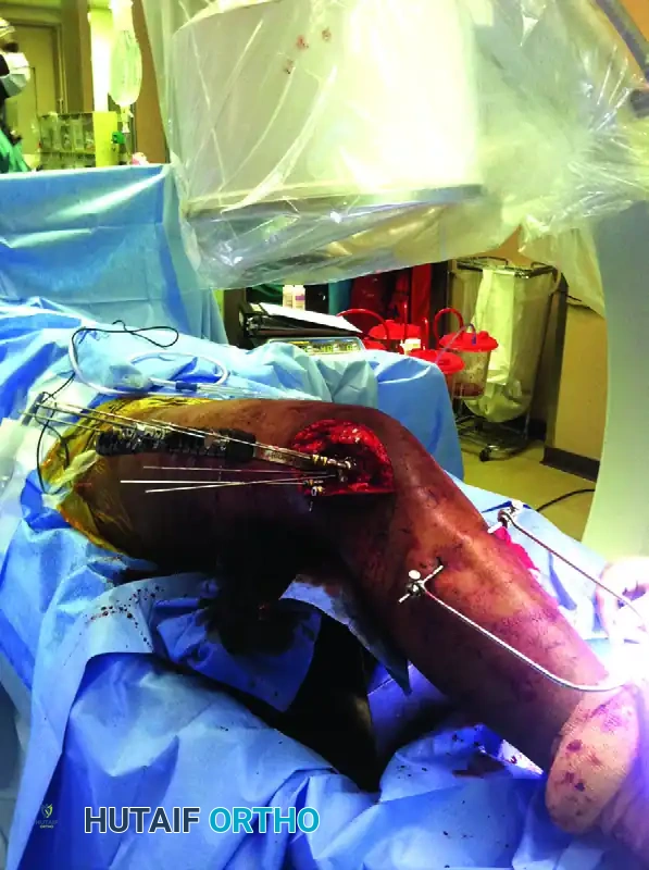
Once the articular block is rigidly reconstituted, the procedure transitions to Minimally Invasive Plate Osteosynthesis (MIPO) for the metaphyseal fixation, as illustrated in the surgical setup above. To restore axial length and correct rotational alignment without devastating the fracture hematoma, skeletal traction is applied via a proximal tibial traction pin and a sterile traction bow. The selected Locking Condylar Plate (LCP) is then introduced submuscularly, sliding it deep to the elevated vastus lateralis from the distal articular exposure proximally along the lateral femoral shaft. Specially designed radiolucent outriggers (targeting jigs) are attached to the distal end of the plate, permitting precise, percutaneous insertion of the proximal diaphyseal screws. Intraoperative fluoroscopy is utilized extensively throughout this phase to verify the restoration of the mechanical axis, confirm the plate sits flush against the lateral condyle, and ensure that the proximal percutaneous screws are centrally located within the femoral diaphysis. This hybrid technique marries the absolute stability of open articular reduction with the biological preservation of MIPO, optimizing the environment for rapid secondary bone healing.
Complications, Incidence Rates, and Salvage Management
Despite meticulous surgical technique and advanced implant technology, the management of complex distal femur fractures via the Swashbuckler approach carries a significant complication profile. The high-energy nature of the initial trauma, combined with the extensile nature of the surgical exposure, creates a precarious environment for both mechanical failure and biological compromise. The surgeon must be acutely aware of these potential pitfalls, aggressively work to prevent them during the primary procedure, and possess the advanced reconstructive skills necessary to manage them should they arise. The balance between achieving rigid fixation and preserving the soft-tissue envelope is razor-thin, and deviations in either direction can lead to catastrophic outcomes.
Loss of fixation and subsequent nonunion represent the most mechanically devastating complications. These typically occur at the metaphyseal-diaphyseal junction rather than within the articular block itself. The etiology is frequently multifactorial, stemming from technical errors such as selecting a plate that is too short (inadequate working length), creating a construct that is too stiff (excessive screw density preventing necessary micromotion), or performing overly aggressive soft-tissue stripping that devitalizes the comminuted fracture fragments. When a nonunion occurs, it is often accompanied by hardware failure, such as plate breakage or screw pullout. Salvage management requires a massive revision surgery. This entails complete removal of the failed hardware, aggressive debridement of the nonunion site down to bleeding bone, and re-stabilization. Revision fixation often necessitates dual plating (adding a medial plate for enhanced biomechanical stability) coupled with the application of autologous iliac crest bone graft (ICBG) or orthobiologics to stimulate osteogenesis.
Knee stiffness and profound arthrofibrosis are the most common functional complications following the Swashbuckler approach. The extensive soft-tissue dissection, the intra-articular nature of the fracture, and the inevitable postoperative hemarthrosis create a perfect storm for the development of dense intra-articular adhesions and capsular contractures. Furthermore, improper closure of the lateral retinaculum or failure to repair the iliotibial band anatomically can lead to severe patellar maltracking, anterior knee pain, and extensor lag. Prevention is paramount and relies on meticulous, layered closure and immediate, aggressive postoperative mobilization. If severe arthrofibrosis develops and is refractory to aggressive physical therapy, salvage management may require a formal arthroscopic or open lysis of adhesions, coupled with a manipulation under anesthesia (MUA) or, in severe cases, a targeted quadricepsplasty to restore functional excursion of the extensor mechanism.
Deep surgical site infection (SSI) is a catastrophic complication that threatens both the limb and the life of the patient. The risk is elevated in high-energy open fractures, patients with significant medical comorbidities (e.g., diabetes, obesity), and cases with prolonged operative times. Infection in the presence of massive orthopedic hardware is notoriously difficult to eradicate due to the formation of bacterial biofilms on the implant surfaces. Management requires a highly aggressive, multidisciplinary approach. Acute infections may be salvaged with emergent, radical irrigation and debridement (I&D), implant retention (if the fixation remains absolutely rigid), and prolonged culture-directed intravenous antibiotic therapy. However, chronic infections or infections associated with loose hardware mandate the complete removal of all implants, radical resection of infected bone (often creating a segmental defect), placement of an antibiotic-impregnated cement spacer, and eventual staged reconstruction once the infection is definitively eradicated.
| Complication | Estimated Incidence Rate | Primary Etiology / Risk Factors | Salvage Management Strategy |
|---|---|---|---|
| Knee Stiffness / Arthrofibrosis | 15% - 30% | Prolonged immobilization, severe initial trauma, intra-articular hematoma, poor patient compliance. | Aggressive PT, Manipulation Under Anesthesia (MUA), Arthroscopic or Open Lysis of Adhesions. |
| Nonunion / Hardware Failure | 5% - 10% | Inadequate plate length, excessive construct stiffness, biological stripping of metaphysis, smoking. | Hardware removal, nonunion takedown, autologous bone grafting (ICBG), revision osteosynthesis (often dual plating). |
| Deep Surgical Site Infection | 2% - 5% | Open fractures, prolonged operative time, diabetes, obesity, massive soft tissue dead space. | Radical I&D, biofilm disruption, IV antibiotics. If hardware loose: removal, antibiotic spacer, staged reconstruction. |
| Patellar Maltracking / Extensor Lag | 3% - 8% | Over-tightening of lateral retinaculum during closure, malrotation of articular block, quadriceps scarring. | Targeted physical therapy, lateral retinacular release, revision of articular malreduction if severe. |
| Vascular Injury (Perforators) | < 1% (Intra-op) | Avulsion of profunda femoris perforators during vastus lateralis elevation. | Emergent exploration, direct clamping and secure ligation of retracted vessels to prevent compartment syndrome. |
Phased Post-Operative Rehabilitation Protocols
The postoperative rehabilitation protocol following a Swashbuckler approach for a distal femur fracture is as critical to the final functional outcome as the surgical execution itself. The rehabilitation philosophy must navigate a delicate, often contradictory balance: the mechanical necessity of protecting the metaphyseal fracture fixation from premature physiological loading versus the biological necessity of early, aggressive joint mobilization to prevent devastating arthrofibrosis and cartilage degradation. This requires a highly structured, phased, and individualized approach, closely coordinated between the operating surgeon and a skilled orthopedic physical therapist. The protocol is heavily dictated by the quality of the patient's bone, the rigidity of the articular fixation, and the degree of metaphyseal comminution.
Phase 1: Maximum Protection and Early Motion (Weeks 0-6)
The immediate postoperative phase focuses on wound healing, edema
