Intramedullary Nailing for Distal Femoral Fractures: Biomechanics, Techniques, and Outcomes
Key Takeaway
Intramedullary nailing provides a biological, load-sharing fixation strategy for distal femoral fractures. While offering superior soft tissue preservation compared to traditional plating, it requires meticulous attention to biomechanical principles to prevent hardware failure. This guide details the indications, deforming forces, surgical positioning, and step-by-step techniques for both antegrade and retrograde approaches, ensuring optimal outcomes and minimizing complications such as malunion or patellar impingement.
Comprehensive Introduction and Patho-Epidemiology
The management of distal femoral fractures has evolved significantly over the past several decades, with intramedullary nailing receiving increased attention and widespread adoption in modern orthopedic traumatology. Historically, the treatment of these complex metaphyseal and intra-articular injuries was fraught with a high incidence of devastating complications. Axial malalignment, posttraumatic arthritis, severe knee stiffness, and profound construct instability were commonplace, particularly following nonoperative management in traction or the use of early, inadequate fixation devices such as flexible rods or non-locking condylar plates. The paradigm shift toward modern intramedullary fixation represents one of the most critical advancements in lower extremity trauma surgery, driven by a deeper understanding of fracture biology and implant biomechanics.
Distal femoral fractures account for approximately 3% to 6% of all femoral fractures and present with a classic bimodal epidemiological distribution. The first peak occurs in young, predominantly male patients involved in high-energy trauma, such as motor vehicle collisions or falls from significant heights. These injuries are frequently associated with severe soft tissue compromise, open fractures, and polytrauma, necessitating a damage-control approach followed by definitive stabilization. The second, rapidly expanding demographic peak involves elderly, predominantly female patients who sustain low-energy falls. In this osteoporotic population, the presence of pre-existing total knee arthroplasties (TKA) has led to a dramatic rise in periprosthetic distal femoral fractures, presenting unique challenges regarding implant selection, bone stock, and the necessity for immediate postoperative mobilization to prevent catastrophic medical complications.
Today, intramedullary devices are favored for their ability to provide "biological" osteosynthesis. Unlike traditional rigid plating constructs, which act as load-sparing devices and historically required extensive surgical approaches with devastating periosteal stripping, intramedullary nails function as load-sharing implants. This fundamental biomechanical advantage offers greater soft tissue preservation, maintains the critical fracture hematoma, and significantly reduces the need for supplemental autologous or allogeneic bone grafting. By utilizing minimally invasive percutaneous or mini-open techniques, surgeons can bridge the zone of comminution, thereby preserving the precarious extraosseous blood supply to the metaphyseal fragments.
However, the application of intramedullary nails in the distal femur is not without distinct biomechanical and technical challenges. The distal metaphyseal flare creates a significant mismatch between the diameter of the nail and the surrounding endosteal cortex, limiting the intrinsic stability of the implant within the canal. Consequently, meticulous preoperative planning, a profound understanding of deforming muscular forces, and precise surgical execution are paramount to achieving union, restoring the mechanical axis, and returning the patient to their pre-injury level of limb function.
Detailed Surgical Anatomy and Biomechanics
Surgical Anatomy and Deforming Forces
A comprehensive mastery of the distal femoral anatomy is non-negotiable for the orthopedic trauma surgeon. The distal femur transitions from a cylindrical diaphyseal tube to a widened, trapezoidal metaphyseal flare, terminating in the medial and lateral femoral condyles. This geometric transition results in a relatively thin cortical shell surrounding a vast expanse of cancellous bone, which is particularly susceptible to comminution and crushing in osteoporotic individuals. The mechanical axis of the lower extremity normally passes through the center of the knee joint, while the anatomical axis of the femur lies in approximately 5 to 7 degrees of valgus relative to the knee joint line. Failure to recreate this precise valgus alignment during intramedullary nailing inevitably leads to altered contact stresses across the tibial plateau and accelerated posttraumatic arthropathy.
The distal femur is enveloped by powerful musculature that exerts predictable, formidable deforming forces on the fracture fragments. The gastrocnemius muscles, originating on the posterior aspect of the medial and lateral condyles, exert a strong posterior pull on the distal articular block. This unyielding force consistently produces an extension deformity (apex posterior angulation or recurvatum) of the distal fragment. Simultaneously, the adductor magnus, inserting on the adductor tubercle, pulls the distal fragment medially, creating a varus malalignment. Proximally, the unopposed pull of the quadriceps and hamstring muscle groups leads to axial shortening and overriding of the fracture fragments. Recognizing and neutralizing these forces intraoperatively is the cornerstone of successful closed reduction and subsequent intramedullary nailing.
The neurovascular proximity to the distal femur further complicates surgical intervention. The superficial femoral artery traverses the adductor hiatus to become the popliteal artery, lying intimately close to the posterior cortex of the distal femur. Apex posterior fracture displacement, or the errant posterior placement of retractors, drills, or locking screws, places the popliteal artery and vein at extreme risk of catastrophic iatrogenic injury.
Biomechanics of Intramedullary Fixation
The primary biomechanical distinction between intramedullary nails and extramedullary plates lies in the distribution of mechanical stress. Intramedullary nails, positioned centrally within the medullary canal and along the mechanical axis of the femur, share axial loads with the surrounding bone cortex. This load-sharing phenomenon minimizes stress shielding and actively stimulates secondary bone healing via robust cartilaginous callus formation. Conversely, traditional plates act as load-sparing devices, shielding the underlying bone from physiological stress. While modern locked plating functions more as an internal fixator, it still creates an asymmetric, eccentric load profile that can occasionally lead to delayed union, nonunion, or catastrophic implant fatigue if the fracture fails to consolidate rapidly.
Despite the profound biological advantages of intramedullary nailing, rigorous biomechanical testing has demonstrated that nail fixation provides less rigid stabilization of distal-third femoral fractures compared to dual-plate or modern locked-plate fixation, particularly in the presence of severe metaphyseal comminution. Hardware failure has been reported in up to 15% of distal femoral fractures treated with early-generation antegrade interlocking nailing, particularly when utilizing older, slotted nail designs that lacked adequate fatigue strength. The incidence of hardware failure increases exponentially if the fracture line propagates within 5 cm of the most proximal screw hole, creating a critical stress riser that localizes bending moments directly over the interlocking holes.
To mitigate the risk of implant failure, several absolute biomechanical principles must be strictly adhered to during the procedure. First, subchondral purchase is imperative; the nail must be driven as distally as possible, anchoring securely into the dense subchondral bone of the intercondylar notch and femoral condyles. Second, implant selection is critical; utilizing solid or robustly designed non-slotted nails with increased wall thickness significantly enhances the fatigue life of the implant. Finally, the strategic placement of multiplanar distal locking screws is essential to control torsional and shear forces, maximizing the working length of the construct while preventing toggling of the distal fragment around the nail.
Construct Stability and Comparative Biomechanics
Extensive mechanical testing has compared antegrade nailing, retrograde nailing, and extramedullary devices under various loading conditions. In femoral shaft fractures with stable configurations (e.g., transverse fractures with excellent cortical contact), no significant difference in stability exists between antegrade and retrograde insertion techniques. However, in unstable, comminuted fracture configurations, the size (diameter) of the nail, rather than the method of insertion, is the primary determinant of overall construct stability. A larger diameter nail exponentially increases both bending and torsional stiffness, highlighting the importance of adequate canal reaming to accommodate the largest templated implant.
When comparing retrograde nails to locked plates, the dynamic condylar screw (DCS) and modern lateral locked plates exhibit greater torsional stiffness due to their broad footprint and dispersed screw patterns. Conversely, the supracondylar intramedullary nail, utilizing a grouped distal screw configuration, absorbs significantly more energy during axial loading. Ito et al. conducted pivotal studies comparing the supracondylar nail with a condylar blade plate, concluding that the supracondylar nail provides stability equal to a plate in axial and bending forces, falling short only when subjected to extreme, non-physiological torsional loads. This biomechanical profile makes the retrograde nail an exceptional load-sharing device for early weight-bearing in appropriately selected fracture patterns.
Exhaustive Indications and Contraindications
The decision to utilize an intramedullary nail for a distal femoral fracture hinges on a meticulous evaluation of the fracture morphology, articular involvement, bone quality, and patient-specific factors. Retrograde intramedullary nailing has become the gold standard for the vast majority of extra-articular distal femoral fractures (OTA/AO type 33A). The technique is particularly advantageous in obese patients, where lateral plating requires massive soft tissue dissection, and in polytrauma patients, where the supine position allows for simultaneous management of concomitant thoracic, abdominal, or contralateral extremity injuries.
Simple intra-articular fractures (OTA/AO type 33C1), characterized by a non-comminuted sagittal split between the condyles, are also excellent indications for retrograde nailing. In these cases, the articular block must first be anatomically reduced and independently stabilized with lag screws positioned anterior or posterior to the anticipated path of the nail. Furthermore, periprosthetic fractures superior to a total knee arthroplasty (TKA) are increasingly managed with retrograde nails, provided the femoral component possesses an open intercondylar box design that permits the passage of the nail. Pathological fractures secondary to metastatic disease also benefit immensely from intramedullary nailing, as the nail protects the entire length of the femur from subsequent lesions and provides immediate, load-sharing stability.
Conversely, absolute contraindications to intramedullary nailing must be respected to prevent catastrophic surgical failures. Active infection, either systemic or localized to the knee joint or previous hardware, absolutely precludes the insertion of an intramedullary device, which would disseminate the infection throughout the entire femoral canal. Severe intra-articular comminution (OTA/AO type 33C2 and 33C3) is a major contraindication. In these highly complex patterns, the intercondylar notch is often obliterated, making it impossible to establish a stable entry point or achieve adequate distal fixation with interlocking screws. Additionally, patients with pre-existing femoral deformity, retained hardware in the proximal or mid-shaft femur, or an exceptionally narrow medullary canal that cannot be safely reamed may require alternative fixation strategies.
Indications and Contraindications Table
| Clinical Parameter | Indications for Intramedullary Nailing | Contraindications for Intramedullary Nailing |
|---|---|---|
| Fracture Morphology (OTA/AO) | Extra-articular (33A1, 33A2, 33A3); Simple articular split (33C1). | Complex multi-fragmentary articular (33C2, 33C3); Coronal shear (Hoffa) requiring extensive plating. |
| Periprosthetic Status | TKA with open-box design allowing nail passage; Supracondylar femur fracture above stable implant. | TKA with closed-box or constrained design blocking the notch; Loose femoral component requiring revision. |
| Soft Tissue Envelope | Severe soft tissue injury laterally (making plating high-risk); Obese habitus. | Active knee sepsis; Overlying traumatic arthrotomy with gross contamination. |
| Bone Quality & Anatomy | Osteoporotic bone (load-sharing preferred); Pathological lesions (prophylactic or therapeutic). | Retained proximal hardware blocking canal; Severe preexisting femoral bowing or dysplasia. |
| Polytrauma Context | Bilateral femur fractures; Concomitant pelvic/spinal trauma requiring supine positioning. | Hemodynamic instability precluding reaming (Damage Control Orthopedics - use external fixation instead). |
Pre-Operative Planning, Templating, and Patient Positioning
Radiographic Evaluation and Templating
Flawless surgical execution begins long before the patient enters the operating theater. Comprehensive radiographic evaluation is mandatory, consisting of full-length anteroposterior (AP) and lateral views of the femur, as well as dedicated orthogonal views of the knee joint. In the modern era, a fine-cut Computed Tomography (CT) scan with sagittal and coronal reconstructions is considered the standard of care for all distal femoral fractures. CT imaging is critical for identifying occult intra-articular extension, particularly coronal plane fractures (Hoffa fragments) of the posterior condyles, which occur in up to 38% of distal femoral fractures and drastically alter the surgical approach and fixation strategy.
Digital templating is an indispensable component of preoperative planning. The surgeon must determine the anticipated nail diameter, length, and the required radius of curvature. The nail length should be templated to end at or just proximal to the lesser trochanter for retrograde nails, ensuring the proximal interlocking screws bypass the subtrochanteric stress riser. Careful attention must be paid to the anterior bow of the femur; utilizing a straight nail or a nail with a mismatching radius of curvature in a highly bowed femur can lead to anterior cortical perforation or iatrogenic fracture during insertion. The diameter of the nail is determined by measuring the narrowest point of the isthmus, planning to over-ream the canal by 1.0 to 1.5 mm to facilitate smooth, unincarcerated nail passage.
Patient Positioning and Deforming Forces
Patient positioning is arguably the most critical step in the procedure, as it directly dictates the ability to achieve and maintain a closed reduction. Tornetta and Tiburzi conducted pivotal research on the profound effects of patient positioning during the intramedullary nailing of distal fractures. When nailing is performed with the patient in the lateral decubitus position, the unsupported weight of the leg inevitably causes valgus angulation at the fracture site, leading to a high rate of malunion. Therefore, the supine position on a radiolucent flat Jackson or standard operating table is universally preferred.
While the supine position eliminates gravity-assisted valgus, it exacerbates the unopposed pull of the gastrocnemius, leading to a severe apex posterior (recurvatum) deformity. To counteract this deforming force, a padded sterile triangle or a rolled blanket bump must be placed strategically under the distal femur—specifically under the metaphyseal segment, not the popliteal fossa. This flexes the knee to approximately 30 to 50 degrees, relaxing the gastrocnemius muscle and aiding in the reduction of the extension deformity.
Furthermore, the entire lower extremity from the iliac crest to the toes must be prepped and draped free to allow for dynamic manipulation, intraoperative traction, and unrestricted fluoroscopic imaging. The fluoroscopy unit (C-arm) should be positioned on the contralateral side of the table, ensuring it can easily sweep between true AP and lateral projections of the proximal and distal femur without compromising the sterile field.
Step-by-Step Surgical Approach and Fixation Technique
Preparation and Provisional Reduction
The operation commences with an attempt at closed reduction using manual traction and gross manipulation. If the fracture remains malaligned, the surgeon must not proceed with opening the knee joint or breaching the canal. Instead, percutaneous reduction techniques must be aggressively employed. Schanz pins (5.0 mm) can be inserted percutaneously into the distal and proximal fragments to serve as joysticks, allowing the surgeon to correct varus/valgus, flexion/extension, and rotational malalignment.
The strategic placement of Poller (blocking) screws is an advanced and highly effective technique for managing wide metaphyseal canals. Placed adjacent to the anticipated path of the nail, Poller screws functionally narrow the medullary canal, directing the reamer and the nail into the correct trajectory and preventing the nail from sliding into the concavity of the deformity. For example, to correct a varus deformity, a Poller screw is placed on the lateral side of the distal fragment's medullary canal.
Articular Reconstruction
If an intra-articular split is present (OTA 33C1), it must be anatomically reduced and definitively fixed before the medullary canal is breached. The articular block is provisionally held with large pointed reduction forceps or K-wires. Definitive fixation is achieved using 6.5-mm or 7.3-mm cannulated cancellous lag screws. It is of paramount importance that these screws are placed either anteriorly or posteriorly within the condyles to avoid obstructing the central path of the retrograde nail. Intraoperative fluoroscopy must confirm that the intercondylar notch is completely free of hardware before proceeding with the awl.
Surgical Approach and Entry Point
A 3- to 5-cm midline longitudinal incision is made centered over the inferior pole of the patella and extending to the tibial tubercle. The approach can be trans-tendinous (splitting the patellar tendon in line with its fibers) or via a medial parapatellar arthrotomy. The trans-tendinous approach provides direct, inline access to the intercondylar notch but carries a theoretical risk of patellar tendinopathy. The medial parapatellar approach avoids the tendon but requires lateral retraction of the extensor mechanism, which can be challenging in muscular individuals.
The starting point is the most critical determinant of eventual coronal and sagittal alignment. On the AP fluoroscopic view, the entry point must be dead center in the intercondylar notch. A medial starting point will drive the femur into varus, while a lateral starting point will drive it into valgus. On the lateral fluoroscopic view, the entry point should be positioned just anterior to Blumensaat's line, at the zenith of the intercondylar notch. An awl or a rigid guide pin is inserted at this precise location, and the entry portal is opened with a rigid opening reamer.
Reaming and Nail Insertion
Following the creation of the entry portal, a ball-tipped guide wire with a slight bend at the tip is passed across the fracture site and advanced into the proximal femur, terminating just distal to the greater trochanter. Sequential flexible reaming is then performed. The surgeon must feel for "cortical chatter" to ensure adequate canal contact. Reaming is typically carried out to 1.0 to 1.5 mm larger than the selected nail diameter to prevent incarceration of the implant.
The selected retrograde nail is then mounted on the insertion jig and advanced manually into the canal. Mallets should be used sparingly; excessive force during insertion is a harbinger of iatrogenic comminution or cortical blowout. The distal end of the nail must be meticulously countersunk at least 2 to 4 mm beneath the articular cartilage of the intercondylar notch. Failure to adequately countersink the nail is a well-documented complication, reported in up to 12% of cases, leading to catastrophic patellar impingement, severe anterior knee pain, and cartilage destruction during knee flexion.
Distal and Proximal Locking
Distal locking is performed first utilizing the outrigger targeting guide attached to the insertion handle. In osteoporotic metaphyseal bone, a minimum of two, and preferably three or four, multiplanar locking screws must be inserted to maximize construct rigidity and prevent toggle. Modern nails offer multiplanar screw trajectories, including oblique and spiral configurations, which vastly improve pull-out strength in the condyles.
Proximal locking is subsequently performed via a freehand "perfect-circle" technique under continuous fluoroscopy. The C-arm is adjusted until the proximal locking hole appears as a perfectly round circle on the monitor. A radiolucent drill drive or a scalpel is used to localize the skin incision, and a trocar is advanced to the bone. The cortex is drilled, and the appropriate length locking screw is inserted. Depending on the fracture pattern, the surgeon may choose to lock the nail statically or dynamically.
Complex Intra-Articular Fractures: Nailing vs. Submuscular Plating
While intramedullary nailing is highly effective, severe comminution with complex intra-articular extension (OTA 33C2, 33C3) often precludes the use of a nail. In such cases, the intercondylar notch is destroyed, and open reduction is absolutely necessary to obtain an anatomical articular reconstruction. Attempting to force a retrograde nail through a shattered articular block will result in catastrophic displacement of the condyles and guaranteed failure.
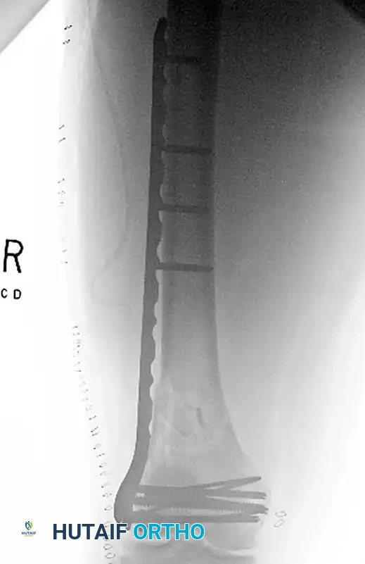
In patients with osteoporotic bone or severe comminution, a buttress plate or modern locked plate is necessary to prevent cephalad migration of the condyles and to bridge the metaphyseal defect. The following imaging sequence demonstrates a highly comminuted distal femur fracture managed with an open articular reduction and submuscular plating, highlighting the alternative approach when nailing is contraindicated by articular complexity:
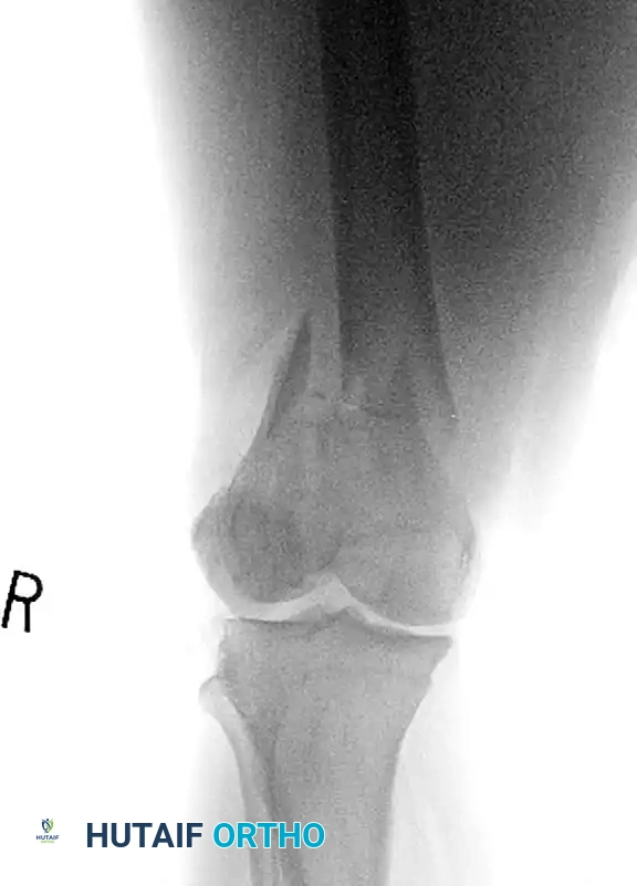
FIGURE A: Comminuted fracture of the distal femur with severe intra-articular extension. The complexity of the articular block makes standard retrograde nailing biomechanically unfavorable without first achieving absolute articular stability.
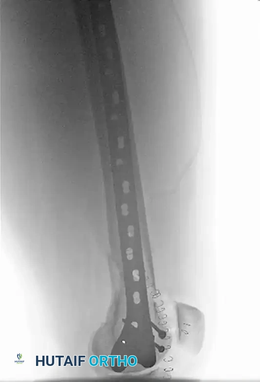
FIGURE B: AP radiograph demonstrating Open Reduction Internal Fixation (ORIF). A lateral parapatellar arthrotomy was utilized to directly view and achieve anatomical reduction of the articular component, converting the complex C-type fracture into a manageable extra-articular equivalent.
FIGURE C: Lateral radiograph showing the submuscular plate, which was positioned percutaneously following the articular reconstruction, bridging the metaphyseal comminution and providing rigid angular stability.
Complications, Incidence Rates, and Salvage Management
Despite the excellent clinical outcomes associated with retrograde nailing, surgeons must remain profoundly vigilant regarding potential complications. The biological and mechanical environment of the distal femur is unforgiving, and technical errors during the index procedure frequently necessitate complex salvage operations. A thorough understanding of these complications is essential for rapid identification and appropriate intervention.
Malunion is arguably the most common complication, with incidence rates ranging from 0% to 8% in modern series. The classic deformity is a combination of apex posterior angulation (recurvatum) and valgus. This is almost universally a result of failing to recognize and counteract the deforming forces of the gastrocnemius and the unsupported weight of the leg prior to reaming. Minor malunions may be tolerated in low-demand elderly patients, but significant mechanical axis deviation in young patients requires corrective osteotomy and revision fixation to prevent rapid unilateral compartment arthropathy of the knee.
Nonunion and hardware failure are intimately linked, occurring in 4% to 10% of cases. These complications are most frequently related to premature weight-bearing in highly comminuted fractures where the nail is forced to act as a pure load-bearing device over a long working length. Alternatively, utilizing nails with inadequate diameters or failing to achieve sufficient subchondral distal purchase can lead to fatigue fracture of the interlocking screws or the nail itself. Salvage of a nonunion typically involves exchange nailing (reaming to a larger diameter to stimulate biology and inserting a thicker nail), plate augmentation (leaving the nail in situ and applying a lateral locked plate for rotational control), and the application of autologous bone graft or orthobiologics.
Infection following retrograde nailing is a devastating complication, though fortunately rare, with rates between 0% and 4%. The risk is minimized by meticulous soft tissue handling, limiting the size of the arthrotomy, and strictly adhering to sterile technique. Deep infections involving the medullary canal require immediate hardware removal, aggressive intramedullary reaming and irrigation (Reamer-Irrigator-Aspirator systems), and the placement of antibiotic-impregnated cement spacers or nails. In elderly patients with catastrophic failure, severe bone loss, or intractable infection, salvage may ultimately require a distal femoral replacement (megaprosthesis) to restore immediate weight-bearing capacity and limb viability.
Complications and Salvage Strategies Table
| Complication | Incidence Rate | Primary Etiology | Salvage / Management Strategy |
|---|---|---|---|
| Malunion (Apex Posterior / Valgus) | 0% - 8% | Failure to control gastrocnemius pull; lateral starting point. | Corrective distal femoral osteotomy; revision fixation with locked plating. |
| Nonunion / Delayed Union | 4% - 10% | Excessive stripping; inadequate construct stability; smoking. | Exchange nailing; plate augmentation; autologous iliac crest bone grafting. |
| Hardware Failure (Nail/Screw Breakage) | 4% - 10% | Premature weight-bearing in comminuted fractures; undersized nail. | Hardware extraction; revision to larger diameter nail or dual plating. |
| Deep Infection / Osteomyelitis | 0% - 4% | Open fractures; prolonged surgical time; poor soft tissue envelope. | Hardware removal; aggressive intramedullary debridement; antibiotic nail/spacer. |
| Anterior Knee Pain / Patellar Impingement | 10% - 15% | Failure to countersink the nail below the articular cartilage. | Nail removal once fracture is completely united; arthroscopic debridement. |
Phased Post-Operative Rehabilitation Protocols
The postoperative rehabilitation protocol is not a monolithic pathway; rather, it must be meticulously tailored to the specific fracture pattern, the patient's intrinsic bone quality, and the intraoperative assessment of fixation rigidity. The ultimate goal of rehabilitation is to restore full, painless range of motion to the knee joint while protecting the healing fracture from catastrophic mechanical failure. Communication between the orthopedic surgeon and the physical therapy team is critical to ensure adherence to weight-bearing restrictions and functional milestones.
Phase I: Immediate Post-Operative (Weeks 0-6)
The primary objectives in the immediate postoperative phase are edema control, prevention of deep vein thrombosis, and the restoration of early knee kinematics. Continuous Passive Motion (CPM) or active-assisted range of motion exercises should begin on postoperative day one. Early mobilization is absolutely critical to prevent arthrofibrosis, intra-articular adhesions, and quadriceps tethering, which are notoriously difficult to treat once established.
Weight-bearing status is heavily dependent on fracture stability. For stable fracture patterns (e.g., transverse or short oblique fractures with excellent cortical contact), touch-down weight-bearing (TDWB) or even partial weight-bearing is initiated immediately. However, in highly comminuted fractures where the nail is functioning purely as a bridging device (load-bearing rather than load-sharing), the patient must remain strictly non-weight-bearing or TDWB to prevent cyclical fatigue of the distal locking screws.
Phase II: Intermediate Rehabilitation (Weeks 6-12)
At the 6-week mark, comprehensive radiographic evaluation is performed to assess for early callus formation and the maintenance of alignment. If bridging callus is visible on orthogonal views, the patient may progressively advance their weight-bearing status. For stable constructs, this means transitioning to full weight-bearing as tolerated. For comminuted fractures, partial weight-bearing (approximately 50% of body weight) is initiated.
Physical therapy during this phase shifts focus toward aggressive quadriceps and hamstring strengthening. Closed kinetic chain exercises, such as partial wall squats and leg presses, are introduced to stimulate osteoblastic activity through controlled axial loading. Modalities to address patellofemoral tracking and soft tissue mobilization around the surgical incisions are heavily utilized.
Phase III: Advanced Strengthening and Return to Function (Months 3+)
By 12 weeks, the majority of distal femoral fractures treated with intramedullary nailing will demonstrate robust radiographic union. Once union is confirmed, all weight-bearing restrictions are lifted. Rehabilitation focuses on advanced proprioceptive training, gait normalization, and the restoration of explosive muscle power. Patients are guided through sport-specific or occupation-specific drills. It is important to counsel patients that maximal functional recovery, particularly regarding terminal knee flexion and quadriceps bulk, may take up to 12 to 18 months following the index injury. Symptomatic hardware, particularly prominent distal locking screws causing iliotibial band irritation, can be considered for removal only after a minimum of 12 months and definitive proof of solid bony consolidation.
Summary of Landmark Literature and Clinical Guidelines
The evolution of intramedullary nailing for distal femoral fractures is deeply rooted in robust clinical research and biomechanical studies. Historically, flexible intramedullary implants, such as the Zickel supracondylar device, Ender rods, and Rush rods, were utilized with some success to treat distal femoral fractures. These devices relied entirely on canal fill and three-point bending principles rather than rigid interlocking. While they provided rapid, minimally invasive stabilization for elderly patients with minimally comminuted fractures, their inability to control length and rotation led to unacceptable rates of shortening and malrotation. Today, the advent of rigid, multiplanar interlocking nails has effectively antiquated the use of flexible devices in adult distal femoral traumatology.
Modern literature heavily supports the use of retrograde nailing. The pivotal biomechanical work by Ito et al. established that retrograde nails provide equivalent axial and bending stability to traditional 95-degree condylar blade plates, validating their use as
