Clinical Case Study: Comminuted Intra-articular Distal Femur Fracture (AO/OTA 33-C3)
Key Takeaway
Evaluating a distal femur fracture involves thorough clinical examination for pain, deformity, and neurovascular status, especially in osteoporotic patients. Definitive diagnosis requires initial AP/lateral radiographs followed by CT with 3D reconstructions to characterize comminution, articular involvement (e.g., AO/OTA 33-C3), and guide preoperative planning.
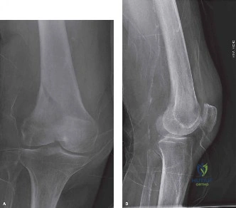
A 72-year-old female presents with an isolated right knee injury following a low-energy fall at home. She is unable to weight-bear. On examination, there is significant thigh swelling, varus deformity, and shortening. Distal pulses are palpable. You are provided with the initial lateral radiograph. Describe your findings and the immediate management priorities.
Candidate: The radiograph shows a complex distal femur fracture with extension into the knee joint. It is comminuted with posterior displacement of the distal fragment. I would assess the patient for polytrauma, check distal neurovascular status, and obtain a CT scan for surgical planning.
Failing to classify the injury or missing the severity of the articular component. Candidates often jump straight to "I will fix this with a plate" without mentioning the necessity of a CT scan for 3D mapping, or failing to acknowledge the implications of the patient's comorbidities (osteoporosis/diabetes) on implant choice.
Describe the fracture systematically: "The lateral radiograph demonstrates a comminuted, intra-articular supracondylar distal femur fracture with posterior angulation of the distal segment. Given the age and mechanism, I suspect poor bone quality. My immediate priority is a formal ATLS assessment, thorough neurovascular documentation (specifically the peroneal nerve and popliteal artery), and definitive imaging via a CT scan with 3D reconstructions. This is likely an AO/OTA 33-C3 fracture, which will require pre-operative templating for a locking plate system to address both articular congruity and metaphyseal-diaphyseal stability."
You have classified this as an AO/OTA 33-C3 fracture. What are the key principles of surgical management for this specific injury pattern, and why is the choice of implant critical here?
Candidate: The goal is to restore the joint surface and the mechanical axis. I would use a long locking plate. Locking plates are better than standard plates because they provide angular stability, which is essential in osteoporotic bone where screw purchase is poor.
Forgetting the 'biological' aspect of the fixation. A candidate who only focuses on 'rigid fixation' and ignores the importance of preserving the soft tissue envelope via minimally invasive plate osteosynthesis (MIPO) will score lower.
The management follows three pillars: 1. Anatomical Articular Reduction: Direct visualization to restore the joint surface to <2mm step-off. 2. Mechanical Axis Restoration: Correcting length, rotation, and coronal/sagittal alignment. 3. Biological Fixation: Utilizing MIPO to preserve the periosteal blood supply. The choice of a locking compression plate (LCP) is vital because it creates a fixed-angle construct, essentially acting as an internal 'ex-fix'. This prevents 'toggle' in osteoporotic bone, allowing for early weight-bearing and motion while bridging the comminuted metaphyseal zone.
The surgery is completed. The patient is now post-operative day 2. What are your specific concerns regarding the rehabilitation phase for this elderly patient?
Candidate: I would start range-of-motion exercises immediately using a CPM machine. I will also make sure she is on DVT prophylaxis and start physical therapy to work on muscle strengthening, while keeping her non-weight bearing for 6 to 12 weeks.
Ignoring the metabolic health of the patient. Simply focusing on the knee joint forgets that the patient has osteoporosis and Type 2 Diabetes; failure to mention bone health optimization and multidisciplinary care is a significant oversight.
My rehabilitation plan is multifaceted: 1. Stiffness Prevention: Early mobilization with CPM and aggressive physiotherapy. 2. Protected Loading: Strict non-weight-bearing protocol for 6-12 weeks, with progression guided by radiographic evidence of callus. 3. Systemic Optimization: Given her diabetes and osteoporosis, I would involve an endocrine/geriatric team to optimize glycemic control (HbA1c) and ensure she is on bone-strengthening therapy (e.g., Bisphosphonates or PTH analogs) to mitigate hardware failure and future fragility fractures. 4. Prophylaxis: Continued LMWH as she remains at high risk for VTE during the immobilization period.
