Achieve Lasting Results: Uncemented Tapered Fluted Hip Reimplantation
Key Takeaway
This article provides essential research regarding Achieve Lasting Results: Uncemented Tapered Fluted Hip Reimplantation. Hip reimplantation is the surgical insertion of a new prosthesis following removal of an original, infected one, typically performed as a single- or two-staged procedure. Prosthesis selection depends on factors like bone defects and host bone quality, often incorporating designs such as an uncemented tapered fluted femoral stem for optimal osseointegration. Eradicating infection is crucial for successful outcomes.
Introduction and Epidemiology
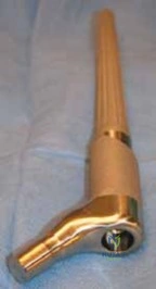
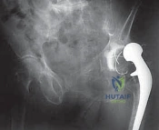
Hip reimplantation refers to the complex surgical insertion of a revision prosthesis following the removal of an original, infected total hip arthroplasty (THA) implant. The procedure is traditionally executed as the second stage of a two-stage exchange arthroplasty for periprosthetic joint infection (PJI), although single-stage exchanges are utilized in highly selected patient populations. The definitive reconstruction may utilize cemented or uncemented components; however, uncemented tapered fluted stems have become the gold standard for managing the substantial femoral bone loss frequently encountered during reimplantation.
The pathogenesis of PJI necessitates meticulous surgical and medical management. Prosthetic reimplantation is typically performed following a resection arthroplasty or first-stage revision arthroplasty where an antibiotic-loaded cement spacer was placed. Eradication of the underlying infection is the absolute prerequisite for the success of hip reimplantation surgery, regardless of the method of hip reconstruction chosen. The organisms most commonly isolated in infected total hip replacements include Staphylococcus aureus, Staphylococcus epidermidis, and various gram-negative bacteria. There is a well-documented increasing prevalence of antibiotic-resistant bacteria, including Methicillin-resistant Staphylococcus aureus (MRSA) and Vancomycin-resistant Enterococcus (VRE), which further complicates eradication protocols.
Epidemiologically, PJI occurs in approximately 1% to 2% of primary THAs and up to 4% to 6% of revision THAs. The economic and physiological burden of PJI is immense, carrying significant morbidity and a notable mortality rate that rivals certain malignancies. The natural history of a successfully treated PJI culminates in reimplantation; however, persistent or recalcitrant infection—suggested by persistently elevated inflammatory markers and confirmed by tissue or fluid culture—remains a strict contraindication to definitive reimplantation. In such scenarios, the clinical trajectory deviates toward chronic suppressive antibiotic therapy, repeat first-stage explantation, or definitive resection arthroplasty (Girdlestone procedure).
Surgical Anatomy and Biomechanics
A profound understanding of the altered surgical anatomy in the revision setting is paramount. The posterolateral approach remains the most versatile and widely utilized approach in hip reimplantation surgery due to its extensile nature and preservation of the abductor mechanism.

Neurologic Considerations in the Revision Setting
The sciatic nerve is the major neurologic structure most commonly at risk during the posterolateral approach to the hip. In the setting of a two-stage exchange, the anatomical planes are typically obliterated by dense fibrotic scar tissue induced by the infection, the previous surgeries, and the presence of the antibiotic spacer. In patients with severe scarring, it is frequently necessary to perform a formal neurolysis. The surgeon must meticulously expose the sciatic nerve as it emerges deep and inferior to the piriformis muscle and courses superficial to the obturator internus muscle.
Conversely, if a direct lateral (transgluteal) approach is utilized, the primary neurologic concern shifts to the superior gluteal nerve. The function of the hip abductors may be severely compromised if sufficient care is not taken to avoid injury to this nerve, which is predictably located approximately 5 cm proximal to the tip of the greater trochanter. Denervation of the anterior portion of the gluteus medius and the entirety of the gluteus minimus leads to a catastrophic postoperative Trendelenburg gait and profound instability.
Biomechanics of Uncemented Tapered Fluted Stems
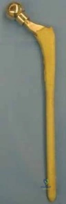
The biomechanical rationale for utilizing an uncemented tapered fluted stem in reimplantation hinges on the requirement to bypass proximal femoral bone defects (e.g., Paprosky Types II, IIIa, IIIb, and IV) to achieve stable diaphyseal fixation. These stems operate on the Wagner principle. The conical taper (typically 2 to 3 degrees) provides immediate axial stability by wedging into the diaphyseal cortical bone, thereby resisting subsidence. Concurrently, the longitudinal flutes engage the endosteal cortex to provide exceptional rotational stability.
This design relies on achieving a minimum of 4 to 5 centimeters of intimate "scratch fit" within the intact diaphysis, distal to any bone defects or osteotomies. The modularity available in modern tapered fluted stems allows the surgeon to independently establish diaphyseal fixation and subsequently adjust leg length, offset, and version via the proximal body, uncoupling fixation from hip biomechanics.
Acetabular Safe Zones
For acetabular reconstruction during reimplantation, the bone stock is often compromised, necessitating the use of highly porous multi-hole jumbo cups or augments. Screw fixation is almost universally necessary to achieve initial rigid stability for osseointegration. The safest zone for the insertion of acetabular screws remains the posterior superior quadrant (the ilium). Screws placed in the anterior superior quadrant risk injury to the external iliac vessels, while those in the anterior inferior quadrant endanger the obturator neurovascular bundle. The posterior inferior quadrant risks injury to the sciatic nerve and inferior gluteal vessels.
Indications and Contraindications
The decision to proceed with the second stage of a two-stage exchange arthroplasty requires a multidisciplinary approach involving orthopedic surgery and infectious disease specialists. The primary indication for reimplantation is the clinical and serological eradication of the periprosthetic joint infection.

Persistent or recalcitrant infection is an absolute contraindication to reimplantation surgery. This is clinically suggested by persistently raised C-reactive protein (CRP) and erythrocyte sedimentation rate (ESR) values, and definitively confirmed by positive tissue or fluid cultures obtained via aspiration or biopsy. The options in such a scenario are to continue with targeted intravenous antibiotics, repeat the first-stage procedure (spacer exchange), or perform a definitive resection arthroplasty.
Operative vs Non Operative Management Parameters
| Clinical Scenario | Recommended Management Strategy | Rationale and Considerations |
|---|---|---|
| Normalized ESR/CRP, Negative Aspirate, Healed Soft Tissues | Definitive Reimplantation (Second Stage) | Infection is considered eradicated; host bone is ready for uncemented tapered fluted stem osseointegration. |
| Equivocal ESR/CRP, Negative Aspirate, Good Clinical Exam | Proceed with caution; consider intraoperative frozen section or Nuclear Medicine Imaging | Rule out false-negative aspirate. Intraoperative histology showing <5 PMNs per high-power field supports reimplantation. |
| Persistently Elevated ESR/CRP, Positive Aspirate | Repeat First-Stage (Spacer Exchange) or Resection Arthroplasty | Active infection remains. Placing definitive uncemented implants will result in immediate reinfection and catastrophic failure. |
| Medically Unfit Patient, High Perioperative Mortality Risk | Chronic Suppressive Antibiotics or Definitive Spacer Retention | Surgical risk outweighs the functional benefit of reimplantation. |
| Massive Unreconstructible Bone Loss (Pelvic Discontinuity, Femoral Destruction) | Proximal Femoral Replacement or Custom Triflange / Cup-Cage | Standard uncemented tapered fluted stems or jumbo cups are insufficient for the degree of bone loss. |
Pre Operative Planning and Patient Positioning
Thorough preoperative assessment is the cornerstone of successful hip reimplantation. The initial assessment should begin with a comprehensive general medical examination to optimize the patient for a major physiological stressor.
Patient History and Physical Findings
The main presenting symptom of patients with an active periprosthetic infection is pain, particularly constant, unrelenting pain while the patient is at rest. In the context of evaluating a patient with an antibiotic spacer, pain must be carefully differentiated between mechanical spacer pain (which is common with mobility) and infectious pain (which is constant).
Delayed wound healing, persistent wound drainage, and a history of superficial wound infection after the primary procedure are highly suggestive of ongoing infection. Risk factors for infection must be optimized, including a history of diabetes mellitus (targeting HbA1c < 7.0%), chronic skin lesions, the use of systemic corticosteroids, malnutrition, and any type of immunocompromise.
The hip wound is examined for warmth, erythema, fluctuance, discharging sinuses, and the presence of any hematoma. An erythematous, warm wound with draining sinuses indicates persistent, ongoing infection, precluding reimplantation. Defects in the underlying fascia are often palpable and may indicate a higher risk of wound dehiscence postoperatively, necessitating advanced soft tissue coverage planning. The abductors should be palpated and their function assessed, as abductor deficiency will dictate the need for constrained liners or dual mobility constructs. A full neurologic examination and palpation of distal pulses are mandatory. Preoperative weakness of the leg extensors or a partial foot drop may indicate existing scarring around the sciatic nerve, which must be documented and addressed intraoperatively.
Imaging and Diagnostic Studies
Infection exclusion is driven by serial assessment of the ESR (normal < 30 mm/hr) and CRP (normal < 10 mg/L). Usually, this requires the patient to demonstrate a downward trend or normalization of these values following the cessation of systemic antibiotics (typically a 2 to 4 week antibiotic holiday). Hip aspiration for cell count, differential, and culture is mandatory. Occasionally, test results may be equivocal; in such cases, nuclear medicine imaging (such as a combined leukocyte-marrow scan) or next-generation sequencing (NGS) of synovial fluid is required to confirm the absence of infection.
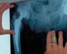
Preoperative radiographic templating is critical for the selection of prostheses. The surgeon must evaluate orthogonal radiographs of the femur to assess the Paprosky defect classification. For uncemented tapered fluted stems, the surgeon must identify the true isthmus of the femur and template for a stem that will achieve at least 4 to 5 cm of diaphyseal engagement distal to any planned Extended Trochanteric Osteotomy (ETO) or existing cortical defects.

Patient Positioning
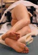
The patient is typically positioned in the lateral decubitus position on a rigid beanbag or peg-board system. Meticulous padding of all bony prominences (e.g., the peroneal nerve at the fibular head of the down leg, the axilla) is required. The pelvis must be rigidly secured to allow for accurate intraoperative assessment of acetabular version and inclination. The surgical field is prepped and draped in a wide, sterile fashion to allow for extensile exposures, including distal extension to the knee if a transfemoral approach or extensive diaphyseal exposure becomes necessary.
Detailed Surgical Approach and Technique
The surgical execution of hip reimplantation using an uncemented tapered fluted stem demands precision, patience, and adherence to strict oncologic-like principles for infection eradication.
Surgical Exposure and Extended Trochanteric Osteotomy
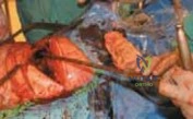
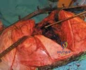
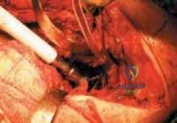
The posterolateral approach is utilized, often extending the previous incision. Deep dissection proceeds through the fascia lata and the gluteus maximus. An Extended Trochanteric Osteotomy (ETO) is frequently required in reimplantation surgery. The ETO provides unparalleled exposure to the acetabulum, facilitates the safe extraction of a well-fixed antibiotic cement spacer, allows for direct visualization of the femoral canal for reaming, and permits controlled advancement of the abductor mechanism to restore resting tension.
The ETO is performed by identifying the posterior border of the vastus lateralis and elevating it anteriorly. A controlled osteotomy is created using an oscillating saw and osteotomes, typically measuring 12 to 15 cm from the tip of the greater trochanter, ensuring it remains proximal to the planned diaphyseal fixation zone of the tapered stem. The osteotomized fragment, retaining the continuity of the gluteus medius, minimus, and vastus lateralis, is reflected anteriorly.
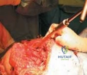
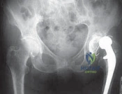
Spacer Extraction and Debridement
The antibiotic spacer is carefully extracted. In cases where the spacer was articulated or heavily cemented, care must be taken to avoid iatrogenic fracture of the thinned host bone. Following extraction, a radical debridement of all pseudocapsule, fibrotic scar tissue, and retained cement is performed. Five distinct tissue samples are sent for aerobic, anaerobic, acid-fast bacilli, and fungal cultures, alongside a sample for frozen section histology to rule out acute inflammation (>5 polymorphonuclear leukocytes per high-power field).

Acetabular Reconstruction
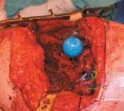
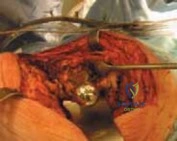
The acetabulum is exposed circumferentially. The selection of prostheses for acetabular reconstruction is determined by the quality and quantity of remaining host bone. Hemispherical reaming is performed to bleeding subchondral bone. A highly porous titanium or tantalum cup is impacted into place. Screw fixation is often necessary to ensure absolute initial stability. Screws are directed into the posterior superior quadrant (the ilium) to maximize purchase and avoid neurovascular injury. In cases of severe bone loss, trabecular metal augments or a cup-cage construct may be required.
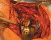
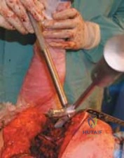
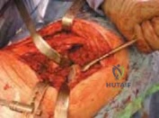
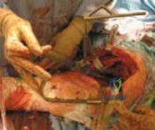
Femoral Preparation and Stem Insertion
The femoral canal is assessed. The key to success with an uncemented tapered fluted stem is achieving rigid diaphyseal fixation.
- Canal Clearing: Flexible reamers or rigid hand reamers are used to clear residual cement and fibrous tissue from the diaphysis.
- Prophylactic Cable: Prior to aggressive reaming or broaching, a prophylactic cerclage cable is placed just distal to the ETO or the lowest bony defect to prevent propagation of a longitudinal fracture during stem impaction.
- Sequential Reaming: Conical reamers are introduced sequentially. The surgeon must feel for the "chatter" that indicates engagement of the cortical bone in the diaphysis. The reaming depth must correspond to the templated size, ensuring at least 4 cm of scratch fit.
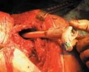
- Stem Impaction: The definitive fluted tapered stem is impacted. The flutes cut into the endosteal cortex, providing rotational stability. The axial seating is confirmed by a change in pitch during impaction and visual confirmation against a fixed bony landmark.
- Modularity and Trialing: If a modular stem is utilized, the proximal body trial is placed. The hip is reduced, and trialing is performed to optimize leg length, offset, and stability. The hip is taken through a full range of motion to ensure absence of impingement and resistance to dislocation.
Closure
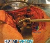
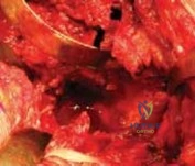
Once the definitive proximal body and head are impacted, the hip is reduced. The ETO is reduced and secured using multiple cerclage cables. Care is taken to ensure rigid compression of the osteotomy site to promote bone healing. The soft tissues are closed in layers over closed suction drains. The fascia lata must be closed meticulously to prevent herniation and ensure a watertight seal.
Complications and Management
Revision hip arthroplasty, particularly in the setting of reimplantation for infection, carries a significantly higher complication profile than primary THA. The surgeon must be prepared to identify and manage these complications intraoperatively and postoperatively.
Intraoperative Complications
Intraoperative fractures are a primary concern when utilizing uncemented tapered fluted stems. The hoop stresses generated by the wedging effect of the conical taper can easily split the compromised diaphyseal bone. This is mitigated by the routine use of prophylactic cerclage cables placed distal to the anticipated fixation zone prior to reaming and impaction. If a fracture occurs, the stem must bypass the most distal aspect of the fracture by at least two cortical diameters, which may necessitate extracting the stem and utilizing a longer implant.

Postoperative Complications
Subsidence is a known complication of tapered fluted stems, particularly if the initial scratch fit was suboptimal or if the bone quality is exceedingly poor. While minor subsidence (1-3 mm) may stabilize as the stem wedges further into the taper, progressive subsidence requires revision. Dislocation is another major risk due to compromised soft tissues, abductor deficiency, and altered biomechanics.
Summary of Complications and Salvage Strategies
| Complication | Incidence Rate | Management and Salvage Strategies |
|---|---|---|
| Intraoperative Femoral Fracture | 5% - 15% | Prophylactic cabling. Bypass fracture by 2 cortical diameters with a longer stem. Strut allografts for severe comminution. |
| Stem Subsidence (>5mm) | 3% - 10% | Observation if asymptomatic and stable. Revision to a larger diameter or longer stem if progressive or causing severe leg length discrepancy. |
| Dislocation | 5% - 20% | Closed reduction and bracing. Revision to a dual mobility articulation or constrained liner if recurrent. Address any component malposition. |
| Recurrent PJI | 5% - 15% | Suppressive antibiotics if implants are stable and patient is a poor surgical candidate. Repeat two-stage exchange or definitive resection arthroplasty. |
| ETO Nonunion / Migration | 2% - 8% | Observation if asymptomatic. Revision internal fixation with cables/claw plate if causing severe abductor weakness or pain. |
| Sciatic Nerve Palsy | 1% - 4% | Ankle-foot orthosis (AFO) for foot drop. Gabapentinoids for neuropathic pain. Most partial palsies recover within 12-18 months. |
Post Operative Rehabilitation Protocols
Rehabilitation following an uncemented tapered fluted hip reimplantation must be carefully tailored to the intraoperative findings, the stability of the implant, and the status of the ETO.

Weight Bearing Status
Unlike primary THA where immediate full weight-bearing is encouraged, the rehabilitation protocol for a tapered fluted stem with an ETO is typically more conservative. Patients are generally restricted to toe-touch or partial weight-bearing (approximately 20 to 30 lbs) on the operative extremity for the first 6 to 8 weeks. This restriction minimizes axial loading forces that could induce subsidence of the stem before osseointegration occurs, and protects the ETO from superior migration caused by the pull of the abductor musculature.
Physical Therapy and Precautions
Strict posterior hip precautions are maintained for 12 weeks to prevent dislocation. Active abduction is generally prohibited for 6 to 8 weeks to protect the ETO repair. Physical therapy focuses on passive and active-assisted range of motion within the safe zones, isometric quadriceps and gluteal sets, and upper extremity strengthening to assist with walker or crutch use.
Deep vein thrombosis (DVT) prophylaxis is mandatory, typically utilizing low molecular weight heparin, direct oral anticoagulants, or aspirin, depending on the patient's risk stratification. Routine radiographic follow-up at 2 weeks, 6 weeks, 3 months, and 1 year is essential to monitor for subsidence, ETO healing, and signs of osseointegration (such as spot welds).
Summary of Key Literature and Guidelines
The management of PJI and the utilization of uncemented tapered fluted stems are heavily guided by established literature and international consensus guidelines.

The Musculoskeletal Infection Society (MSIS) and the International Consensus Meeting (ICM) on PJI provide the definitive criteria for diagnosing PJI and determining the timing for reimplantation. The criteria emphasize the importance of major criteria (sinus tract communicating with the prosthesis, or two positive cultures of the same organism) and minor criteria (elevated serum CRP/ESR, elevated synovial WBC count, elevated synovial PMN percentage, positive histological analysis). Adherence to these guidelines ensures that reimplantation is only performed when the joint is sterile.