Flexor Hallucis Longus Tenosynovitis & Os Trigonum Syndrome: A Ballet Dancer Case Study
Key Takeaway
Flexor Hallucis Longus (FHL) tenosynovitis and os trigonum syndrome are often diagnosed by posterior ankle pain exacerbated by hallux flexion and passive dorsiflexion. Clinical examination reveals FHL tenderness, crepitus, and positive impingement tests. Radiographs may show an os trigonum, while MRI confirms FHL sheath inflammation and soft tissue pathology, crucial for elite athletes like ballet dancers.
A 32-year-old professional ballet dancer presents with a 14-month history of insidious, right posterior ankle pain. It is worse with 'en pointe' work and jumping. She has failed 6 months of conservative management including physio and ultrasound-guided steroid injection. Physical examination reveals tenderness at the posteromedial ankle and a painful posterior impingement test. Given this presentation, what is your primary clinical suspicion and how would you investigate it?
Candidate: I suspect a combination of posterior ankle impingement syndrome and flexor hallucis longus (FHL) tenosynovitis. I would order weight-bearing radiographs, specifically a lateral view to look for an os trigonum. I would also order an MRI of the ankle to assess for bone marrow edema in the posterior talus and fluid in the FHL sheath.
Focusing only on the os trigonum and ignoring the FHL. Failing to mention that the MRI should be non-contrast or failing to emphasize the need for dynamic imaging (like ultrasound) to confirm the "snapping" or "triggering" component of the FHL pathology.
Structure the answer by anatomy and pathology. "This presentation is classic for 'Dancer’s Tendinitis,' involving posterior impingement (often due to an os trigonum) and FHL stenosing tenosynovitis. Investigations must be two-fold: First, weight-bearing radiographs (AP/Lateral) to evaluate the os trigonum/Stieda process. Second, MRI to assess the synchondrosis (bone marrow edema) and the FHL tendon sheath (tenosynovitis). Finally, I would mention dynamic ultrasound, as it is highly sensitive for identifying FHL 'triggering' and provides functional correlation to the patient’s clinical symptoms."
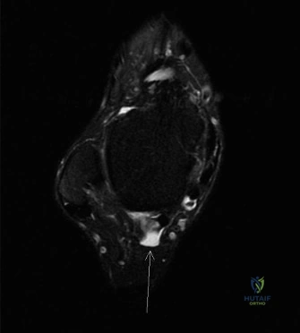
Based on the MRI findings below, discuss the surgical management. How would you approach this case, and what are the critical anatomical landmarks you must respect during the procedure?
Candidate: I would recommend posterior ankle endoscopy for excision of the os trigonum and release of the FHL fibro-osseous tunnel. The critical landmark is the FHL tendon itself. All dissection must remain lateral to the tendon to protect the tibial neurovascular bundle which lies medially.
Suggesting an open approach without justification. Ignoring the risk to the sural nerve when creating the posterolateral portal, or failing to mention dynamic intraoperative testing.
Clearly state the preference for endoscopy due to faster recovery and reduced scarring. Structure the "Safety" component: 1) Posterolateral portal placement is at risk to the sural nerve (requires blunt dissection). 2) The FHL tendon is the 'compass'—all work remains lateral to it. 3) The tibial neurovascular bundle is medial to the FHL. 4) Emphasize the requirement for dynamic intraoperative testing: move the hallux to ensure free excursion and plantarflex the ankle to confirm complete bony resection.
