Diagnosing UBC From Aneurysmal: Avoid Pitfalls in Orthopedics
Key Takeaway
This article provides essential research regarding Diagnosing UBC From Aneurysmal: Avoid Pitfalls in Orthopedics. A unicameral bone cyst (UBC) is a common, serous fluid-filled bone lesion typically seen in patients under 20. Its natural history is to fill with bone as the patient reaches skeletal maturity. Distinguishing ubc from aneurysmal bone cysts, UBCs rarely contain blood unless a fracture is present, whereas aneurysmal cysts are often filled with blood.
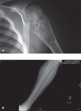
A 12-year-old boy presents to your clinic with persistent dull pain in the left proximal humerus. He has had no trauma. Radiographs show a lytic, central, metaphyseal lesion. You suspect a Unicameral Bone Cyst (UBC). What are the critical radiographic features you would look for to distinguish this from an Aneurysmal Bone Cyst (ABC), and what further imaging would you request?
Candidate: I would look for the location and margins. A UBC is typically central and metaphyseal with a sclerotic rim. An ABC is usually eccentric and more expansile, often with a "blown-out" or "soap-bubble" appearance. I would definitely order an MRI to look for fluid-fluid levels, which are pathognomonic for an ABC.
Candidates often stop at mentioning the "soap-bubble" appearance or simple X-rays. They fail to mention the "fallen leaf sign" (pathognomonic for fractured UBC) or the necessity of MRI for fluid-fluid levels, which is the high-stakes differentiator. Failing to mention MRI as the gold standard for differentiation is a critical oversight.
To distinguish these, I focus on growth and architecture: UBCs are typically central and unilocular. I look for the "fallen leaf sign" if a fracture has occurred. ABCs are eccentric, expansile, and show a thin periosteal shell. The gold standard for differentiation is an MRI, where I specifically look for fluid-fluid levels, representing blood products of different ages. I would also note that ABCs show enhancing septa on T2-weighted MRI, whereas UBCs appear as simple, non-enhancing fluid-filled cavities.
The MRI confirms a lesion consistent with an ABC in the proximal femur. The patient is 14 years old and active in sports. He is currently asymptomatic, but the lesion appears large and the cortex is thinned. How would you structure your decision-making regarding surgical intervention versus conservative management?
Candidate: I would consider the risk of fracture. If the cortex is significantly thinned or the lesion is in a weight-bearing zone like the proximal femur, I would likely recommend surgery. I'd mention curettage and bone grafting, and potentially internal fixation if the structural integrity is compromised.
Candidates often jump straight to surgery. They fail to quantify "large" or "weak." A top-tier candidate must reference established risk criteria: cortical thinning >50% or the lesion occupying >2/3 of the bone diameter. Also, they frequently miss mentioning the need for pre-operative arterial embolization for large, vascularized ABCs.
My decision-making follows a structured hierarchy: 1. Structural Risk: I assess cortical thinning (>50%) and lesion size (>2/3 bone diameter). Given this is the proximal femur (weight-bearing), the threshold for surgery is lower. 2. Lesion Type: ABCs are aggressive; therefore, observation is rarely appropriate unless the lesion is tiny and asymptomatic. 3. Surgical Planning: If surgery is indicated, I must consider pre-operative selective arterial embolization to reduce intraoperative hemorrhage, as ABCs are highly vascular. 4. Technique: I would plan for aggressive intralesional curettage with high-speed burring and adjuvants (like phenol or cryotherapy) to minimize the high recurrence rates associated with ABCs.
During your fellowship, you notice a high recurrence rate of ABCs after simple curettage and bone grafting. What specific intraoperative steps or "adjuvants" would you employ to reduce this risk in your next case?
Candidate: I would use a high-speed burr to ensure all septa and cyst lining are removed. I could also use adjuvants like phenol or liquid nitrogen for cryotherapy to kill any residual abnormal cells.
Candidates often mention these adjuvants without acknowledging the severe risks involved. Using phenol or cryotherapy near neurovascular structures or growth plates without appropriate protection is a recipe for iatrogenic disaster. They fail to mention the critical need for meticulous local soft tissue protection.
Aggressive surgical clearance is the baseline. To reduce recurrence: 1. Mechanical: Use of a high-speed burr to remove the "rim" of the cavity, extending beyond the visible cyst wall. 2. Chemical/Thermal Adjuvants: I would use phenol (followed by alcohol neutralization) or cryotherapy (liquid nitrogen). 3. Safety: These are high-risk maneuvers. I would ensure meticulous protection of adjacent neurovascular structures and soft tissues using barrier drapes or saline-soaked sponges. 4. Biological: If the cavity is massive, I would consider filling it with autologous bone graft mixed with DBM or synthetic substitutes to provide a scaffold for healing, while confirming the diagnosis with USP6 gene translocation testing to rule out other giant cell-rich mimics.
