Part of the Master Guide

Femoral Neck Fractures: Epidemiology, Surgical Anatomy, Classification, & Management

Femoral Neck Fracture Case Study: Comprehensive Clinical and Diagnostic Approach

17 Apr 2026 20 min read 169 Views
Illustration of case femoral neck - Dr. Mohammed Hutaif

Key Takeaway

Femoral neck fractures are diagnosed through detailed clinical examination revealing pain, inability to bear weight, limb shortening, and external rotation, confirmed by plain radiographs. They are classified using systems like Garden (e.g., Type III for displaced fractures with valgus impaction) and Pauwels (e.g., Type II for significant shear forces) to guide treatment and prognosis.
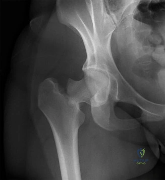
Patient Presentation and History

Mechanism of Injury and Biomechanics

A 78-year-old female presented to the emergency department following a mechanical fall from a standing height. The patient reported tripping over a rug and landing directly onto her right lateral hip. This mechanism is the classic vector for proximal femur fractures in the geriatric population, where a direct lateral impact to the greater trochanter bypasses the energy-absorbing mechanisms of the lower extremity musculature. The kinetic energy is transmitted directly through the trochanteric region into the femoral neck. She immediately experienced severe pain localized to the right groin and hip articulation, rendering her entirely non-ambulatory and unable to bear weight.

Demographics and Preinjury Functional Status

Understanding the patient's baseline physiological reserve and functional demands is paramount in orthopedic trauma, particularly in geriatric hip fractures. Her pre-injury functional status was relatively high, documented as a Modified Harris Hip Score of 85. She lives independently, manages her activities of daily living without assistance, and utilizes a single-point cane solely for community ambulation outdoors, remaining unassisted indoors. This level of baseline activity categorizes her as a community ambulator, which heavily influences the algorithm for surgical intervention, specifically pushing the decision matrix toward arthroplasty constructs that allow immediate full weight-bearing and restore high functional capacity.

Comorbidities and Pharmacologic Considerations

Her medical history is significant for well-controlled essential hypertension and Type 2 Diabetes Mellitus managed with oral hypoglycemic agents. The presence of diabetes necessitates strict perioperative glycemic control to mitigate the risk of surgical site infections (SSIs) and optimize wound healing, given the known microvascular complications associated with the disease.

Furthermore, she has a documented history of osteoporosis, currently managed with alendronate, a bisphosphonate. While bisphosphonates increase bone mineral density by inhibiting osteoclast-mediated bone resorption, long-term use can alter bone turnover dynamics. Although her presentation is classic for an osteoporotic fragility fracture rather than an atypical subtrochanteric femur fracture (which is classically associated with prolonged bisphosphonate therapy), her altered bone metabolism must be factored into implant selection, particularly regarding the choice between cemented versus cementless femoral stems. She is also on aspirin 81mg daily for primary cardiovascular prevention. Current orthopedic trauma guidelines generally support proceeding with urgent hip fracture surgery without delaying for aspirin cessation, as the morbidity of delay significantly outweighs the hemorrhagic risks associated with low-dose aspirin. She reports no prior history of falls, suggesting this was an isolated mechanical event rather than a manifestation of underlying syncope, arrhythmia, or progressive neuromuscular decline.

Clinical Examination Findings

Visual Inspection and Deformity

Initial assessment in the trauma bay revealed a patient in moderate distress secondary to acute nociceptive pain. Inspection of the right lower extremity demonstrated the classic deformity associated with displaced proximal femur fractures: the limb was subtly shortened and positioned in fixed external rotation compared to the contralateral side.

This characteristic deformity is biomechanically driven. The shortening is a direct result of the proximal migration of the distal femoral segment, driven by the unopposed longitudinal pull of the robust thigh musculature, primarily the rectus femoris, adductor complex, and the hamstrings. The external rotation is dictated by two primary factors: gravity acting on the unanchored lower limb, and the unopposed pull of the iliopsoas muscle on the lesser trochanter, which exerts an anterior and superior vector, causing the femoral shaft to externally rotate when structural continuity with the femoral head is lost. No obvious open wounds, ecchymosis, or significant abrasions were present, indicating a closed injury. Mild diffuse swelling was observed around the right hip joint, though contained within the robust fascial compartments of the thigh.

Palpation and Provocative Maneuvers

Palpation elicited significant, focal tenderness over the right greater trochanter and deep within the femoral triangle, correlating with the intracapsular hematoma and periosteal disruption. Systematic palpation of the anterior superior iliac spine, pubic symphysis, iliac crests, and sacroiliac joints did not elicit tenderness, effectively decreasing the clinical suspicion for concomitant pelvic ring injuries.

Range of Motion Assessment

Active range of motion of the right hip was severely restricted and excruciatingly painful in all planes, effectively zero. Passive range of motion assessment revealed a fixed external rotation deformity. Any attempt to passively internally rotate or flex the hip exacerbated the pain significantly, confirming capsular distension and osseous instability. A notable pseudo-flexion was observed upon attempted passive hip extension, a compensatory mechanism due to the loss of the normal fulcrum of the hip joint.

Neurological and Vascular Status

A meticulous neurovascular examination is critical to rule out concomitant neurovascular compromise, although rare in isolated, low-energy femoral neck fractures. Sensation to light touch and pinprick was intact and symmetric throughout the right lower extremity across all dermatomes (L2-S1). Motor strength was challenging to quantify accurately due to pain inhibition (muscle guarding), but was grossly intact in ankle dorsiflexion (deep peroneal nerve), plantarflexion (tibial nerve), and extensor hallucis longus function. Sciatic and femoral nerve motor and sensory distributions appeared grossly normal.

Vascular assessment demonstrated strong and symmetric distal pulses, including the femoral, popliteal, dorsalis pedis, and posterior tibial arteries. Capillary refill was brisk (under two seconds) in the right foot, and the extremity was warm and well-perfused. The clinical presentation was unequivocally highly consistent with a displaced right femoral neck fracture.

Imaging and Diagnostics

Standard Radiographic Evaluation

Standard trauma protocols dictate immediate plain radiographic evaluation. An anteroposterior (AP) radiograph of the pelvis and a dedicated cross-table lateral view of the right hip were obtained in the emergency department. The standard AP hip view with 15 degrees of internal rotation to profile the femoral neck was not attainable due to the patient's severe pain and fixed external rotation deformity.

The AP Pelvis radiograph demonstrated a displaced intracapsular fracture of the right femoral neck. The fracture line propagated obliquely, extending from the subcapital region inferomedially toward the basicervical junction. The femoral head was positioned in relative valgus with respect to the femoral shaft, and there was approximately 1 cm of superior displacement of the femoral shaft relative to the head. The greater and lesser trochanters appeared structurally intact, confirming the fracture was isolated to the intracapsular neck region.

The cross-table lateral hip radiograph was instrumental in confirming the true extent of displacement and provided critical multi-planar visualization of the fracture morphology. It revealed significant posterior comminution of the femoral neck and extension of the fracture into the basicervical region. The lateral view is essential as AP radiographs alone can severely underestimate the degree of posterior tilt and displacement in femoral neck fractures.

Image

Advanced Imaging Modalities

While not routinely indicated for straightforward, clearly displaced femoral neck fractures that are destined for arthroplasty, advanced imaging such as Computed Tomography (CT) or Magnetic Resonance Imaging (MRI) can be invaluable in specific, nuanced clinical scenarios.

A CT scan with fine 1mm cuts and 3D reconstruction is indicated when there is suspicion of associated acetabular fractures, pelvic ring disruptions, or when evaluating the exact degree of comminution in a younger patient where joint-preserving internal fixation is being considered. In this elderly patient with a clearly displaced fracture, a CT scan would not alter the surgical management plan and was therefore deferred to minimize radiation exposure and expedite surgical intervention.

MRI remains the gold standard for diagnosing occult femoral neck fractures. In patients presenting with a classic clinical picture of a hip fracture but negative or equivocal initial plain radiographs, an MRI utilizing T1-weighted and Short Tau Inversion Recovery (STIR) sequences should be obtained urgently. T1 sequences will demonstrate a hypointense fracture line, while STIR sequences will highlight hyperintense localized bone marrow edema.

Preoperative Templating

Preoperative templating is a mandatory step in the diagnostic and preparatory phase. Utilizing digital templating software calibrated with a radiographic marker, the contralateral, uninjured hip was templated to determine the native center of rotation, femoral offset, and leg length.

In this patient, templating focused on sizing the appropriate femoral stem and acetabular component (or bipolar head). The medullary canal diameter was assessed to determine the appropriate stem size and to evaluate the cortical thickness (Dor type bone classification). Given her history of osteoporosis, her bone morphology leaned towards Dorr Type B or C, characterized by a widened metaphyseal canal and thinner diaphyseal cortices, which strongly influences the decision toward a cemented femoral stem to ensure immediate macroscopic interlock and prevent subsidence.

Differential Diagnosis

When evaluating an elderly patient with acute hip pain following a fall, the differential diagnosis must encompass various osseous and soft tissue injuries. A systematic approach is required to differentiate between intracapsular and extracapsular proximal femur fractures, as well as pelvic ring injuries, as the surgical management algorithms diverge significantly.

Diagnosis Mechanism of Injury Clinical Presentation Radiographic Hallmarks Management Strategy
Femoral Neck Fracture (Intracapsular) Low-energy lateral fall on greater trochanter. Shortened, externally rotated limb. Pain in groin. No ecchymosis. Fracture line proximal to the intertrochanteric line. Disruption of Shenton's line. Arthroplasty (Hemi or THA) in elderly. ORIF in young patients or non-displaced fractures.
Intertrochanteric Femur Fracture (Extracapsular) Low-energy fall. Often involves torsional forces. Shortened, severely externally rotated limb. Significant thigh swelling and ecchymosis. Fracture line extending between greater and lesser trochanters. Extracapsular. Cephalomedullary nailing or Sliding Hip Screw (DHS). Arthroplasty rarely indicated.
Subtrochanteric Femur Fracture High-energy trauma in young; low-energy or bisphosphonate-related in elderly. Deformity of proximal thigh. Proximal fragment flexed, abducted, externally rotated. Fracture line within 5cm distal to the lesser trochanter. Often transverse or short oblique if atypical. Long cephalomedullary nailing. Open reduction often required due to severe deforming muscle forces.
Lateral Compression Pelvic Ring Injury Lateral impact to the iliac wing or greater trochanter. Groin or buttock pain. Leg length usually equal. Pain with pelvic compression. Sacral impaction fractures, superior/inferior pubic rami fractures. Non-operative (weight-bearing as tolerated) for stable patterns. Percutaneous fixation for unstable patterns.
Occult Hip Fracture Low-energy fall. Groin pain with weight-bearing. Minimal to no resting deformity. Normal or equivocal plain radiographs. Requires MRI (T1/STIR) for definitive diagnosis. Prophylactic in situ fixation (cannulated screws) to prevent displacement.

Surgical Decision Making and Classification

Fracture Classification Systems

Accurate classification of femoral neck fractures is critical for standardizing communication, predicting the risk of avascular necrosis (AVN), and guiding surgical decision-making. Two primary classification systems are utilized: the Garden classification and the Pauwels classification.

Based on the AP pelvis radiograph, this patient's fracture was classified as a Garden Type III. The Garden classification evaluates the degree of displacement based on the alignment of the compressive trabeculae in the femoral head relative to the femoral neck.
* Garden I: Incomplete, valgus-impacted fracture.
* Garden II: Complete fracture, non-displaced.
* Garden III: Complete fracture, partially displaced. The trabecular pattern of the femoral head does not align with the acetabulum.
* Garden IV: Complete fracture, fully displaced. The femoral head loses contact with the femoral neck and aligns itself within the acetabulum.

While historically significant, modern orthopedic trauma often simplifies the Garden classification into "non-displaced" (Garden I/II) and "displaced" (Garden III/IV) due to poor inter-observer reliability and because the treatment algorithm primarily hinges on this binary distinction.

The fracture angle, estimated at approximately 50-60 degrees to the horizontal on the AP radiograph, placed it in Pauwels Type II. The Pauwels classification is based on the biomechanical angle of the fracture line relative to the horizontal plane.
* Pauwels I: < 30 degrees. Predominantly compressive forces, highly stable.
* Pauwels II: 30-50 degrees. Increasing shear forces, decreased stability.
* Pauwels III: > 50 degrees. Predominantly shear forces, highly unstable with a high rate of nonunion and varus collapse if treated with internal fixation.

Biomechanical Considerations and Vascular Anatomy

The surgical decision-making process for femoral neck fractures is heavily dictated by the unique vascular anatomy of the proximal femur. The primary blood supply to the femoral head is derived from the medial femoral circumflex artery (MFCA), specifically its lateral epiphyseal branches, which traverse the femoral neck intracapsularly. A displaced fracture (Garden III/IV) reliably disrupts these retinacular vessels, leading to a high risk of avascular necrosis (AVN) and subsequent structural collapse of the femoral head.

Furthermore, intracapsular fractures lack a cambium layer of the periosteum, meaning healing relies entirely on endosteal callus formation. The presence of synovial fluid within the joint space further inhibits osteogenesis. The combination of precarious blood supply, lack of periosteal healing, and high shear forces (Pauwels II/III) results in unacceptable rates of nonunion and AVN (ranging from 20% to 40%) when displaced fractures are treated with internal fixation in the elderly population.

Treatment Algorithm Formulation

Given the patient's age (78 years), the displaced nature of the fracture (Garden III), and the high shear angle (Pauwels II), joint-preserving internal fixation (e.g., multiple cannulated screws or a dynamic hip screw) is contraindicated due to the prohibitively high risk of fixation failure, nonunion, and AVN. The definitive treatment pathway mandates arthroplasty.

The secondary decision involves choosing between a Hemiarthroplasty and a Total Hip Arthroplasty (THA).
* Hemiarthroplasty (Unipolar or Bipolar): Involves replacing only the femoral side. It is a shorter operation with a lower dislocation rate, suitable for lower-demand, older patients. However, it carries a risk of progressive acetabular wear and groin pain over time.
* Total Hip Arthroplasty (THA): Involves replacing both the femoral head and the acetabulum. It provides superior functional outcomes, better pain relief, and eliminates the risk of acetabular erosion. It is the procedure of choice for active, independent, community ambulators.

Based on landmark studies, including the HEALTH (Hip Fracture Evaluation with Alternatives of Total Hip Arthroplasty versus Hemiarthroplasty) trial, THA provides superior long-term functional outcomes in independent, cognitively intact, active elderly patients. Given this patient's high pre-injury functional status (mHHS 85), independent living situation, and community ambulation, a Total Hip Arthroplasty (THA) is the most appropriate surgical intervention to restore her baseline function and provide a durable, pain-free joint.

Regarding stem fixation, given her documented osteoporosis and anticipated Dorr Type B/C bone morphology, a cemented femoral stem is highly recommended. Cemented stems in the elderly have been definitively shown to reduce the risk of intraoperative and postoperative periprosthetic femur fractures compared to cementless stems, without compromising long-term survivorship.

Surgical Technique and Intervention

Anesthesia and Patient Positioning

The patient was evaluated by the anesthesia team and deemed appropriate for a combined spinal-epidural anesthesia with conscious sedation. Regional anesthesia is preferred in geriatric hip fractures as it reduces intraoperative blood loss, minimizes the risk of deep vein thrombosis, and avoids the cognitive sequelae associated with general anesthesia.

The patient was positioned in the lateral decubitus position on a radiolucent operating table, with the right hip facing superiorly. Rigid pelvic positioners were utilized anteriorly at the pubic symphysis and posteriorly at the sacrum to ensure strict orthogonal alignment of the pelvis, which is critical for accurate assessment of acetabular component version and inclination. All bony prominences, particularly the contralateral peroneal nerve at the fibular head, were meticulously padded. The surgical site was prepped and draped in a standard sterile fashion, allowing for full manipulation of the operative leg.

Surgical Approach Selection

A standard Posterior Approach (Moore or Southern approach) to the hip was selected. This approach provides excellent, extensile exposure to both the acetabulum and the proximal femur, which is highly advantageous in the trauma setting. While the direct anterior approach is gaining popularity, the posterior approach remains the workhorse for trauma arthroplasty due to its reliability and the ability to easily extend the incision distally if a periprosthetic fracture occurs.

Soft Tissue Dissection and Capsulotomy

A 15 cm curvilinear incision was made centered over the posterior aspect of the greater trochanter. The subcutaneous tissues were dissected down to the fascia lata. The fascia lata and the gluteus maximus aponeurosis were sharply incised in line with the skin incision. The gluteus maximus fibers were bluntly split, exposing the underlying short external rotators and the posterior border of the gluteus medius.

The sciatic nerve was identified by palpation in the subgluteal space and protected throughout the procedure. The piriformis, superior gemellus, obturator internus, and inferior gemellus tendons were identified, tagged with non-absorbable heavy sutures, and sharply released from their insertion at the greater trochanteric fossa. The capsule was completely exposed.

A T-shaped capsulotomy was performed, with the vertical limb parallel to the femoral neck and the transverse limb along the acetabular rim. The capsular flaps were tagged for later meticulous repair.

Femoral Head Extraction and Acetabular Preparation

Upon opening the capsule, the fracture hematoma was evacuated. The femoral neck fracture was identified. A corkscrew extractor was inserted into the displaced femoral head. Due to the complete disruption of the ligamentum teres and the retinacular vessels, the head was easily extracted. The femoral head was measured with calipers to confirm preoperative templating.

Attention was then turned to the acetabulum. The labrum was excised, and the pulvinar was removed from the cotyloid fossa to expose the true medial wall. The acetabulum was sequentially reamed, starting with a smaller reamer and progressing until bleeding subchondral bone was achieved concentrically, ensuring complete removal of the articular cartilage. A highly cross-linked polyethylene acetabular liner housed in a titanium hemispherical shell was impacted into place. The component was positioned in approximately 40 degrees of abduction and 20 degrees of anteversion. Excellent press-fit stability was achieved, augmented with two cancellous screws placed in the posterosuperior quadrant (safe zone) for additional immediate fixation.

Femoral Canal Preparation and Cementing Technique

The operative leg was internally rotated, flexed, and adducted to deliver the proximal femur into the surgical field. The remaining femoral neck was resected at the level determined by preoperative templating, typically 1-1.5 cm proximal to the lesser trochanter, utilizing an oscillating saw.

The femoral canal was sequentially broached. Given the plan for a cemented stem, the canal was broached to a size slightly larger than the intended implant to allow for a uniform 2mm cement mantle. A restrictive distal cement plug was inserted into the diaphysis at a depth 1-2 cm distal to the planned tip of the stem.

The canal was thoroughly irrigated with pulsatile lavage to remove all marrow, fat, and debris, and then meticulously dried with laparotomy sponges and a suction catheter. This step is critical to ensure optimal micro-interlock between the bone cement and the cancellous bone.

Polymethylmethacrylate (PMMA) bone cement was mixed under vacuum to reduce porosity and increase tensile strength. The cement was injected into the canal in a retrograde fashion using a cement gun, starting from the distal plug and withdrawing the nozzle as the canal filled, to prevent air entrapment. The cement was then pressurized to force it into the cancellous interstices.

Implant Insertion and Closure

The selected polished, double-tapered femoral stem was inserted into the pressurized cement mantle in neutral alignment and appropriate anteversion (approximately 15 degrees). The stem was held rigidly in place until the cement fully polymerized.

Once the cement cured, trial reductions were performed using various modular neck lengths and head sizes to optimize soft tissue tension, leg length equality, and component stability. The hip was taken through a full range of motion, confirming no impingement or tendency for dislocation in extreme flexion/internal rotation or extension/external rotation.

The definitive cobalt-chrome femoral head was impacted onto the trunnion. The hip was reduced, and stability was re-confirmed. The surgical site was copiously irrigated. A meticulous, layered closure was performed. The posterior capsule and the tagged short external rotators were securely repaired through transosseous drill holes in the greater trochanter to minimize the risk of postoperative posterior dislocation. The fascia, subcutaneous tissue, and skin were closed in standard fashion. A sterile occlusive dressing was applied.

Post Operative Protocol and Rehabilitation

Immediate Postoperative Management

The patient was transferred to the Post-Anesthesia Care Unit (PACU) and subsequently to the orthopedic ward. Immediate postoperative management in geriatric hip fractures is governed by Enhanced Recovery After Surgery (ERAS) protocols, which emphasize early mobilization, multimodal analgesia, and the prevention of systemic complications.

Pain management utilized a multimodal, opioid-sparing approach. A fascia iliaca compartment block was administered preoperatively and supplemented postoperatively with scheduled oral acetaminophen, NSAIDs (if renal function permits), and gabapentinoids. Opioids were reserved for severe breakthrough pain to minimize the risk of postoperative delirium, a frequent and highly morbid complication in this demographic.

Venous Thromboembolism (VTE) Prophylaxis

Hip arthroplasty for trauma carries a profound risk of deep vein thrombosis (DVT) and pulmonary embolism (PE). Chemical VTE prophylaxis was initiated postoperatively, balancing the risk of thrombosis against the risk of surgical site bleeding. Given her baseline use of aspirin, she was transitioned to a low-molecular-weight heparin (LMWH), specifically enoxaparin 40mg subcutaneously daily, initiated 12 hours postoperatively, and continued for 35 days. Mechanical prophylaxis with sequential compression devices (SCDs) was utilized continuously while the patient was in bed.

Physical Therapy and Weight Bearing

A critical advantage of arthroplasty (and cemented stems in particular) is the ability to permit immediate, unrestricted weight-bearing. Physical therapy was consulted on postoperative day zero. The patient was instructed to bear weight as tolerated (WBAT) on the operative extremity. Early mobilization is the most effective intervention to prevent atelectasis, pressure ulcers, and functional decline. She was educated on posterior hip precautions (avoiding hip flexion past 90 degrees, adduction across the midline, and internal rotation) to mitigate the risk of dislocation during the initial 6-12 weeks of soft tissue healing.

Long Term Surveillance and Secondary Prevention

Postoperative radiographs (AP pelvis and lateral hip) were obtained in the PACU, confirming anatomic component positioning, restoration of leg length and offset, and an excellent, uniform cement mantle without evidence of periprosthetic fracture.

Crucially, this index fracture represents a sentinel event for underlying metabolic bone disease. The patient's osteoporosis management must be optimized. While she is currently on alendronate, a comprehensive metabolic bone workup (including Vitamin D, Calcium, PTH levels, and a DEXA scan) is indicated postoperatively. Transitioning to an anabolic agent (e.g., teriparatide or romosozumab) may be warranted to significantly reduce the risk of a contralateral hip fracture or subsequent vertebral compression fractures.

Clinical Pearls and Pitfalls

Diagnostic Pearls

  • The Occult Fracture Trap: Never dismiss severe groin pain and inability to bear weight in an elderly patient following a fall, even if AP and lateral radiographs are read as "negative." Up to 10% of femoral neck fractures are radiographically occult. A high index of suspicion mandates immediate cross-sectional imaging (MRI T1/STIR is the gold standard; CT is an acceptable alternative if MRI is contraindicated or unavailable) to prevent a non-displaced fracture from displacing, which drastically alters the surgical intervention from a simple percutaneous pinning to a major arthroplasty.
  • Evaluate the Entire Femur: Always ensure imaging visualizes the entire femoral shaft to rule out concomitant lesions, particularly in patients on long-term bisphosphonates who are at risk for atypical subtrochanteric or diaphyseal fractures.

Surgical Pitfalls

  • Bone Cement Implantation Syndrome (BCIS): When utilizing cemented stems, surgeons and anesthesiologists must be hyper-vigilant for BCIS. This phenomenon is characterized by acute intraoperative hypoxia, hypotension, cardiac arrhythmias, and potentially cardiovascular collapse, occurring precisely during cement pressurization and stem insertion. It is believed to be caused by the embolization of fat and marrow elements into the venous system. Mitigation strategies include thorough pulsatile lavage of the canal, meticulous drying, and the use of a distal restrictor plug to limit intramedullary pressure.
  • Intraoperative Periprosthetic Fractures: The geriatric femur is brittle. Aggressive broaching, over-sizing of cementless stems, or excessive force during hip dislocation/reduction can easily result in a calcar split or diaphyseal fracture. If a cementless stem is chosen, the surgeon must accept a slightly undersized stem to prioritize avoiding fracture over achieving a flawless radiographic press-fit, or default to a cemented stem, which significantly lowers this risk.

Complication Management

  • Dislocation: Posterior dislocation is the most common mechanical complication following a posterior approach THA. Meticulous repair of the posterior capsule and short external rotators is mandatory. If dislocation occurs, closed reduction under conscious sedation is the first line of treatment, followed by rigid adherence to hip precautions and potentially the use of an abduction brace. Recurrent instability requires revision surgery to correct component malposition or augment soft tissue tension.
  • Infection: Periprosthetic joint infection (PJI) is a catastrophic complication. Meticulous sterile technique, systemic prophylactic antibiotics (e.g., cefazolin) administered within one hour prior to incision and continued for 24 hours postoperatively, and optimization of the patient's preoperative glycemic control (HbA1c < 7.0%) are critical preventative measures.

Dr. Mohammed Hutaif
Medically Verified Content by
Prof. Dr. Mohammed Hutaif
Consultant Orthopedic & Spine Surgeon
Chapter Index